

 PDF
PDF ePub
ePub Citation
 Print
Print


Abstract
Hallux valgus, or a ‘bunion’, is a deformity characterized by lateral deviation of the big toe. Surgery is indicated when conservative treatments have failed to result in improvement of symptoms. Operative techniques include simple bunionectomy, distal soft tissue procedure, phalangeal osteotomy, metatarsal osteotomy (distal, shaft, or proximal), arthrodesis (metatarsophalangeal or tarsometatarsal), or resection arthroplasty. Good results are expected when the selection of operative technique is based on the correct treatment principle.
Go to : 
References
1. Robinson AH, Limbers JP. Modern concepts in the treatment of hallux valgus. J Bone Joint Surg Br. 2005; 87:1038–45.

2. Coughlin MJ, Mann RA. Hallux valgus. Coughlin MJ, Mann RA, Saltzman CL, editors. Surgery of the foot and ankle. 8th ed.Philadelphia: Mosby;2006. p. 183–362.
3. Austin DW, Leventen EO. A new osteotomy for hallux valgus: a horizontally directed "V" displacement osteotomy of the metatarsal head for hallux valgus and primus varus. Clin Orthop Relat Res. 1981; 157:25–30.
4. Chou LB, Mann RA, Casillas MM. Biplanar chevron osteotomy. Foot Ankle Int. 1998; 19:579–84.
5. Ahn JH, Choy WS, Kim HY, Lee DH, Bae KW. Treatment of hallux valgus with distal chevron metatarsal osteotomy. J Korean Foot Ankle Soc. 2009; 13:124–8.
6. Deorio JK, Ware AW. Single absorbable polydioxanone pin fixation for distal chevron bunion osteotomies. Foot Ankle Int. 2001; 22:832–5.
7. Choi YR, Lee HS, Jeong JJ, Kim SW, Jeon IH, Lee DH, et al. Hallux valgus correction using transarticular lateral release with distal chevron osteotomy. Foot Ankle Int. 2012; 33:838–43.
8. Resch S, Stenström A, Gustafson T. Circulatory disturbance of the first metatarsal head after Chevron osteotomy as shown by bone scintigraphy. Foot Ankle. 1992; 13:137–42.
9. Jones KJ, Feiwell LA, Freedman EL, Cracchiolo A 3rd. The effect of chevron osteotomy with lateral capsular release on the blood supply to the first metatarsal head. J Bone Joint Surg Am. 1995; 77:197–204.
10. Madjarevic M, Kolundzic R, Matek D, Smigovec I, Crnkovic T, Trkulja V. Mitchell and Wilson metatarsal osteotomies for the treatment of hallux valgus: comparison of outcomes two decades after the surgery. Foot Ankle Int. 2006; 27:877–82.
11. Mann RA, Rudicel S, Graves SC. Repair of hallux valgus with a distal soft-tissue procedure and proximal metatarsal osteotomy. A longterm follow-up. J Bone Joint Surg Am. 1992; 74:124–9.
12. Markbreiter LA, Thompson FM. Proximal metatarsal osteotomy in hallux valgus correction: a comparison of crescentic and chevron procedures. Foot Ankle Int. 1997; 18:71–6.
13. McCluskey LC, Johnson JE, Wynarsky GT, Harris GF. Comparison of stability of proximal crescentic metatarsal osteotomy and proximal horizontal "V" osteotomy. Foot Ankle Int. 1994; 15:263–70.
14. Sammarco GJ, Brainard BJ, Sammarco VJ. Bunion correction using proximal Chevron osteotomy. Foot Ankle. 1993; 14:8–14.
15. Easley ME, Kiebzak GM, Davis WH, Anderson RB. Prospective, randomized comparison of proximal crescentic and proximal chevron osteotomies for correction of hallux valgus deformity. Foot Ankle Int. 1996; 17:307–16.
16. Ahn JH, Kim WJ, Kim HY, Choy WS, Kang SI. Treatment of moderate hallux valgus with proximal chevron metatarsal osteotomy and distal soft tissue procedure. J Korean Foot Ankle Soc. 2007; 11:39–44.
17. Borton DC, Stephens MM. Basal metatarsal osteotomy for hallux valgus. J Bone Joint Surg Br. 1994; 76:204–9.
18. Trnka HJ, Mühlbauer M, Zembsch A, Hungerford M, Ritschl P, Salzer M. Basal closing wedge osteotomy for correction of hallux valgus and metatarsus primus varus: 10- to 22-year followup. Foot Ankle Int. 1999; 20:171–7.
19. Chiodo CP, Schon LC, Myerson MS. Clinical results with the Ludloff osteotomy for correction of adult hallux valgus. Foot Ankle Int. 2004; 25:532–6.
20. Beischer AD, Ammon P, Corniou A, Myerson M. Three-dimensional computer analysis of the modified Ludloff osteotomy. Foot Ankle Int. 2005; 26:627–32.
21. Barouk LS. Scarf osteotomy for hallux valgus correction. Local anatomy, surgical technique, and combination with other forefoot procedures. Foot Ankle Clin. 2000; 5:525–58.
22. Coughlin MJ, Carlson RE. Treatment of hallux valgus with an increased distal metatarsal articular angle: evaluation of double and triple first ray osteotomies. Foot Ankle Int. 1999; 20:762–70.
23. Lapidus PW. Operative correction of the metatarsus varus primus in hallux valgus. Surg Gynecol Obstet. 1934; 58:183.
24. Coughlin MJ, Shurnas PS. Hallux valgus in men. Part II: first ray mobility after bunionectomy and factors associated with hallux valgus deformity. Foot Ankle Int. 2003; 24:73–8.
25. Faber FW, Mulder PG, Verhaar JA. Role of first ray hypermobility in the outcome of the Hohmann and the Lapidus procedure. A prospective, randomized trial involving one hundred and one feet. J Bone Joint Surg Am. 2004; 86:486–95.
26. Myerson M, Allon S, McGarvey W. Metatarsocuneiform arthrodesis for management of hallux valgus and metatarsus primus varus. Foot Ankle. 1992; 13:107–15.
27. Coetzee JC, Resig SG, Kuskowski M, Saleh KJ. The Lapidus procedure as salvage after failed surgical treatment of hallux valgus: a prospective cohort study. J Bone Joint Surg Am. 2003; 85:60–5.
28. Coughlin MJ, Grebing BR, Jones CP. Arthrodesis of the first metatarsophalangeal joint for idiopathic hallux valgus: intermediate results. Foot Ankle Int. 2005; 26:783–92.
29. Richardson EG. Keller resection arthroplasty. Orthopedics. 1990; 13:1049–53.
30. Schneider W, Knahr K. Keller procedure and chevron osteotomy in hallux valgus: five-year results of different surgical philosophies in comparable collectives. Foot Ankle Int. 2002; 23:321–9.
Go to :
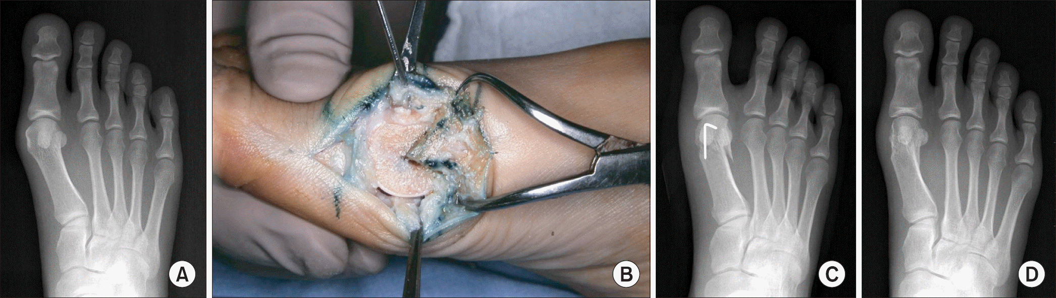 | Figure 1.(A) The radiograph of right foot shows mild hallux valgus deformity with a hallux valgus angle of 26o and the 1st intermetatarsal angle of 12o. (B) This photo demonstrates the distal chevron metatarsal osteotomy. The metatarsal head fragment was laterally displaced and fixed with a Kirschner-wire. (C) The deformity was adequately corrected with distal chevron metatarsal osteotomy. Postoperative hallux valgus angle was 8o and the 1st intermetatarsal angle was 7o. (D) The radiograph at postoperative 1 year shows that the correction of the deformity has been well maintained. |
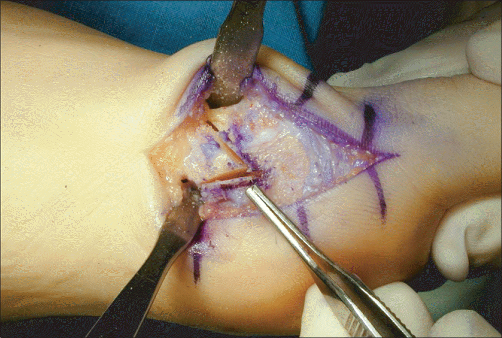 | Figure 2.This photo demonstrates the biplanar distal chevron metatarsal osteotomy with excision of medial wedge to decrease the distal metatarsal articular angle. |
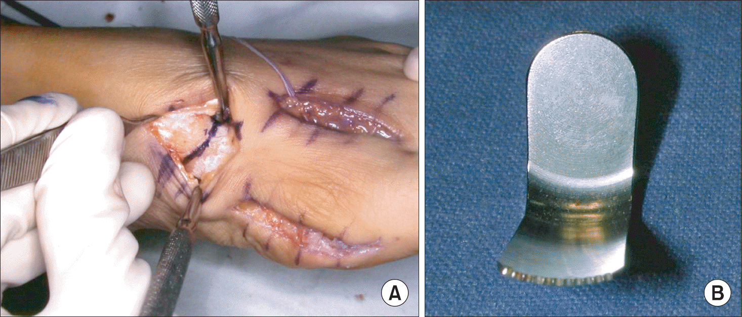 | Figure 3.(A) This photo shows the proximal crescentic metatarsal osteotomy through the dorsal approach. (B) A special saw-blade is needed for the proximal crescentic osteotomy. |
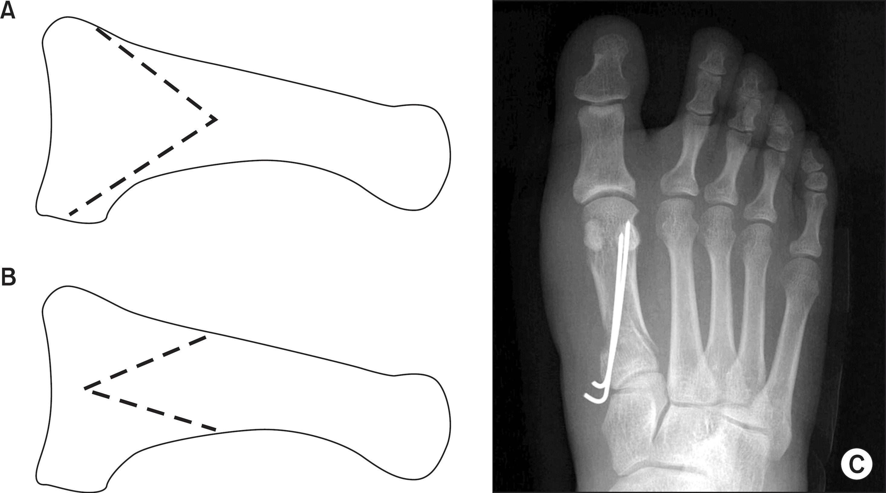 | Figure 4.These illustrations demonstrate two types of proximal chevron metatarsal osteotomy. (A) The apex of osteotomy is located 2 cm distally from the metatarso-cuneiform joint. (B) The apex of osteotomy is located proximally, and the base is located 2 to 2.5 cm distally from the metatarso-cuneiform joint. (C) Postoperative radiograph shows the correction of deformity with proximal chevron metatarsal osteotomy. |
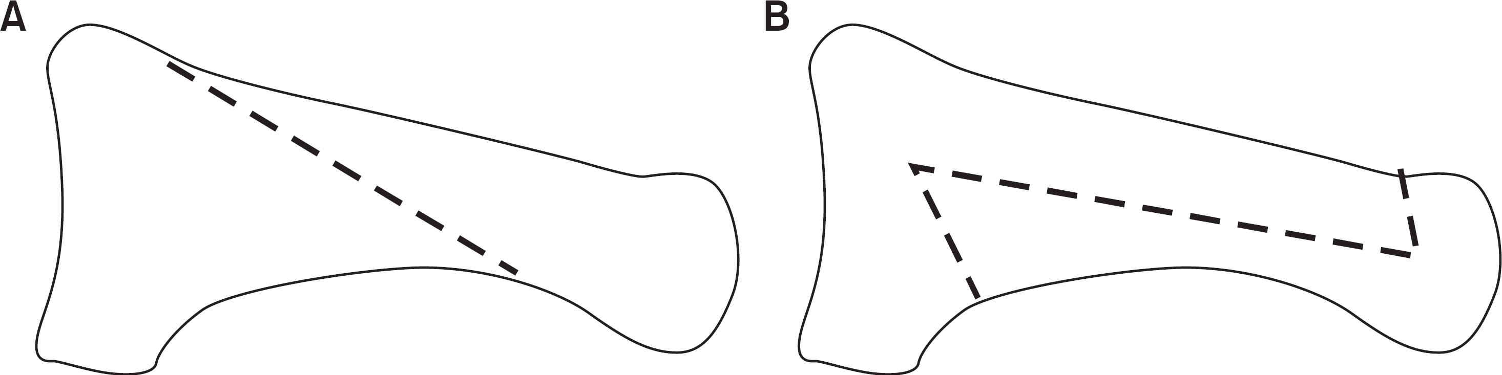 | Figure 5.These illustrations demonstrate two types of diaphyseal metatarsal osteotomy. One is the Ludloff oblique osteotomy (A), which is characterized by an oblique cut through the shaft, and the other is the scarf osteotomy (B), which consists of a transverse cut through the shaft with two chevron cuts proximally and distally. |
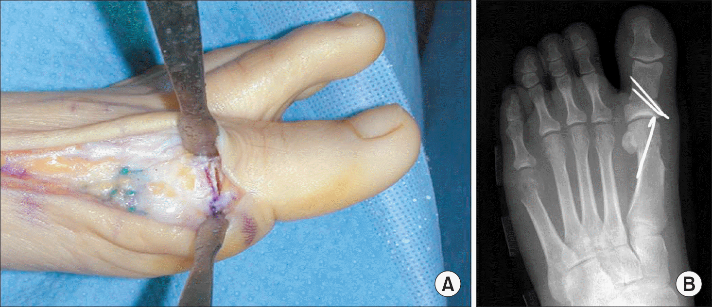 | Figure 6.(A) This photo shows an Akin phalangeal osteotomy with excision of medial wedge to decrease hallux valgus interphalangeus. (B) The Akin phalangeal osteotomy is usually combined with a distal or proximal chevron metatarsal osteotomy. |
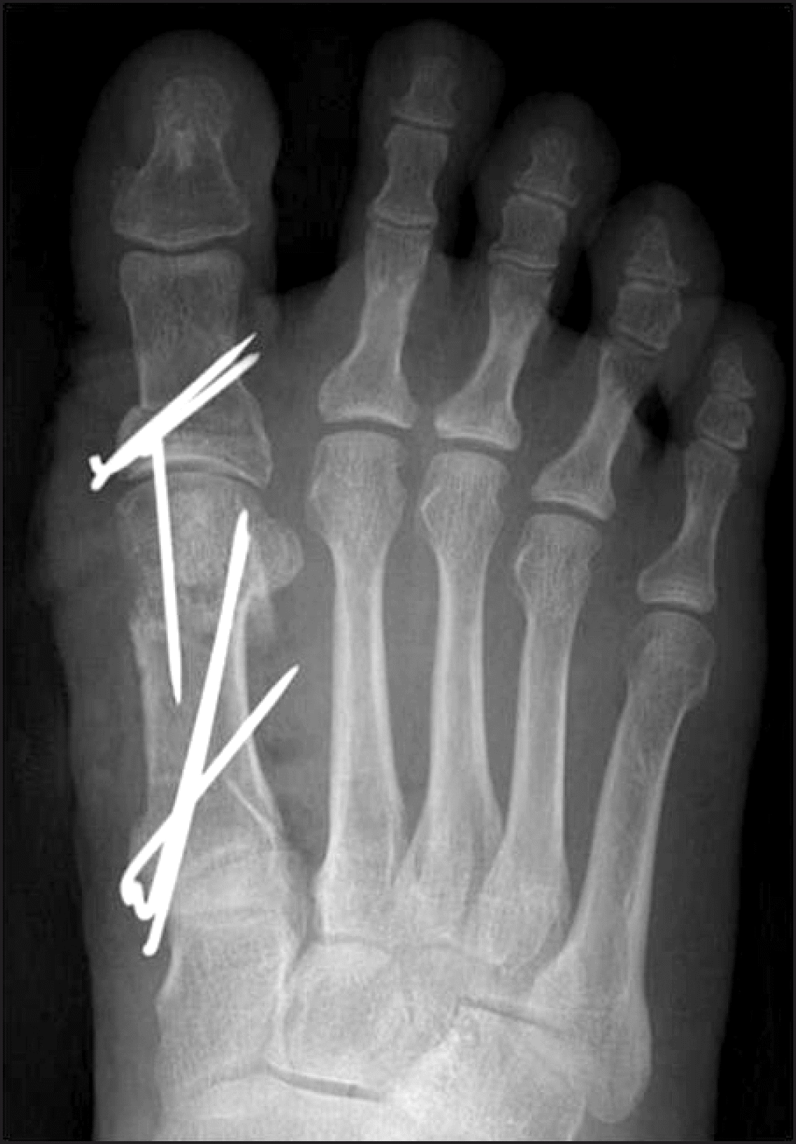 | Figure 7.This radiograph shows the correction of hallux valgus deformity with triple osteotomy, which consists of proximal chevron metatarsal osteotomy, biplanar distal chevron metatarsal osteotomy and Akin phalangeal osteotomy. |
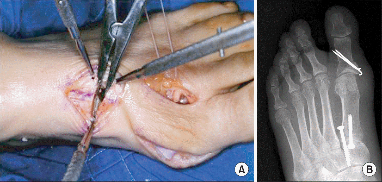 | Figure 8.(A) This photo demonstrates an excision of laterally-based wedge in the first metatarso-cuneiform joint for the correction of metatarsus varus deformity during the Lapidus procedure. (B) The radiograph at postoperative 4 weeks shows two cross screws to fix the first metatarso-cuneiform joint. The metatarsus primus varus deformity is well corrected. |
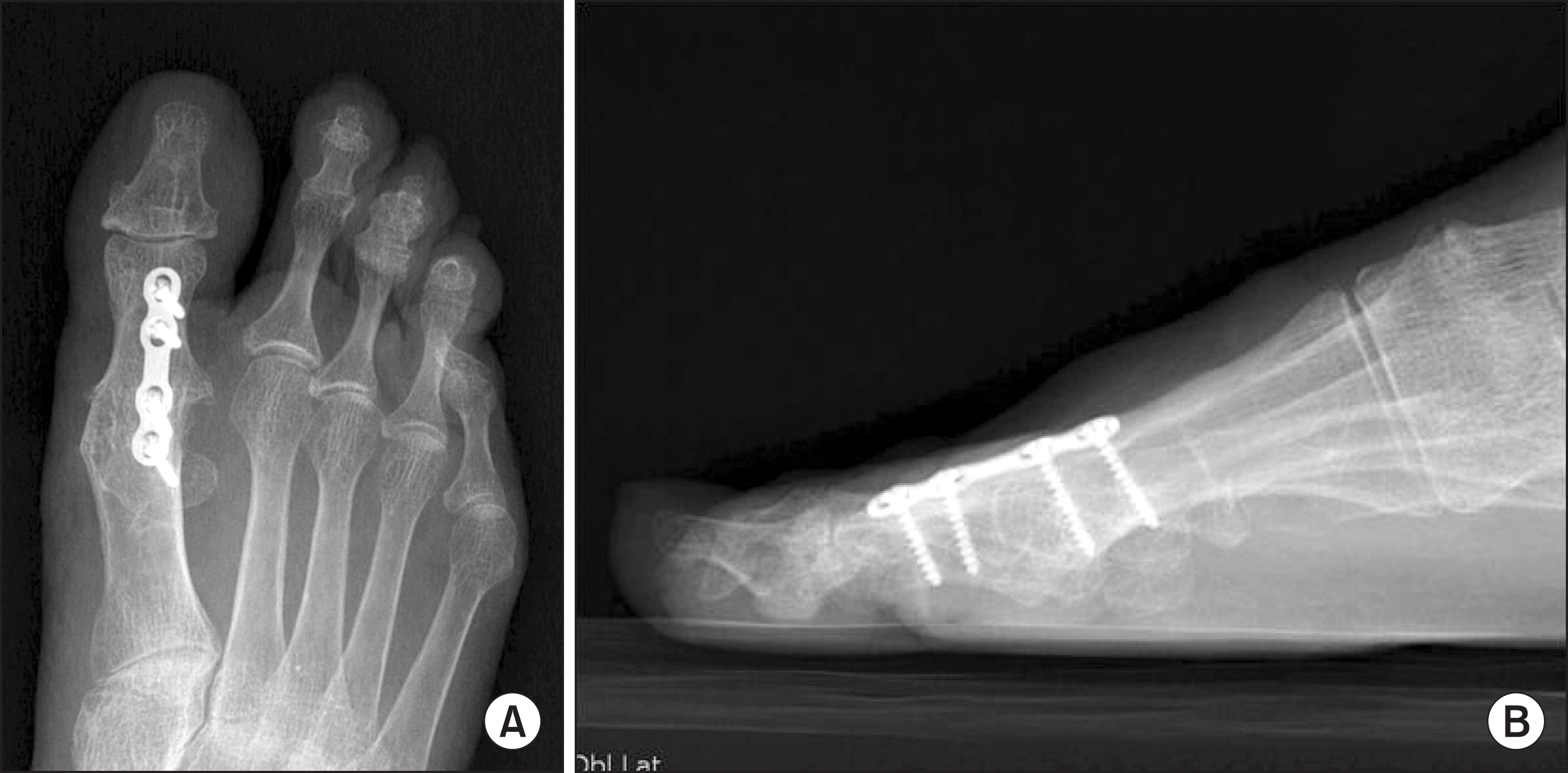 | Figure 9.(A) The right foot of a 57-year-old female with severe hallux valgus deformity was treated with a metatarsophalangeal joint fusion using a dorsal plate. (B) The sagittal angle of the first metatarsophalangeal joint should be around 20 degrees to ensure adequate function of the foot during the gait. |
 XML Download