

 PDF
PDF ePub
ePub Citation
 Print
Print


Abstract
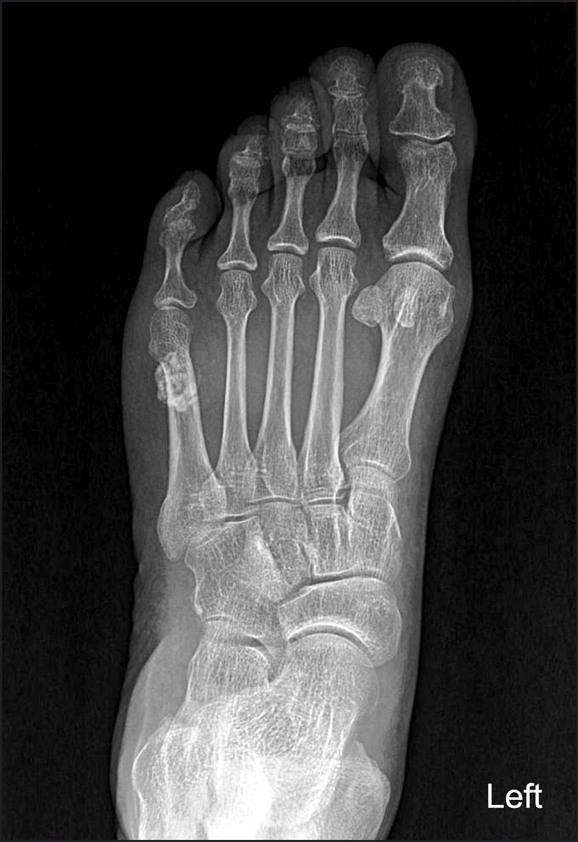
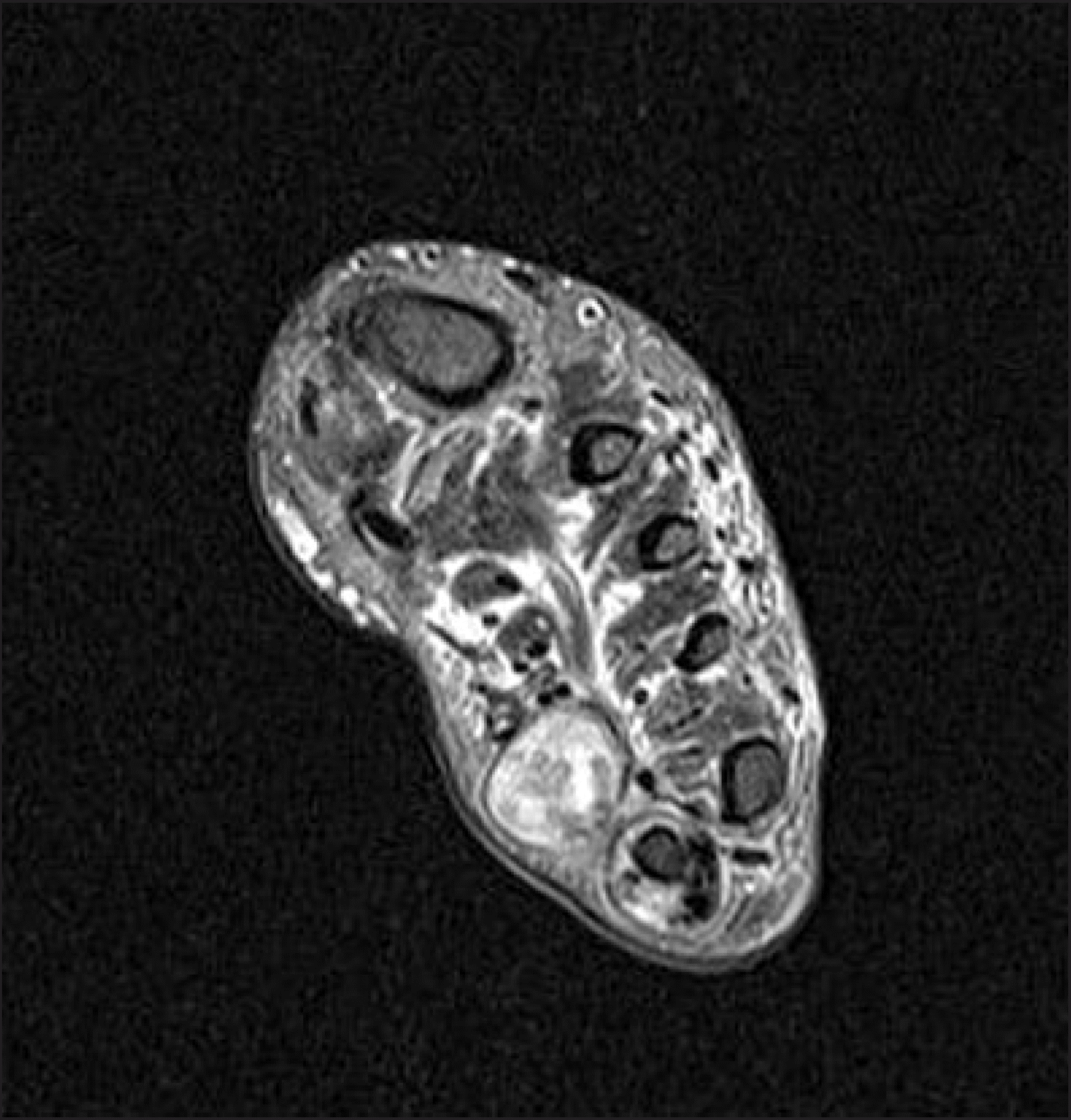
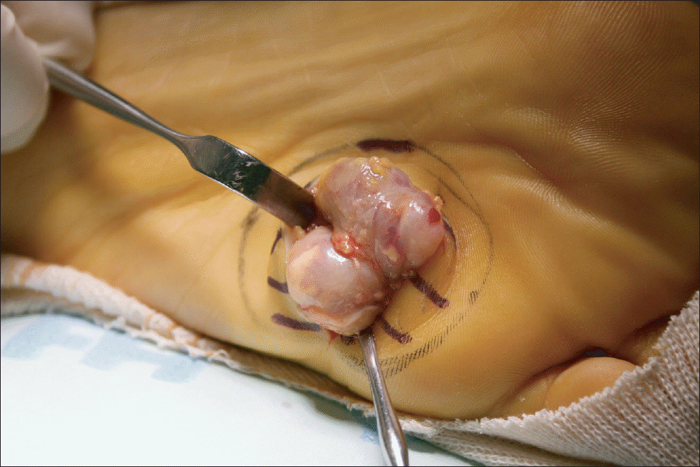
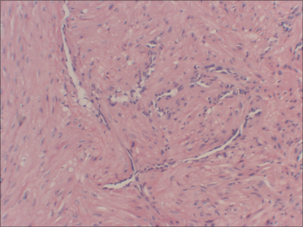
Angioleiomyomas are relatively uncommon benign tumors originating from smooth cells of a blood vessel. Although curative by surgical excision, they are rarely diagnosed definitely before surgery. We report on a case of calcified angioleiomyoma occurring on the sole, which was treated by surgical excision without recurrence and a review of literature is presented.
Go to : 
References
1. Hachisuga T, Hashimoto H, Enjoji M. Angioleiomyoma. A clinicopathologic reappraisal of 562 cases. Cancer. 1984; 54:126–30.

2. Kirby EJ, Shereff MJ, Lewis MM. Soft-tissue tumors and tumorlike lesions of the foot. An analysis of eighty-three cases. J Bone Joint Surg Am. 1989; 71:621–6.
3. Ramesh P, Annapureddy SR, Khan F, Sutaria PD. Angioleiomyoma: a clinical, pathological and radiological review. Int J Clin Pract. 2004; 58:587–91.
4. Tsoutsouris G. Vascular leiomyoma. J Foot Surg. 1982; 21:37–40.
5. Gajanthodi S, Rai R, Chaudhry RK. Vascular leiomyoma of foot. J Clin Diagn Res. 2013; 7:571–2.
6. Hwang JW, Ahn JM, Kang HS, Suh JS, Kim SM, Seo JW. Vascular leiomyoma of an extremity: MR imaging-pathology correlation. AJR Am J Roentgenol. 1998; 171:981–5.
7. Ha YC, Kim SR, Choi GM, Cho SW, Koo KH. Angioleiomyoma in the hand: a report of two cases. J Korean Orthop Assoc. 2002; 37:571–3.
8. Freedman AM, Meland NB. Angioleiomyomas of the extremities: report of a case and review of the Mayo Clinic experience. Plast Reconstr Surg. 1989; 83:328–31.
9. Seong SC, Lee JH, Lee MC, Seong IH, Kim TK. Angioleiomyoma occuring around the common peroneal nerve-1 case report-. J Korean Bone Joint Tumor Soc. 1998; 4:161–5.
10. Herren DB, Zimmermann A, Büchler U. Vascular leiomyoma in an index finger undergoing malignant transformation. J Hand Surg Br. 1995; 20:484–7.
Go to :
 XML Download