Abstract
Tarsal tunnel syndrome is defined as a compressive neuropathy of the posterior tibial nerve in the tarsal canal. Schwannoma is a benign tumor that arises from the peripheral nerve sheath. It presents as a discrete, often tender, and palpable nodule associated with neurogenic pain or paresthesia when compressed or traumatized. The growth rate is usually slow, and these lesions seldom exceed 2 cm in diameter. In addition, local recurrence occurs less than 5%. We report on a case of tarsal tunnel syndrome caused by a large recurred space-occupying lesion measuring 4.3×2.7×2.7 cm3.
References
1. Boya H, Ozcan O, Oztekin HH. Tarsal tunnel syndrome associated with a neurilemoma in posterior tibial nerve: a case report. Foot (Edinb). 2008; 18:174–7.

2. Grossman MR, Mandracchia VJ, Urbas WM, Mandracchia DM. Neurilemmoma of the posterior tibial nerve with an uncommon case presentation. J Foot Surg. 1992; 31:219–24.
3. Miranpuri S, Snook E, Vang D, Yong RM, Chagares WE. Neurilemoma of the posterior tibial nerve and tarsal tunnel syndrome. J Am Podiatr Med Assoc. 2007; 97:148–50.

4. Kwag SY, Yoon BS, Choi CS, Kim YJ. Tarsal tunnel syndrome caused by neurilemoma of the posterior tibial nerve. J Korean Orthop Assoc. 1978; 13:57–9.
5. Kwon JH, Yoon JR, Kim TS, Kim HJ. Peripheral nerve sheath tumor of the medial plantar nerve without tarsal tunnel syndrome: a case report. J Foot Ankle Surg. 2009; 48:477–82.

6. Belding RH. Neurilemoma of the lateral plantar nerve producing tarsal tunnel syndrome: a case report. Foot Ankle. 1993; 14:289–91.

7. Bailie DS, Kelikian AS. Tarsal tunnel syndrome: diagnosis, surgical technique, and functional outcome. Foot Ankle Int. 1998; 19:65–72.

8. Varma DG, Moulopoulos A, Sara AS, Leeds N, Kumar R, Kim EE, et al. MR imaging of extracranial nerve sheath tumors. J Comput Assist Tomogr. 1992; 16:448–53.

9. Choi JO, An JH, Cho KH, Park BH, Hwang MS, Lee JK, et al. US findings of neurilemmoma of the extremities: pathologic correlation. J Korean Soc Med Ultrasound. 1999; 18:221–6.
Figure 1.
The sagittal (A) and coronal (B) sonogram shows an oval shaped well-defined hypoechoic mass with inhomogeneous echo texture. (C) Color-coded Doppler scan shows flow signals in the mass.

Figure 2.
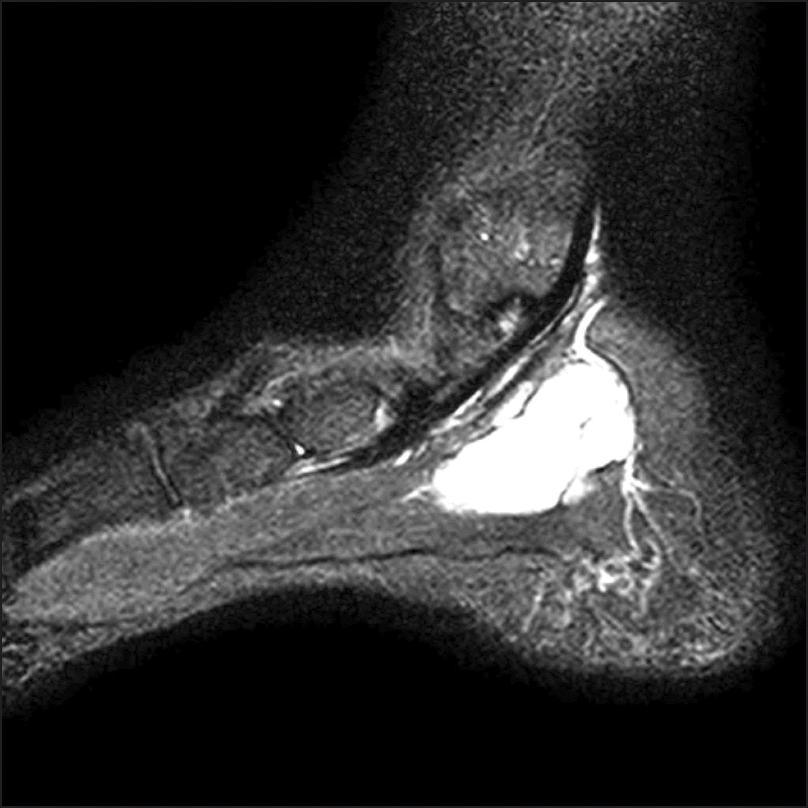
Sagittal T2-weighted magnetic resonance imaging image with high signal intensity on the total mass without target sign.


 PDF
PDF ePub
ePub Citation
 Print
Print


 XML Download