Abstract
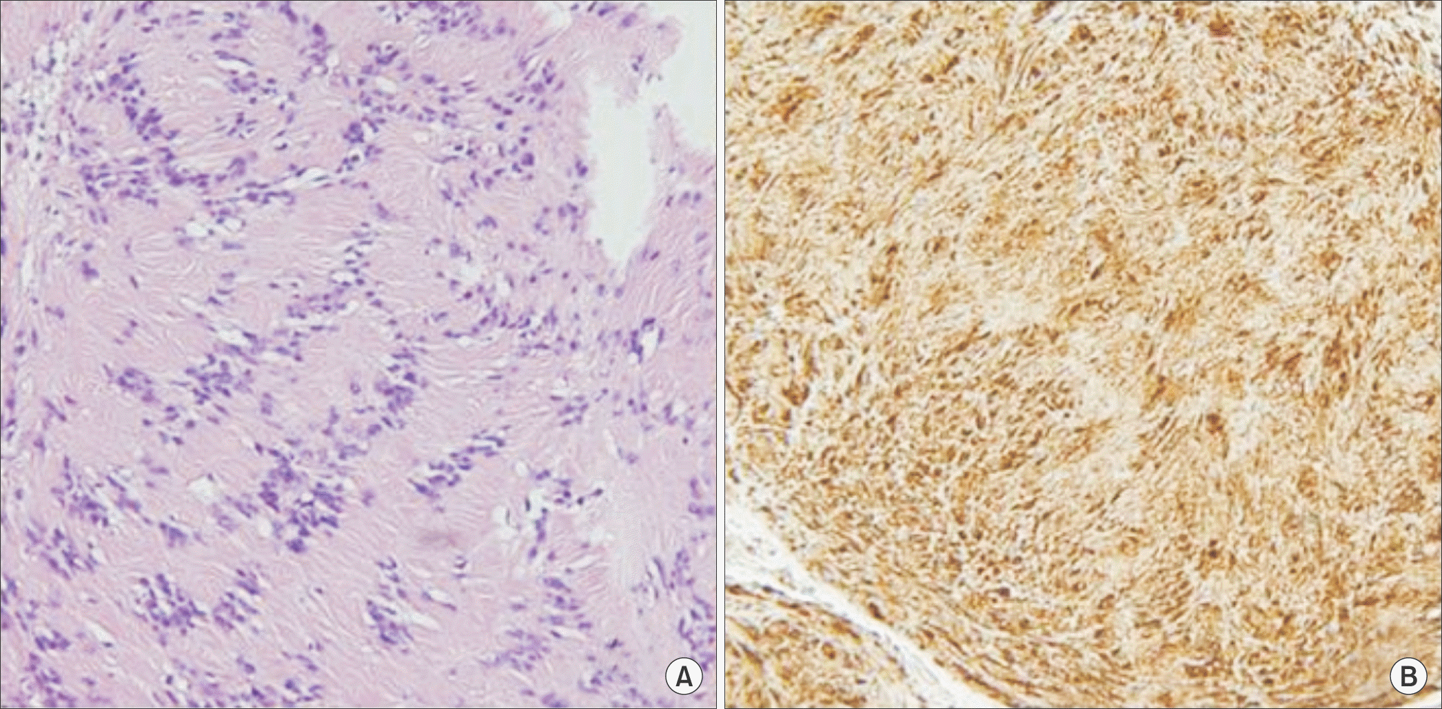
A schwannoma is a benign neurogenic tumor derived from Schwann cells. A rare case of a large painful schwannoma in the foot with metatarsal deformity was presented. Due to suspicion of malignancy, amputation had been recommended previously. We report on a rare case of a large forefoot schwannoma causing pain and paresthesia of the forefoot.
REFERENCES
1.Enzinger FM., Weiss SW. Benign tumors of peripheral nerves. Enzinger FM, Weiss SW, editors. editors.Enzinger and Weiss’s soft tissue tumors. 5th ed.St Louis: Mosby;2008. p. 853–69.
2.Weiss SW., Langloss JM., Enzinger FM. Value of S-100 protein in the diagnosis of soft tissue tumors with particular reference to benign and malignant Schwann cell tumors. Lab Invest. 1983. 49:299–308.
3.Mangrulkar VH., Brunetti VA., Gould ES., Howell N. Unusually large pedal schwannoma. J Foot Ankle Surg. 2007. 46:398–402.

4.Pyun YS., Kim SR., Joh YR. Surgical treatment of the neurilemoma in extremities. J Korean Bone Joint Tumor Soc. 1998. 4:88–93.
5.Das Gupta TK., Brasfield RD., Strong EW., Hajdu SI. Benign solitary Schwannomas (neurilemomas). Cancer. 1969. 24:355–66.
6.Odom RD., Overbeek TD., Murdoch DP., Hosch JC. Neurilemoma of the medial plantar nerve: a case report and literature review. J Foot Ankle Surg. 2001. 40:105–9.

7.Rettenbacher T., Sögner P., Springer P., Fiegl M., Hussl H., zur Ned-den D. Schwannoma of the brachial plexus: cross-sectional imaging diagnosis using CT, sonography, and MR imaging. Eur Radiol. 2003. 13:1872–5.

8.Lee SH., Jung HG., Lee HK. Neurilemoma of trunk and extremities. J Korean Orthop Assoc. 1996. 31:556–63.

9.Flores Santos F., Pinheiro M., Felicíssimo P. Large foot schwannoma with bone invasion: a case report. Foot Ankle Surg. 2014. 20:e23–6.
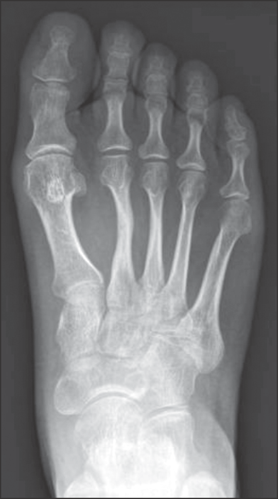
Figure 1.
Anteroposterior radiograph shows lateral angulation and sclerosis of second, third metatarsal shaft.
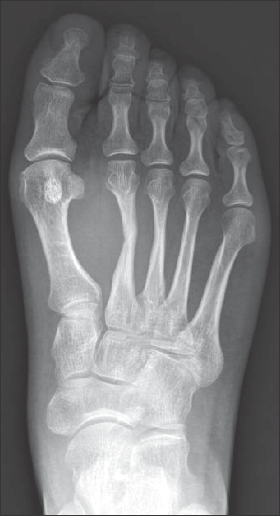
Figure 2.
The axial T2-weighted magnetic resonance image shows a heterogenous cystic mass with low to hyperdense signal intensity, with dorsal and plantar extension.
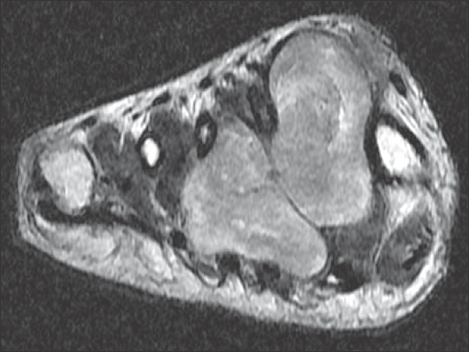
Figure 3.
(A) A excised mass was removed through plantar incision. (B) Clinical photograph after mass excision shows both a deep peroneal nerve and a common proper digital nerve derived from medial plantar nerve.
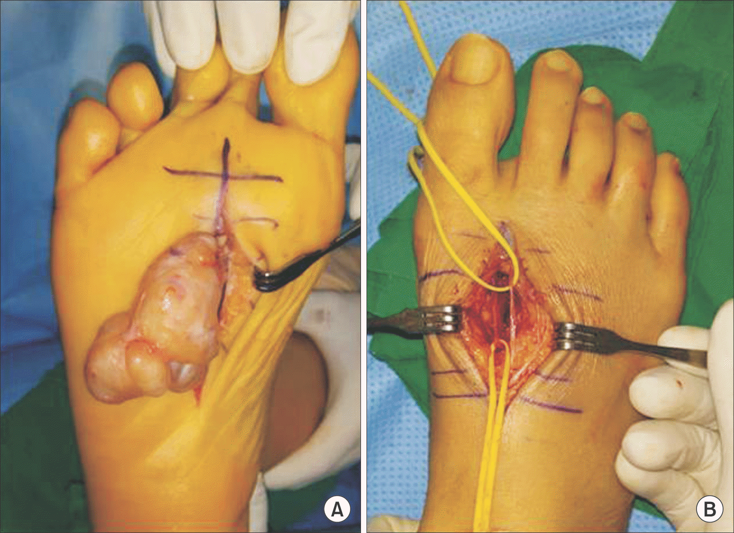
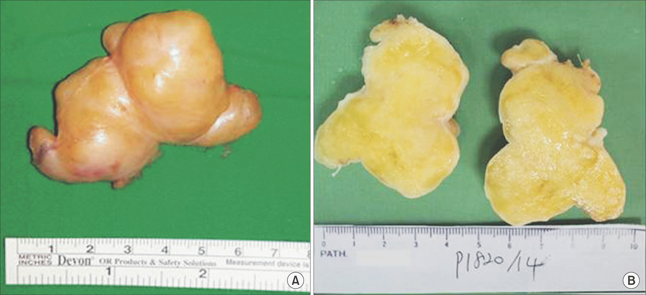
Figure 4.
(A) Gross photograph shows a well circumscribed and encapsulated mass. (B) The cross section of mass appears diffusely myxoid and grayish yellow cut surfaces without hemorrhage or necrosis.


 PDF
PDF ePub
ePub Citation
 Print
Print


 XML Download