

 PDF
PDF ePub
ePub Citation
 Print
Print


Abstract
Purpose
The purpose of this study is to evaluate the clinical and radiographic results of symptomatic bunionette treated with a diaphyseal oblique osteotomy.
Materials and Methods
We retrospectively reviewed 12 feet of nine patients diagnosed as symptomatic bunionette and treated with diaphyseal oblique osteotomy. All patients were female and the average age at the time of surgery was 48 years. We checked the foot standing anteroposterior, oblique, and lateral images pre- and post-operatively. We measured the fourth intermetatarsal angle and fifth metatarsophalangeal angle and evaluated the clinical results using the American Orthopaedic Foot and Ankle Society (AOFAS) lesser metatarsophalangeal-interphalangeal (MTP-IP) scale preoperatively and six months postoperatively.
Results
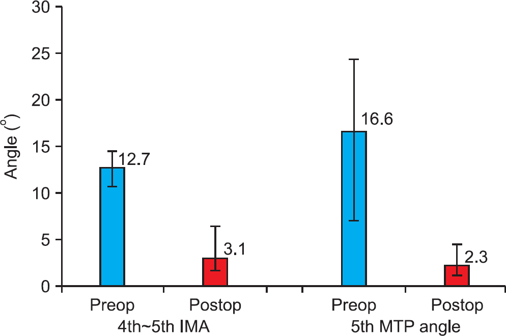
Of the nine patients, hallux valgus was combined with symptomatic bunionette in seven feet of five patients. In all of our cases, the average AOFAS lesser MTP-IP scale showed improvement after surgery. Painful callosity around the fifth metatarsophalangeal joint disappeared after surgery in all of our cases. The fourth intermetatarsal angle improved from 12.7o to 3.1o and the fifth metatarsophalangeal angle improved from 16.6o to 2.3o.
Go to : 
References
1. Buchbinder IJ. DRATO procedure for tailor's bunion. J Foot Surg. 1982; 21:177–80.
2. Diebold PF, Bejjani FJ. Basal osteotomy of the fifth metatarsal with intermetatarsal pinning: a new approach to tailor's bunion. Foot Ankle. 1987; 8:40–5.

3. Coughlin MJ. Etiology and treatment of the bunionette deformity. Instr Course Lect. 1990; 39:37–48.
4. Koti M, Maffulli N. Bunionette. J Bone Joint Surg Am. 2001; 83:1076–82.
5. Davies H. Metatarsus quintus valgus. Br Med J. 1949; 1:664.
6. Brown JE. Functional and cosmetic correction of metatarsus latus (splay foot). Clin Orthop. 1959; 14:166–70.
7. Sgarlato TE. A compendium of podiatric biomechanics. San Francisco: California College of Podiatric Medicine;1971. p. 381–96.
8. Yancey HA Jr. Congenital lateral bowing of the fifth metatarsal. Report of 2 cases and operative treatment. Clin Orthop Relat Res. 1969; 62:203–5.
9. Kitaoka HB, Holiday AD Jr. Lateral condylar resection for bunionette. Clin Orthop Relat Res. 1992; 278:183–92.
10. Steinke MS, Boll KL. Hohmann-Thomasen metatarsal osteotomy for tailor's bunion (bunionette). J Bone Joint Surg Am. 1989; 71:423–6.
11. Haber JH, Kraft J. Crescentic osteotomy for fifth metatarsal head lesions. J Foot Surg. 1980; 19:66–7.
12. Leach RE, Igou R. Metatarsal osteotomy for bunionette deformity. Clin Orthop Relat Res. 1974; 100:171–5.
13. Sponsel KH. Bunionette correction by metatarsal osteotomy: preliminary report. Orthop Clin North Am. 1976; 7:809–19.
14. Coughlin MJ. Treatment of bunionette deformity with longitudinal diaphyseal osteotomy with distal soft tissue repair. Foot Ankle. 1991; 11:195–203.
15. London BP, Stern SF, Quist MA, Lee RK, Picklesimer EK. Long oblique distal osteotomy of the fifth metatarsal for correction of tailor's bunion: a retrospective review. J Foot Ankle Surg. 2003; 42:36–42.
16. Ahn JH, Kim HY, Kang JW, Choy WS, Kim YI. Treatment of bunionette deformity with diaphyseal oblique osteotomy. J Korean Foot Ankle Soc. 2008; 12:31–5.
17. Diebold PF. Basal osteotomy of the fifth metatarsal for the bunionette. Foot Ankle. 1991; 12:74–9.
18. McKeever DC. Excision of the fifth metatarsal head. Clin Orthop Relat Res. 1959; 13:321–2.
19. Fallat LM, Buckholz J. An analysis of the tailor's bunion by radiographic and anatomical display. J Am Podiatry Assoc. 1980; 70:597–603.
20. Kitaoka HB, Alexander IJ, Adelaar RS, Nunley JA, Myerson MS, Sanders M. Clinical rating systems for the ankle-hindfoot, midfoot, hallux, and lesser toes. Foot Ankle Int. 1994; 15:349–53.
21. Schoenhaus H, Rotman S, Meshon AL. A review of normal intermetatarsal angles. J Am Podiatry Assoc. 1973; 63:88–95.
22. Steel MW 3rd, Johnson KA, DeWitz MA, Ilstrup DM. Radiographic measurements of the normal adult foot. Foot Ankle. 1980; 1:151–8.
23. Gerbert J, Sgarlato TE, Subotnick SI. Preliminary study of a closing wedge osteotomy of the fifth metatarsal for correction of a tailor's bunion deformity. J Am Podiatry Assoc. 1972; 62:212–8.
24. Nestor BJ, Kitaoka HB, Ilstrup DM, Berquist TH, Bergmann AD. Radiologic anatomy of the painful bunionette. Foot Ankle. 1990; 11:6–11.
25. Fallat LM. Pathology of the fifth ray, including the tailor's bunion deformity. Clin Podiatr Med Surg. 1990; 7:689–715.
26. Mann RA, Mann JA. The bunionette deformity. Instr Course Lect. 2004; 53:303–9.
27. Cohen BE, Nicholson CW. Bunionette deformity. J Am Acad Orthop Surg. 2007; 15:300–7.
Go to :
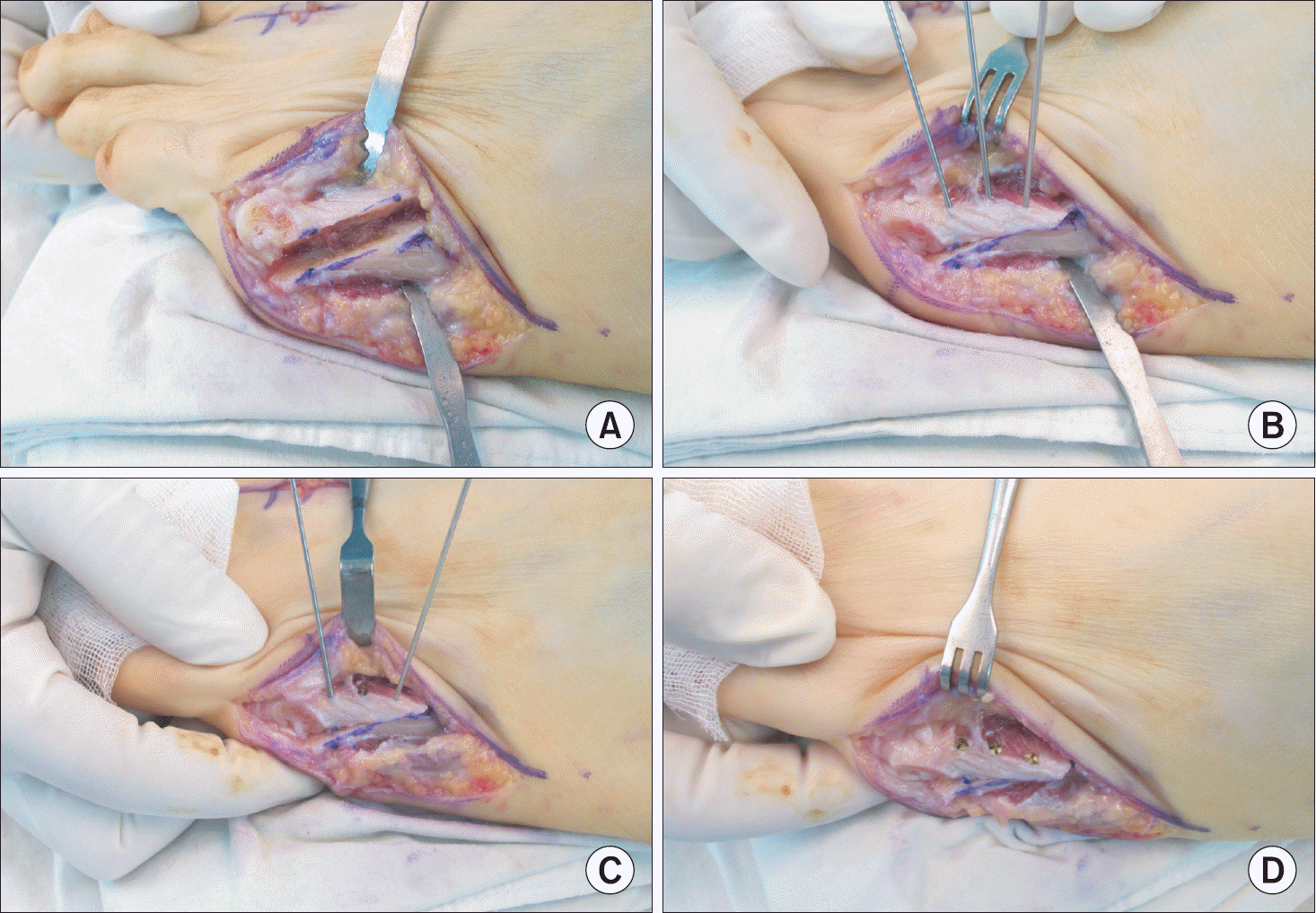 | Figure 1.Surgical techniques of diaphyseal oblique osteotomy. (A) We performed a diaphyseal oblique osteotomy on the 5th metatarsal. The direction of the osteotomy was from the dorsal side of the proximal metatarsal to the plantar side of the distal metatarsal. (B) We corrected the location of the distal fragment under the C-arm fluoroscopy and inserted three 1.1 mm Kirschner wires (K-wires). (C) We removed one K-wire and inserted a 2.3 mm mini screw at the same position one by one. (D) Three mini screws were used for the fixation of the diaphyseal oblique osteotomy of the 5th metatarsal. |
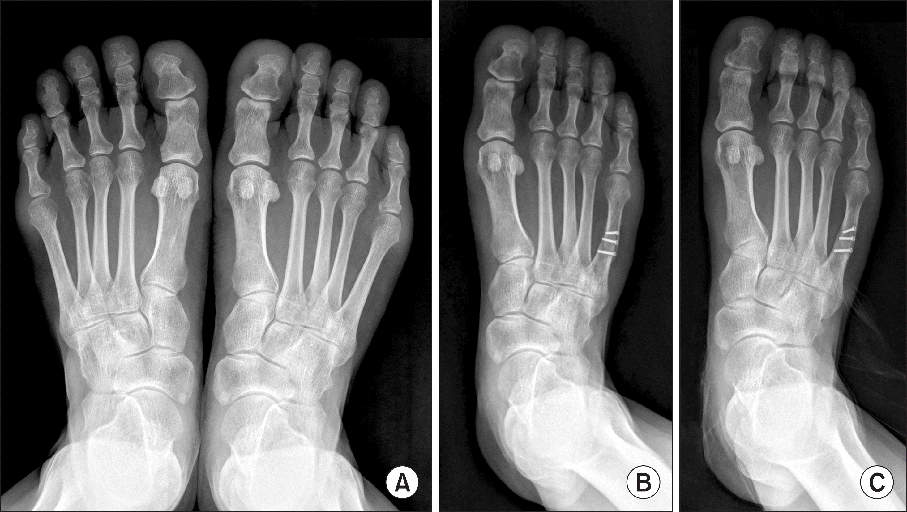 | Figure 2.A 52-year-old woman with painful plantar callosity under the 5th metatarsal head on the right side. (A) Initial both foot standing antero-posterior images show increased 4th intermetatarsal angle (13.6o) and the 5th metatarsophalangeal angle (17.6o) on the right side. (B) The bunionette deformity was corrected with diaphyseal oblique osteotomy and fixed with three screws. (C) Bony union was achieved and the correction of the bunionette deformity was well maintained 6 months postoperatively. |
 XML Download