

 PDF
PDF ePub
ePub Citation
 Print
Print


Abstract
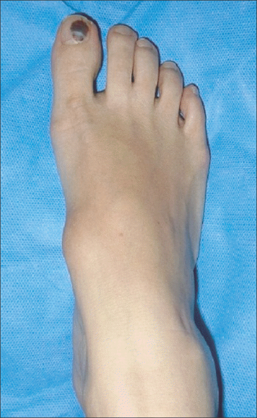
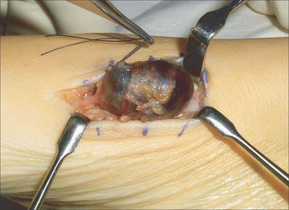
Intravascular papillary endothelial hyperplasia (IPEH) has appeared in the literature under a variety of names, including Masson’s tumor, Masson’s hemangioma, and Masson’s pseudoangiosarcoma. It is a benign lesion of the skin and subcutaneous tissue characterized by reactive proliferation of vascular endothelial cells with papillary formations. The clinical picture is not specific and the lesion resembles malignant angiosarcoma clinically and histopathologically. Therefore, it is often mistaken for angiosarcoma and a group of other benign and malignant vascular lesions. We report on a case of IPEH adherent to peripheral nerve treated with operative excision.
Go to : 
REFERENCES
1.Hashimoto H., Daimaru Y., Enjoji M. Intravascular papillary endothelial hyperplasia. A clinicopathologic study of 91 cases. Am J Dermatopathol. 1983. 5:539–46.
2.Masson P. Hemangioendotheliome vegetant intravasculaire. Bull Soc Anat (Paris). 1923. 93:517–23.
3.Fink B., Temple HT., Mizel MS. Intravascular papillary endothelial hyperplasia: a pseudotumor presenting on the plantar foot. Foot Ankle Int. 2003. 24:871–4.

4.Papagelopoulos PJ., Mavrogenis AF., Skarpidi E., Nikolaou I., Sou-cacos PN. A 56-year-old woman with a right arm mass. Clin Orthop Relat Res. 2008. 466:2892–8.
5.Miyamoto H., Nagatani T., Mohri S., Nakajima H. Intravascular papillary endothelial hyperplasia. Clin Exp Dermatol. 1988. 13:411–5.
6.Clearkin KP., Enzinger FM. Intravascular papillary endothelial hyperplasia. Arch Pathol Lab Med. 1976. 100:441–4.
7.Amérigo J., Berry CL. Intravascular papillary endothelial hyperplasia in the skin and subcutaneous tissue. Virchows Arch A Pathol Anat Histol. 1980. 387:81–90.
8.Lee SJ., Choo HJ., Park JS., Park YM., Eun CK., Hong SH, et al. Imaging findings of intravascular papillary endothelial hyperplasia presenting in extremities: correlation with pathological findings. Skeletal Radiol. 2010. 39:783–9.
9.Mestdagh H., Lecomte-Houcke M., Reyford H. Intraneural haemangioma of the posterior tibial nerve. J Bone Joint Surg Br. 1990. 72:323–4.
10.Lim R., Tay SC., Yam A. Intravascular papillary endothelial hyperplasia (Masson’s tumour) of the finger presenting as a digital nerve schwannoma. J Hand Surg Eur Vol. 2011. 36:612–3.
Go to :
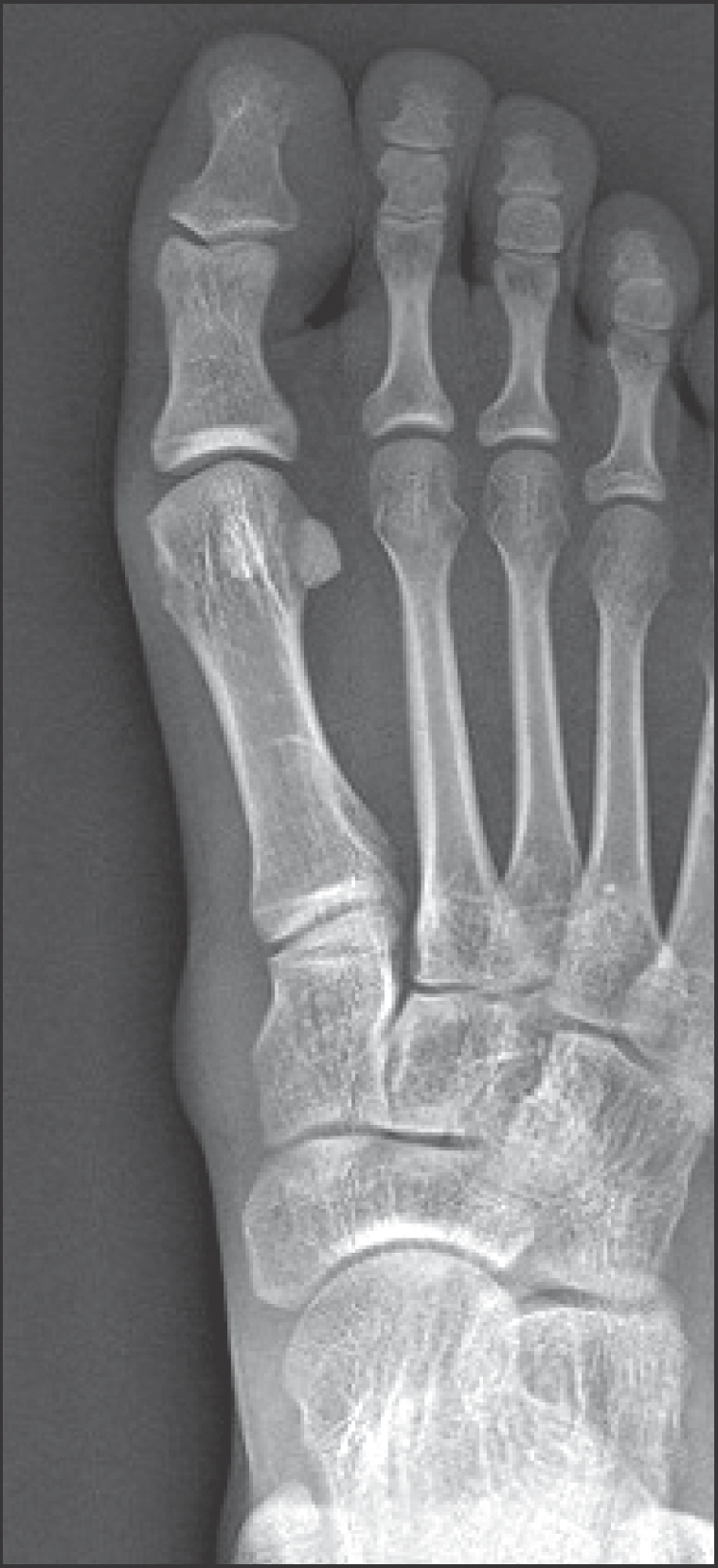 | Figure 2.Plain radiograph shows a round soft tissue lesion on medial aspect of medial cuneiform. There is no bony erosion or calcification. |
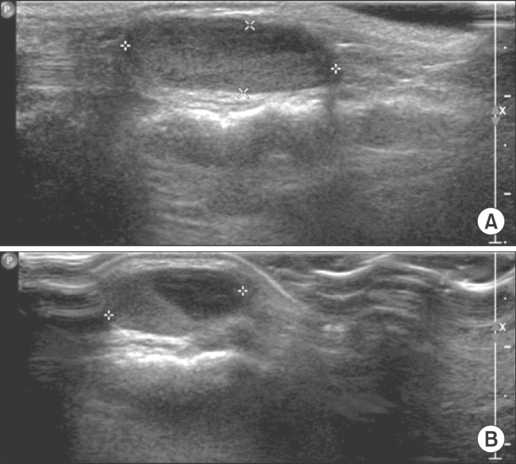 | Figure 3.Ultrasonography shows an ovoid-shape, 2.2×0.7×1.4 cm size cystic mass on dorsomedial aspect of right foot. Sagittal (A) and Axial (B). |
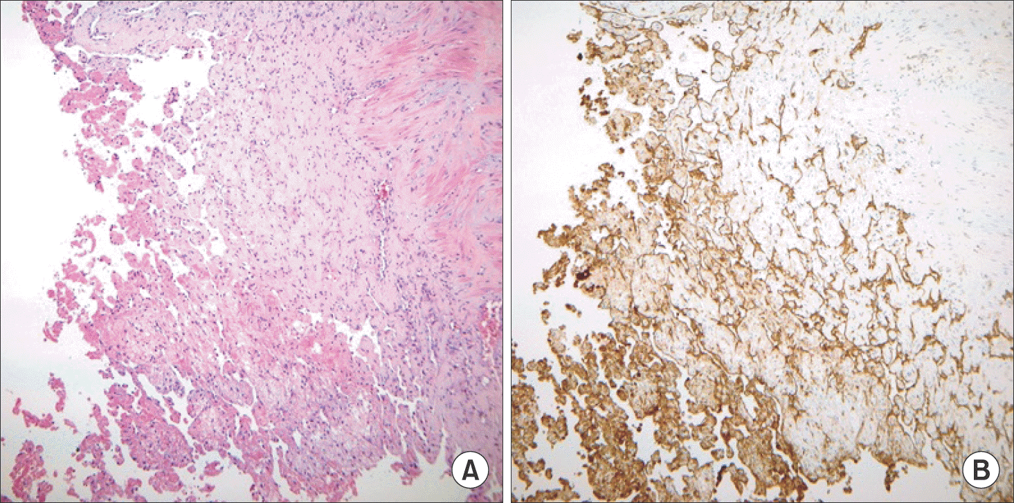 | Figure 6.Pathologic features. (A) Myriad small delicate papillae project into the vascular lumen. The papillae are composed of a single layer of vascular endothelium surrounding a stromal core (H&E stain, ×100). (B) The vascular endothelium of papillae is well demonstrated by positive (brown) immunohistochemical reaction to CD31 antibody (immunohistochemistry stain, ×100). |
 XML Download