

 PDF
PDF ePub
ePub Citation
 Print
Print


Abstract
Myopia is a major public health problem and its prevalence is increasing over time in Korea. The main treatment options of myopia correction are spectacle lenses, contact lenses, refractive surgeries such as photorefractive keratec-tomy (PRK), laser in situ keratomileusis (LASIK), and laser epithelial keratomileusis (LASEK), phakic intraocular lenses and clear lens extraction. Each treatment option has its own indications and contraindications, and not only has some advantages over the others but also some disadvantages. The evidence shows that most therapies for myopia have stable and safe results, although some do not. Customized therapy, real time tracking and sensing, and femtosecond laser-assisted surgeries might be ubiquitous in the near future. This review will discuss current treatment options for myopia and will introduce possible future therapies.
Figures and Tables
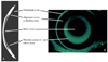 | Figure 1(A) Cross-section of reverse geometry lens. (B) Fluorescein pattern of reverse geometry lens.
|
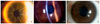 | Figure 2(A) Radial keratotomy scars on the human eye. (B) Epikeratophakia demonstrating a layer of corneal lenticule tissue over the host cornea. (C) Intrastromal corneal ring segments in the corneal stroma.
|
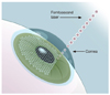
 | Figure 3(A) Photorefractive keratectomy. After removal of the corneal epithelium, the excimer laser is used to reprofile the anterior curvature of the cornea, which changes its refractive power. (B) Laser-assisted stromal in situ keratomileusis. The flap, with its intact epithelium, is folded back, and as it drapes over the modified stromal surface.
|
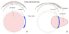
 | Figure 5Phakic intraocular lenses. (A) Verisyse (courtesy of Ophtec). (B) Visian Implantable Collamer Lens (courtesy of STAAR Surgical Co.).
|
References
1. Chung SK, Kang SB, Kim HK, Rho CR, Paik JS, Lee NY. Explanation of ophthalmic terminology. 2010. Goyang: Naewaehaksool;177.

2. Mimura T, Azar DT. Yanoff M, Duker JS, Wiggs JL, Miller D, Azar DT, Goldstein MH, Rosen ES, Rao NA, Augsburger JJ, Sadun AA, Schuman JS, Diamond GR, Dutton JJ, editors. Current concepts, classification, and history of refractive surgery. Ophthalmology. 2009. 3rd ed. [Edinburgh]: Mosby Elsevier;107–117.
3. Bennett AG. An historical review of optometric principles and techniques. Ophthalmic Physiol Opt. 1986. 6:3–21.
4. Fick AE. A contact lens: 1888. Arch Ophthalmol. 1997. 115:120–121.
5. Trokel SL, Srinivasan R, Braren B. Excimer laser surgery of the cornea. Am J Ophthalmol. 1983. 96:710–715.
6. Grosvenor T. A review and a suggested classification system for myopia on the basis of age-related prevalence and age of onset. Am J Optom Physiol Opt. 1987. 64:545–554.
7. Curtin BJ. Physiologic vs pathologic myopia: genetics vs environment. Ophthalmology. 1979. 86:681–691.
8. Coon LJ. Orthokeratology. Part I. Historical perspective. J Am Optom Assoc. 1982. 53:187–195.
9. Swarbrick HA. Orthokeratology review and update. Clin Exp Optom. 2006. 89:124–143.
10. Waring GO 3rd, Lynn MJ, McDonnell PJ. Results of the prospective evaluation of radial keratotomy (PERK) study 10 years after surgery. Arch Ophthalmol. 1994. 112:1298–1308.
11. Kaminski SL, Biowski R, Koyuncu D, Lukas JR, Grabner G. Ten-year follow-up of epikeratophakia for the correction of high myopia. Ophthalmology. 2003. 110:2147–2152.
12. Sher NA, Chen V, Bowers RA, Frantz JM, Brown DC, Eiferman R, Lane SS, Parker P, Ostrov C, Doughman D, Carpel E, Zabel R, Gothard T, Lindstrom RL. The use of the 193-nm excimer laser for myopic photorefractive keratectomy in sighted eyes. A multicenter study. Arch Ophthalmol. 1991. 109:1525–1530.
13. Ditzen K, Anschutz T, Schroder E. Photorefractive keratectomy to treat low, medium, and high myopia: a multicenter study. J Cataract Refract Surg. 1994. 20:Suppl. 234–238.
14. Shortt AJ, Allan BD. Photorefractive keratectomy (PRK) versus laser-assisted in-situ keratomileusis (LASIK) for myopia. Cochrane Database Syst Rev. 2006. (2):CD005135.
15. Azar DT, Ang RT. Laser subepithelial keratomileusis: evolution of alcohol assisted flap surface ablation. Int Ophthalmol Clin. 2002. 42:89–97.
16. Teus MA, de Benito-Llopis L, Alio JL. Mitomycin C in corneal refractive surgery. Surv Ophthalmol. 2009. 54:487–502.
17. Ratkay-Traub I, Juhasz T, Horvath C, Suarez C, Kiss K, Ferincz I, Kurtz R. Ultra-short pulse (femtosecond) laser surgery: initial use in LASIK flap creation. Ophthalmol Clin North Am. 2001. 14:347–355. viii–viix.
18. Kullman G, Pineda R 2nd. Alternative applications of the femtosecond laser in ophthalmology. Semin Ophthalmol. 2010. 25:256–264.
19. Sekundo W, Kunert KS, Blum M. Small incision corneal refractive surgery using the small incision lenticule extraction (SMILE) procedure for the correction of myopia and myopic astigmatism: results of a 6 month prospective study. Br J Ophthalmol. 2011. 95:335–339.
 XML Download