

 PDF
PDF ePub
ePub Citation
 Print
Print


Introduction
Members of the family Birnaviridae have 2-segmented genomes - A and B. This family comprises 3 main genera, including the genus Aquabirnavirus, Avibirnavirus, and Entomobirnavirus [4,19]. The type species of the genus Aquabirnavirus is the infectious pancreatic necrosis virus (IPNV); the genus comprises marine aquatic birnaviruses (MABV) of fish and shellfish [3]. Other members of the family Birnaviridae include infectious bursal disease virus (IBDV) belongs to the genus Avibirnavirus, and Drosophila X virus (DVX) that belongs to the genus Entomobirnavirus. Aquatic birnaviruses are the largest and most diverse group of viruses within the family Birnaviridae. The first reported MABV was isolated from the yellowtail Seriola quinqueradiatain Japan [22], other MABVs have been subsequently isolated from various marine fishes in Korea and Japan, and their characteristics have been investigated [7,8,14,18,23,24]. The genome segment B of birnaviruses encodes the VP1 protein, which is the presumptive virion-associated RNA-dependent RNA polymerase (RdRp) [13,15]. Some researchers reported the characteristics of VP1 and compared the VP1 region among birnaviruses [4,25]. They identified several conserved domains associated with RdRps and GTP-binding proteinsin the IPNV strains; these domains were the same as those in other RNA viruses. However, they also discovered that the typical Gly-Asp-Asp (GDD) motif that is found in all RNA viruses was absent in the VP1 region of some IPNV [4] IBDV, and DXV [2] strains.
The physical, antigenic, and genetic features of the VP2/NS junction region of the aquatic birnavirus GC1 isolated from the rockfish Sebastes (S.) schlegeli, which is the second most important in the aquaculture industry in Korea, has been studied [8,9,20].
In the present study, we investigated the genetic characteristics of the VP1 protein and compared the genetic relationship between aquatic birnaviruses and other genuses within family Birnaviridae.
Materials and Methods
Virus and cell
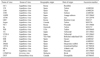
GC1 was isolated from the rockfish S. schlegeli, and it was grown in the Chinook Salmon Embryo-214 cell line supplemented with Eagle's minimum essential medium. The sources of VP1 sequence cited in this study are listed in Table 1.
Viral RNA extraction and primers
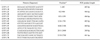
The viral genomic RNA was extracted using the methods described by Joh et al. [8]. Briefly, GC1-infected cells were frozen and thawed 3 times and clarified by centrifugation. Viral dsRNA was then extracted with phenol and chloroform, followed by digestion with proteinase K. Seven primer pairs were used for reverse transcription-polymerase chain reaction (RT-PCR). The oligonucleotide sequences were deduced according to the published dsRNA sequences of the Western Buxton strains (AF078669) (Table 2).
cDNA synthesis by RT-PCR
The RT-PCR procedure used in this study was a modification of the method previously described by Joh et al. [10]. The RT-PCR solution was heated to 95℃ for 3 min and passed through 35 cycles under the following conditions: 1 min at 95℃ for denaturation; 1 min at 54℃ to -58℃ (depending on the primer) to allow annealing; 1 min at 72℃ for extension and final amplification at 72℃ for 3 min. The ethidium bromide-stained PCR products were electrophoresed on a 1.5% agarose gel and were visualized by UV fluorescence.
Construction of recombinant plasmids
Each resulting product was gel purified and then cloned into pCR2.1 TA cloning vectors (Invitrogen, USA) according to the manufacturer's instructions. All the clones were amplified by transformation into competent DH5 α cells. Clones with correct inserts were confirmed by restriction enzyme digestion of the recombinant vectors.
Nucleotide sequencing and analysis of the VP1
Nucleotide sequencing was carried out on an ABI 377 sequencer (Applied Biosystems, USA) by the dideoxynucleotide chain termination method by using T7 DNA and SP6 DNA polymerase. The nucleotide and deduced amino acid sequences were analyzedby Vector NTI ver 10.0 (Hitachi, Japan) and were compared with the corresponding sequences of previously reported cite accession numbers of aquabirnaviruses in Table 1.
Results
Nucleotide and amino acid sequences of the VP1 protein
The nucleotide sequence of GC1 was found to be 2,776 bp long. The VP1 open reading frame (ORF) gene starts at nucleotide 101 and ends with a single TAA termination codon at nucleotide 2,638. The predicted molecular weight of this virus is 94,263 Daltons, and it contains a single large ORF encoding the 846-amino acid VP1 protein. The VP1 sequence starts with the nucleotide sequence 'GGAAA' and contains the inverted terminal repeats 'GGGTCAAGTTGGTGG' and 'GTGCCACCAAC-TGACCC' near the 5' and 3' terminal sequences, respectively.
Characterization of the VP1 protein
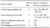
The amino acid composition of VP1 was determined. The VP1 amino acid sequence was scanned for several functional motifs, and the results are summarized in Table 3. We observed that the VP1 sequence contained 6 potential Asn-X-Ser/Thr motifs. These motifs were presumed to contain an N-linked glycosylation site. There were 8 potential Ser phosphorylation sitesand 1 Tyr phophorylation site. The amino acid sequence of VP1 did not contain the GDD motif, which exists commonly in the RdRps of RNA viruses; however, we could identifythe Leu-Lys-Asn (LKN) motif at position 521 (Table 3). Further, we confirmed the 'GLPYIGKT'motif at amino acid position 248; this motif is the putative GTP-binding site that is commonly found in other aquatic birnaviruses.
Comparative studies of nucleotide and amino acid sequences of the VP1 protein
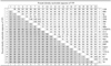
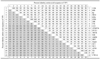
On comparing the nucleotide sequences of VP1 in 22 birnavirus strains, it was found that GC1 shares 97-98% homology with MABVs; 86% homology with the IPNV strains of aquabirnaviruses isolated mainly from the USA, Japan, and Korea; 80-82% homology with the IPNV strains of aquabirnaviruses from Spain; 54-56% homology with the avibirnaviruses; and 46% homology with entomobirnaviruses (Table 4). On comparing the amino acid sequence of VP1, it was found that GC1 shares 97-98% homology with MABVs; 94% homology with the IPNV strains of aquabirnaviruses found mainly in the USA, Japan, and Korea; 87-89% homology with the IPNV strains of aquabirnaviruses from Spain; 46-47% homology with the avibirnaviruses; and 29% homology with the entomobirnaviruses (Table 5).
Phylogenetic relationships
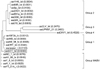
In the phylogenetic cladograms that were based on both nucleotide and amino acid sequences, the genetic relationships among the 22 birnaviruses were established and the viruses, including GC1, were clustered into 5 genogroups that generally correlated with the geographic origin of the viruses and the water environment of the host. The MABV genogroup consisted of strains such as GC1 and NC1 from Korea and YT01A, H1, AY98, and Y6 from Japan. Genogroup 1 mostly consisted of strains from the Pacific coastal nations; DRT is from Korea, WB from the USA, Jasper from Canada, and AM98 from Japan. The isolates of 1146, 88R, 20G1, 2290, and 6B1A from Spain and Sp from Denmark comprised genogroup 2. The 2 avibirnaviruses UPM976/61 from Malaysia and CLV from Vietnamformed genogroup 3, and 1 entomobirnavirus, DVX, formed genogroup 4 (Fig. 1).
Discussion
The viral B segment encodes VP1, which is approximately 90 kDa in weight [2,11-13]. The estimated molecular weight of VP1 ranges from 95 kDa for the Jasper isolate [4] to 89 kDa for the Sp and Ab isolates of IPNV [6]. The molecular weight of GC1 has been estimated as 94 kDa and has been shown to be similar to that of the Jasper strain.
Some researchers have reported that the sequence GXXXXGKS/T is a constant motif in GTP-binding proteins [1,16] and is observed in several viral proteins that have a tentative role in RNA replication [15]. The same motif was present in the IPNV strains [4] and in GC1 between residues 248 and 255 (GLPYIGKT). We believe that this motif represents a potential GTP-binding site in the VP1 protein, and has been conserved in GC1, including aquatic birnaviruses.
As reported previously [1,5,17], the GDD sequence is a highly conserved motif that is present in almost all putative RdRps. Researchers have found that the Asp-Asp (DD) sequence lacking Gly, is conserved in IBDV, and also that IPNV does not contain the typical GDD motif in the corresponding region of its VP1 [4,21]. Some IPNV strains containedthe Leu-Lys-Asp (LKD) or LKN motifs instead of the typical GDD motif [4]. GC1 contains the LKN motif instead of the typical GDD motif, which is present in other aquatic birnaviruses.
The study of genetic relationships using a phylogenetic cladogram revealed that GC1 is more closely related to genogroup 1 than genogroup 2. This result indicatesthat genetic relationships may be influenced by the geographical distributions of the isolates. Aquatic birnaviruses, including GC1 and IPNV, also belong to genogroups that are distinct from those of the avibirnaviruses and Entomo-birnaviruses. This result may thus indicate that the genus Birnavirus has evolved in different ways resulting in the formation of distinct genogroups.
 XML Download