

 PDF
PDF ePub
ePub Citation
 Print
Print


Introduction
Over the past years, development and characterization of mAbs developed against leukocyte differentiation molecules (LDM) in humans has been facilitated by the convening of international workshops to compare the reactivity of mAbs developed in different laboratories [66]. Similar workshops have been convened for characterization of mAbs to LDM in ruminants [29,30,46], pigs [23,38,52,55], horses [33,36], and dogs [8]. However, progress has been much slower owing to limited number of laboratories participating in the workshops and the smaller number of mAbs submitted for analysis. In effort to accelerate identification of important mAbs, investigators have explored the possibility that many of the well characterized mAbs to human LDM might recognize epitopes conserved on orthologous LDM in other species. Although some useful cross reactive mAbs have been identified [56-58], recent results from analysis of a large set of anti-human LDM mAbs submitted to the Animal Homologues Section of the eighth human LDM workshop [54] and results reported in the ruminant and pig workshops [29,30,46,56-58] have shown the probability of finding a mAb that recognizes an epitope conserved on orthologous LDM is greater between closely related species than between distantly related species [4] for example, between cattle, bison, water buffalo, Cape buffalo, goats, sheep, and camelids [28,44,45,47,61]. The most successful approach for identifying mAbs to LDM in the species of interest has remained a focused effort on developing mAbs to LDM in that species, taking advantage of cross reactive mAbs whenever they are found to facilitate characterization of new mAbs [14].
The rabbit is an example of a species where there is a critical need for mAb reagents (NCBI Rabbit Genome Resources, USA). To date, however, only a few mAbs have been developed to meet this need. Efforts to expand the available sets of mAbs with cross reactive mAbs generated against LDM in other species has only yielded a few mAbs. The mAbs found in our sets of mAbs (this report) and mAbs submitted to the Animal Homologues Section of the HLDA8 have been specific for major histocompatibility (MHC) I and II molecules, CD7, CD9, CD14, CD21, CD11a, CD18, CD44, CD45RB, CD49d, CD209 [54]. In light of these findings, it is apparent that a more direct approach will be required to identify mAbs for research in rabbits. As part of our continued effort to develop mAbs critical to our research efforts in ruminants, we have developed a flow cytometric approach for initial identification and characterization of mAbs to LDM [11]. Previous studies have shown that two parameter single fluorescence flow cytometry can be used to cluster mAb that recognize the same or different epitopes on the same LDM, based on the pattern of expression of the molecule on one or more lineages of leukocytes [11,16,35]. Comparative studies have shown this method can also be used to identify and tentatively cluster mAbs that recognize epitopes on orthologous LDM based on the similarity of the pattern of expression of the LDM on leukocytes in different species. Our studies have revealed the pattern of expression of many orthologous LDM has been conserved cross species. This observation has proven useful, especially in the characterization of mAbs specific for LDM in less well studied species [13-15,59,60]. It has also proven useful in determining whether mAbs that cross react with LDM in one or more species recognize an epitope conserved on bona fide LDM orthologues. Specificity has also been documented by cloning and expression of LDM initially identified with cross reactive mAbs [59]. To aid others as well as ourselves, we have also developed a web based program, the Taxonomic Key Program (TKP; College of Veterinary Medicine, Washington State University, USA), to facilitate characterization of mAbs generated against LDM in less well studied species. The program contains a searchable database on known CD molecules and a database containing a catalog of mAbs known to react with LDM in one or more of the less well studied species. In the present report we summarize the results we have obtained thus far, in our efforts to develop mAbs for use in immunological investigations in the rabbit. Information on the mAbs recognizing rabbit LDMs are listed under reactivity of antibodies in the TKP program (NCBI Rabbit Genome Resources, USA).
Materials and Methods
Animals
Rabbits being used in other studies were used as a source of blood and tissues. They were housed and maintained according to the Institutional Animal Care and Use committee guidelines and Association for Assessment and Accreditation of Laboratory Animal Care (USA). Both male and female rabbits were used since initial studies did not reveal any apparent differences in the frequency of leukocyte subsets. The age of the rabbits varied from six months to about two years.
Preparation of leukocytes for flow cytometry
Because of the tendency for T lymphocytes to bind to erythrocytes, separation medium could not be used to isolate leukocytes. Whole blood, collected in anti-coagulant citrate-dextrose (ACD), was used with a fix-lyse solution to obtain leukocytes for analysis. For single color flow cytometry (FC), 50 µl of blood was distributed in conical bottom 96-well microtiter plates (Corning, USA) containing 50 µl of optimally diluted mouse mAbs and then incubated for 15 min on ice. Following centrifugation, the supernatants were removed by aspiration. The lymphocytes were subjected to 3 cycles of centrifugation and washing in FC first wash buffer (FWB, PBS co ntaining 20% ACD and 0.5% horse serum) and then incubated with a second step fluorescein conjugated polyclonal goat anti-mouse IgG/IgM second step reagent (Caltag Laboratories, USA) for an additional 15 min. Following 2 cycles of centrifugation and washing in FC second wash buffer (PBS-20% ACD) the lymphocytes were resuspended in FACS lysing solution (Becton Dickinson, USA) to lyse erythrocytes. The lymphocytes were then centrifuged and resuspended in 2% PBS-buffered formaldehyde and kept in the refrigerator until examined. For multi-color FC, blood was distributed in microtiter plates containing 2 or 3 mAbs and incubated as described. Following centrifugation and 3 cycles of washing, the lymphocytes were incubated with second step reagents. For most of the studies, combinations of mAbs of different isotype were used with isotype specific goat anti-mouse immunoglobulins conjugated with fluorescein (FL), phycoerythrin (PE), PE-Cy5, or Cy5 (Caltag Laboratories, USA). Where the mAbs of interest were the same isotype, Zenon Fab fragments of goat isotype specific anti-mouse antibody, conjugated with different fluorochromes, were used according to the manufacturers' instructions (Invitrogen, USA). One µg of each mAb in 20 µl of FWB were incubated separately with 5 µl of Zenon-Fab reagent conjugated with different fluorochromes (FL, PE, PE-Cy5, or Cy5) for 5 min at room temperature as recommended by the manufacturers. The mixtures were then incubated with 5 µl of blocking reagent (mouse immunoglobulin) for an additional 5 min. The labeled antibodies were then added to the lymphocyte preparations under study. Following 15 min of incubation on ice, the lymphocytes were processed as described and fixed in 2% buffered formaldehyde.
Peripheral blood mononuclear lymphocytes (PBMC) and spleen lymphocytes stimulated with concanavalin A (ConA) were used for immunization and identification of mAbs that recognize molecules upregulated on activated lymphocytes (rabbit activation molecules, RACT). To simplify initial screening of supernatants from primary cultures of hybridomas for the presence of a mAb that recognizes a RACT, spleen lymphocytes stimulated with ConA (5 µg/ml) for 24 to 48 h were incubated with hydroethidine (250 µg/ml in tissue culture medium), a vital dye that is selectively taken up by live cells. Hydroethidine (Polysciences, USA) intercalates into DNA similar to propidium iodide. It is excited at 488 nm and emits at high wave lengths (580 nm and higher). Following 8 min incubation at 37℃, the cells were subjected to 2 cycles of washing by centrifugation and re-suspension in medium and then added to an equivalent concentration of unstimulated cells. The mixed populations of cells were then incubated with tissue culture supernatants on ice as described and prepared for FC. Screening was performed with live cells immediately after labeling.
For further analysis of the pattern of expression of mAb-defined LDM, cells were obtained from thymus, spleen, and appendix at the time of necropsy. Cells from the respective tissues were isolated by mincing the tissues with a scissors and then passing the tissue preparation through a 100 mesh stainless steel sieve and suspended in PBS. Cells were used immediately or cryopreserved for later use. For cryopreservation, 107 to 108 cells were resuspended in bovine calf serum containing 10% DMSO and kept in a liquid nitrogen freezer.
Development of mAbs to rabbit LDM
Five fusions were made with groups of 5 mice hyperimmunized with thymus (RT and RTH), ConA stimulated spleen cells (ISC), resting and ConA stimulated spleen and PBMC (MRB), or ConA stimulated PBMC (RACT) as previously described [22]. The general protocol was to immunize mice 5 times subcutaneously with ~5 × 10⁶ cells per mouse. Seventy two hours before fusion, mice were injected i.v. through the tail vein with approximately 3 × 10⁶ cells. After 72 h, spleen cells were harvested and pooled. 108 lymphocytes were fused with 4 × 107 X63 myeloma cells as previously described [22] and then distributed into ten 96 well culture plates. The rest of the lymphocytes were cryopreserved for use in additional fusions. At 8 days, supernatants were collected and screened by FC for the presence of antibody, using blood or unstimulated and ConA stimulated spleen cells as described above.
Supernatants from primary cultures of hybridomas were screened for the presence of mAb specific for LDM using FC with whole blood. Positive cultures were expanded in 12 well culture plates. Supernatants were collected for further analysis and the cells cryopreserved. Since there was limited information on the pattern of reactivity of known LDM expressed on rabbit leukocytes, all hybridomas producing mAb were cryopreserved. This included hybridomas identified in screening experiments where hydroethidine was used to identify hybridomas producing mAbs to activation molecules.
Antibodies
Cross reactive and new mAbs developed in our laboratory are shown in Table 1. mAbs specific for CD4 (Ken4) [31], CD11b (mAb 198) [65], CD11c (mAb 3/22, no longer listed by AbD Serotec [NC]), CD45 (mAb L12/201) [65], CD58 (VC21) [64] were purchased from AbD Serotec (USA). A mAb thought to react with rabbit CD5 (Ken5; BioSource, USA) [31]. CD8 (12.C7) [18] was purchased from Abcam (USA). mAbs specific for CD11a (Ken11) [31] and CD25 (Kei-α1) [32] were purchased from BD Pharmingen (USA). Fluorescein conjugated anti-rabbit Ig was purchased from Zymed (USA).
Clustering and Characterization of mAbs
All hybridomas producing mAbs to LDM expressed on lymphocytes or granulocytes were cloned. Hybridomas producing mAbs to LDM expressed on multiple lineages of leukocytes were first clustered based on the unique patterns of expression of the molecule on leukocytes, as detected by 2 parameter FC (SSC vs fluorescence). Two or three hybridomas were selected from each distinct cluster for cloning and further analysis. Hybridomas producing mAbs that yielded profiles similar to MHC class I and II molecules were set aside for later analysis. For further characterization, FC dot plot profiles of whole blood preparations of leukocytes labeled with new mAbs were compared to each other and with profiles obtained with the cross reactive mAbs or commercially available mAbs specific for rabbit LDM. Two color FC analysis was performed to determine whether mAbs in a cluster recognized the same or different molecules. Two mAbs were considered to recognize the same molecule if one of the mAb blocked labeling by the other or if the mAbs being compared yielded a diagonal pattern of labeling [11,34,35]. Pairs of mAbs yielding a diffuse pattern of labeling were considered to recognize different molecules on the same population of lymphocytes.
Results
Identification of cross-reactive mAbs that recognize conserved epitopes expressed on orthologous LDM in rabbits
At the initiation of the study, we screened sets of mAbs we developed against LDM in cattle, goats, sheep, horses, pigs, cats, and dogs for mAb that cross reacted with rabbit LDM. We also screened additional sets of mAbs we developed during the course of the study for cross reactivity. Several strategies were used to increase the potential of generating mAbs that react with conserved determinants. These included hyperimmunization with leukocytes from multiple species and then selecting a single species to screen supernatants from primary cultures of freshly prepared hybridomas, hyperimmunization with leukocytes from a single species and screening for mAbs reactive with leukocytes from another species of interest, hyperimmunizing with leukocytes from a single species and screening for all mAbs that reacted with LDM from the same species and then screening for cross reactivity with LDM in other species. Although not used extensively for identification of cross reactive mAbs, simultaneous examination of primary cultures for mAb that recognized epitopes conserved on LDM in two species, using hydroethidine to mark one set of cells, showed that cross reactive mAbs could be identified directly. Cross reactive mAbs to bovine, caprine, and ovine CD4, CD8, CD45R, and CD45R0 were identified by this method [11,28]. Regardless of the strategy used for immunization, the most frequently encountered cross reactive mAbs were specific for MHC class I and II molecules. Other mAbs of interest that were identified by single fluorescence analysis recognized epitopes only conserved on orthologous molecules in closely related species e.g.: epitopes conserved on orthologous LDM in bison, water buffalo, Cape buffalo, goats, and sheep, with highest conservation noted between orthologues in cattle and bison [43]. Some of the epitopes recognized by mAbs were highly conserved and expressed on LDM in closely and distantly related species[12,54] (Table 1).
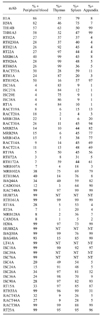
The screening of several hundred mAbs developed in our laboratory yielded 13 mAbs that recognize conserved epitopes expressed on rabbit LDM. The specificity of 10 of the mAbs (RH1A and LT86A [CD9]; HUH73A [CD11a]; CAM36A [CD14]; H20A, BAQ30A, and HUH82A [CD18]; and BAG40A and LT41A [CD44] ) was validated in the Animal Homologues section of the HLDA8 (Table 1, Fig. 1) [12,54]. Two additional mAbs, RACT48A and GBSP71A submitted to the workshop reacted with molecules expressed on multiple lineages of leukocytes in humans and other species. No clear match was obtained with standard panels of human leukocytes or cell lines transfected with known CD molecules. BAQ44A and CADO34A were not submitted to the HLDA8 workshop since they did not react with leukocytes from humans. However, the mAb-defined epitope recognized by BAQ44A is expressed on B lymphocytes in multiple species of ruminants. The epitope recognized by CADO34A is expressed on granulocytes, B lymphocytes and subsets of T lymphocytes in dogs and cats. Multiple mAbs were identified that reacted with rabbit MHC I and II molecules. The best characterized mAbs are listed in Table 1. Analysis of the specificities of TH14B and TH81A5 have shown they recognize epitopes conserved on the orthologues of HLA-DR and HLA-DQ, respectively [1].
Identification of mAbs that recognize LDM expressed on T lymphocytes
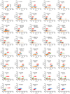
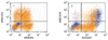
Screening of the mAb sets obtained from the different fusions yielded multiple mAbs that recognize LDM expressed on all lymphocytes or subsets of lymphocytes. These were further analyzed to determine which mAbs detected LDM expressed on T lymphocytes, B lymphocytes, or T and B lymphocytes using 2 color FC. Fluorescein conjugated anti-rabbit Ig was used to identify mAbs recognizing LDM on B lymphocytes. Ken4 (CD4) and Ken5 (pan T) were used to identify mAbs recognizing LDM on T lymphocytes. 12.C7 (CD8) was used to verify specificity of mAbs reacting with CD8 [18]. As summarized in Tables 1 and 2 and fig. 1A, 1B, 8 mAbs were identified that recognize LDM expressed on all T lymphocytes (MRB61A, RT22A, RTH2A, RTH21A, RTH26A, RTH65A, RTH230A, and RACT53A). Cross comparison of the patterns of reactivity of the mAbs using 2 color FC showed RTH2A and RTH230A; RT21A and RTH21A; and RTH26A, RTH65A, and Ken5; recognize Pan T1, Pan T2, and Pan T4 LDM respectively. Zenon second step antibodies were used to demonstrate RTH2A (IgG1) and RTH230A (IgG1) recognize the same LDM (Fig. 2). RACT53A (PanT5) recognizes an additional molecule expressed on all T cells (Fig. 3). Analysis of the reactivity of MRB61A (Pan T3) revealed it detects a LDM expressed on all T lymphocytes and basophils (Fig 1 #7, two color labeling not shown).
Seven mAbs were identified that recognize LDM expressed on T lymphocyte subsets. Comparison of labeling with Ken 4 and 12.C7 demonstrated that RTH1A recognizes CD4 [41] and that ISC16A, ISC27A, ISC29A, ISC38A, and RT1A recognize CD8 [18] (Table 1, Fig. 1 #9 & #10, FC two color comparisons not shown). No information was obtained on whether the CD8 mAbs recognize epitopes on CD8α or CD8β.
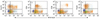
Comparison of labeling with RACT19A (Fig. 1 #12) with PanT1, RTH1A and ISC38A revealed the molecule detected is expressed on a large subset of CD4 and the majority of CD8 lymphocytes (Fig. 4).
Identification of mAbs that recognize LDM expressed on B lymphocytes
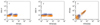
Eleven mAbs were identified that recognize LDM expressed on B lymphocytes (Tables 1 and 2, Fig. 1 #16, #17 & #18). Comparison of labeling with fluorescein conjugated polyclonal anti-rabbit immunoglobulin (Ig), RACT30A (Fig. 5) and PanT5 (Fig. 3) were used to demonstrate that MRB25, MRB29A, and MRB143A recognize one or more molecules expressed on all B lymphocytes (dot plots not shown). Comparison of labeling of MRB107A with MRB25A and BAQ44A demonstrated that MRB107A recognizes a LDM expressed on a subset of B lymphocytes. The molecule detected is only expressed on a subset of MRB25+ B lymphocytes. The whole population is included in the BAQ44A positive population of B lymphocytes (Fig. 6). As shown in Table 2, the level of expression of the pan B mAb-defined LDM(s) were similar in peripheral blood, thymus and spleen. However, other mAbs that recognize LDM expressed on subsets of B lymphocytes exhibited different patterns of expression (Table 2, FC for thymus and spleen not shown). The subset of B lymphocytes detected with RACT14A and RACT21A was in low frequency in peripheral blood and in high frequency in spleen and appendix. The subset detected with RTH72A was low in peripheral blood, thymus, and appendix and high in spleen. The subset detected with mAbs RT19A and RTH172A was low in peripheral blood but high in thymus, spleen, and appendix.
Identification of mAbs that recognize LDM expressed on T and B lymphocytes
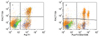
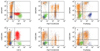
Four mAbs were identified that recognize LDM expressed on T and B lymphocytes, MRB102A, RTH186A, RTH192A, and BAQ44A (a cross reactive mAb) (Fig. 1 #19, #20, #11 & #15, respectively). The level of expression of the LDM detected with MRB102A on lymphocytes was higher than the LDM detected with RTH186A. However, two color analysis showed the level of expression of both LDMs on CD4 and CD8 T and B lymphocyte subsets was similar (FC not shown). The level of expression of the MRB102A-defined LDM was also high on lymphocytes in the thymus, spleen, and appendix. In contrast, the RTH186A defined LDM was only expressed on a few lymphocytes in the thymus and appendix. It was expressed on a large population of lymphocytes in the spleen (Table 2, FC not shown).
The level of expression of the LDM detected with RTH192A and BAQ44A differed on CD4 and CD8 T and B lymphocytes. The level of expression of the RTH192A-defined LDM was variable on PanT 1+ lymphocytes (Fig. 7). It was low on CD4 T and B lymphocytes (Fig. 7). It was high on CD8 T lymphocytes (Fig. 7). It was only expressed on a few thymocytes. It was expressed at a high level on about 50% of lymphocytes in the spleen and essentially all lymphocytes in the appendix (Table 2, FC not shown). The pattern of expression on T and B lymphocytes suggests the LDM detected is CD5 [41,49-51].
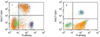
The level of expression of the LDM detected with BAQ44A also differed on CD4 and CD8 T and B lymphocytes (Fig. 8). Simultaneous labeling with Pan T1, CD4, and CD8 mAbs and anti-rabbit Ig demonstrated that the LDM is highly expressed on B lymphocytes, a subset of CD4 and CD8 negative T lymphocytes, a large subset of CD4 T lymphocytes and the majority of CD8 T lymphocytes. The BAQ44A-defined LDM was expressed on large populations of lymphocytes in the thymus, spleen and appendix (Table 2, FC not shown).
Identification of a mAb that recognizes a LDM expressed on granulocytes, T and B lymphocytes
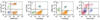
One cross reactive mAb, CADO34A, was identified that recognizes a LDM expressed on granulocytes as well as T and B lymphocytes (Fig. 8). Comparison of labeling with CADO34A to labeling with BAQ44A revealed the pattern of labeling is similar to the labeling pattern obtained with BAQ44A for T and B lymphocytes. Simultaneous labeling with Pan T1, CD4, and CD8 mAbs demonstrated the LDM is expressed on a subset of CD4 and CD8 negative lymphocytes, a large subset of CD4 and the majority of CD8 T lymphocytes. The LDM was only expressed on a few thymocytes. It was expressed on a large population of lymphocytes in the spleen and most of the lymphocytes in the appendix (Table 2, FC not shown).
Identification of a mAbs that recognize a LDM expressed on granulocytes and monocytes, granulocytes, monocytes and basophils, or basophils
Three mAb were identified that detect LDM expressed on granulocytes and monocytes, granulocytes, monocytes and basophils or basophils (CAM36A, MRB120A, and RACT20A, Fig. 1 #26, #22 & #13, respectively and Fig. 9). CAM36A recognizes a conserved epitope expressed on CD14. In contrast to some species, expression of CD14 is high on rabbit granulocytes. Two color analyses showed CD14 is not expressed on basophils (Fig. 9). Two color analyses showed the MRB120A recognizes a LDM expressed on granulocytes, monocytes and basophils while the RACT20A recognizes a LDM only expressed on basophils and a subset of CD4+ lymphocytes (Fig. 9). Expression on CD8 lymphocytes is low or absent. Two color analyses were not performed to determine whether expression of the RACT20A-defined LDM on small populations of cells detected in the thymus, spleen and appendix were basophils or T lymphocytes (Table 2).
Identification of mAbs recognizing LDM expressed on granulocytes
Two mAbs (RACT43A and RACT44A Table 1, Fig. 1 #36) were identified that recognize a LDM expressed on granulocytes. The similarity of the pattern of labeling with the mAbs suggests they may recognize the same LDM.
Identification of mAbs recognizing CD9
Comparison of the labeling patterns with cross reactive RH1A and LT86A (Table 1, Fig. 1 #21) showed they recognize CD9 in rabbits.
Identification of mAbs recognizing CD11a, CD11b, CD11c, and CD18
mAbs that recognize CD11a, CD11b, CD11c, and CD18 (Table 1, Fig. 1 #23, #24, #25 & #27 respectively) were identified by two color FC with mAbs that recognize epitopes conserved on orthologues in one or more species or with commercially available mAbs generated against the rabbit orthologues. Comparison of the patterns of labeling obtained with RACT48A and RTH161A with commercially available anti-CD11a (Ken11) [31] suggested these mAbs recognize CD11a. Subsequent comparative two color FC analysis with HUH73A, a mAb demonstrated to recognize a conserved epitope on the CD11a orthologue in the Animal Homologues section of the HLAD8 workshop [54], verified these mAbs recognize CD11a (Fig. 10). The studies also demonstrated RTH161A recognizes a species restricted epitope. Comparison of the patterns of labeling obtained with RT18A with anti-CD11b (198) and subsequent 2 color FC showed RT18A recognizes CD11b (Fig. 1 #24). Similar studies comparing RT3A with 3/22 showed RT3A recognizes CD11c (Fig. 1 #25). Three mAbs demonstrated to recognize conserved epitopes on orthologues of CD18 (Fig. 1 #27), in 2 or more species were shown to recognize rabbit CD18. The pattern of expression of the rabbit orthologues for each molecule was similar to that noted in other species.
Identification of mAbs that recognize CD44, CD45, and CD58
Screening of a large series of mAbs that recognize conserved epitopes on CD44 in 2 or more species, including humans, showed 25-32 [40], BAG40A, and LT41A (Table 1, Fig. 1 #28) recognize epitopes expressed on rabbit CD44 [14,24,54]. The pattern of expression of rabbit CD44 was similar to that noted in other species. Comparison of the pattern of labeling obtained with L12/201 (CD45) with the panels of mAbs developed against rabbit LDM revealed several mAbs yielded similar patterns of labeling (Table 1, Fig. 1 #29). Two color flow cytometry yielded diagonal patterns of labeling or blocking, indicating the mAbs recognize rabbit CD45 (FC not shown). Comparison of the labeling pattern obtained with VC21 (CD58) revealed two mAbs (RTH32A, RTH33A, Table 1, Fig. 1 #30) yielded similar patterns of labeling. Two color comparisons yielded diagonal patterns of labeling indicating the mAbs recognize CD58 (FC not shown).
Identification of mAbs that recognize LDM expressed on granulocytes and lymphocytes
Six mAbs were identified that recognize LDM expressed on granulocytes and lymphocytes ISC4A, ISC24A, ISC26A, ISC36A, ISC90A, and RT15A (Table 1, Fig. 1 #31, #26, #36, #90 and #35). Two color analysis showed ISC4A and ISC24A recognize the same molecule (FC not shown). The others identified different molecules. The molecules identified with ISC90A and RT15A are also expressed on some monocytes. The molecule identified with RT15A is highly expressed on thymocytes, spleen lymphocytes, and lymphocytes in the appendix (Table 2). Two color analysis with anti-CD4 and -CD8 mAbs indicate none of the mAbs recognize CD45R0. The studies completed thus far indicate there are no clearly defined subsets of CD4 and CD8 negative for these LDM (FC not shown).
Identification of mAbs that recognize LDM expressed on all leukocytes
Three mAbs under investigation identify LDM expressed on all leukocytes RACT38A, RT23A, and GBSP71A (Table 1, FC not shown). Comparison of the flow cytometric profiles with those of known CD molecules has thus far not suggested which molecules are recognized by these mAbs.
Identification of mAbs that recognize LDM expressed on activated lymphocytes
Three mAbs were identified that recognize mAbs upregulated on ConA activated lymphocytes RACT1A, RACT4A, RACT12A (Table 1, Fig. 1 #38, #39 & #40). Two color analysis with mAbs specific for ConA and CD25 [32] demonstrated the mAbs recognize different LDM (FC not shown).
Discussion
Cumulative data obtained from international workshops convened to complete characterization of mAbs to LDM in humans have shown flow cytometry can be used to compare and cluster mAbs that appear to recognize the same LDM for further analysis [34]. Our studies and studies conducted as part of workshops convened to characterize mAb-defined LDM in ruminants, swine, horses, and dogs have shown that the pattern of expression of many orthologous of CD molecules is conserved cross species [11]. These findings have afforded an opportunity to devise a strategy for characterizing mAb to LDM in additional less well studied species. mAb that identify LDM with patterns of expression similar to the patterns of expression of known Human Cell Differentiation Molecules (HCDM) can be clustered for further analysis. The TKP can be used to facilitate determining whether a mAb recognizes a new or known HCDM where flow cytometric data are not conclusive. Where cross reactive mAbs are identified, they can be used to validate the specificity of the mAbs recognizing species restricted epitopes using two color FC. A diagonal pattern of labeling implies the epitopes detected are present on the same molecule. Complete or partial blocking of labeling with one of the mAbs indicates the epitopes detected are sterically close on the same molecule, with the binding of one mAb interfering with the binding of the second mAb. These methods of analysis have been used effectively to develop a set of mAbs for use in alpacas and llamas [14] and, as demonstrated in the present report, rabbits. Cross reactive mAbs allowed us to identify mAbs specific for MHC class I and II and several CD molecules early in the course of the studies. Two color analyses with commercially available mAbs to some rabbit LDM facilitated validation of the specificity of additional mAbs generated in our laboratory. To date, we have identified mAbs to MHC class I and II molecules and 16 known LDM. Additional mAbs to T and B lymphocytes have been identified that require further characterization to determine their relation to known mAb-defined HCDM.
Until now the lack of mAbs to rabbit LDM and MHC has made it difficult to compare the immune system of rabbits to those characterized in other species. Comparative studies have shown the immune systems of different orders and species of vertebrates are similar but not identical. Differences have been noted in the evolution and expression of immunoglobulin genes with expansion of the IgA genes a unique feature of rabbits [41] and the development of IgG heavy chain genes a unique feature of camelids [21,41]. The expression of αβ CD4 and CD8 T cell subsets have appeared similar with an exception in swine. There is a large population of CD4/CD8 double positive cells in peripheral blood. Analysis has shown the proportion of double positive cells increases with age and is correlated with the appearance of the majority of memory T cells in this population [67,68]. The most striking difference noted is in the abundance of γδ T cells in some species [20]. γδ T cells comprise a high proportion of lymphocytes in peripheral blood of chickens [5,9], swine [2,3,17] and ruminants [19,25,39]. The relation between the large population of γδ T cells that have evolved in chickens and the one in pigs and ruminants is not clear. However, recent studies have provided an explanation for the abundance in the latter species. Abundance is attributable to the presence of a unique subset of γδ T cells that has only been found in artiodactyla. The subset is characterized by the expression of a molecule referred to as workshop cluster 1 (WC1) in ruminants [42]. The WC1+ population may comprise 50% or more of T lymphocytes in the blood of young ruminants [62,63]. A WC1-population is also present but comprises ~5% of γδ T cells in blood [10]. The large subset identified in swine expresses the orthologue of WC1 [7,17]. The subset expressing the orthologue has been identified in camelids also [14].
An additional difference noted in the comparative studies is in the expression of MHC II. In humans, mice, and ruminants MHC II is expressed primarily on monocytes and B lymphocytes in blood. MHC II is upregulated on T cells following activation. MHC II is expressed on monocytes, B cells and resting T cells in horses [37], swine [48], dogs [8], and cats (personal observation).
As mentioned, the flow cytometric approach to identifying and characterizing mAbs to rabbit LDMs has shown the pattern of expression of rabbit orthologues of known hCDLDMs have proven, thus far, to be similar. The composition of lymphocyte subsets also appear more similar to that of humans than ruminants and swine. Analysis of T lymphocytes in the rabbit shows αβ T cells are the major population present in blood. Comparison of the percentages of cells expressing LDM present on all T cells with subsets of cells expressing CD4 or CD8 in two color flow cytometry has not revealed any CD4 CD8 double negative subset. Likewise comparison of the percentages of lymphocytes expressing LDM on T cells with those expressing LDM on B cells has not revealed the presence of a clearly defined subset of lymphocytes negative for T or B cell LDM. The findings suggest γδ T cells comprise a small percentage of lymphocytes in blood. Further studies are needed to identify rabbit γδ T cells.
The unique differences that have been noted are with the expression of MHC II on granulocytes, expression of certain mAb-defined LDM, and presence of immunoglobulin on basophils. Multiple mAbs of different isotype to MHC II were used to confirm expression of MHC II on granulocytes. Both IgG1 and IgG2a isotype mAbs yielded identical patterns of labeling. As in humans and mice, expression of MHC II on monocytes and B cells is similar. It is upregulated on activated T cells. Basophils comprise 5% to 30% of blood leukocytes in rabbits. Two mAbs were identified that identify molecules expressed on all T cells and basophils (MRB61A) and basophils and a subset of CD4 T cells (RACT20A). No information has been obtained on the functional activity of these LDM. Two color analyses have demonstrated that immunoglobulin is present on basophils. This finding is in agreement with an earlier study demonstrating the presence of multiple immunoglobulin isotypes on basophils, presumably binding to basophils through Fc receptors [6].
In summary, the use of flow cytometry has provided an approach to the identification and characterization of mAbs to MHC I and MHC II molecules and LDM in less well studied species. mAbs that recognize the same LDM can be clustered based on the similarity of the pattern of expression and further characterized by comparing the pattern of expression with that of known LDM. When available, identity can be verified by comparison with a mAb that recognizes a conserved epitope on orthologous molecules. Where needed, mAb cluster-defined LDM can be subjected to immunoprecipitation and micro sequencing. We have used this approach successfully when participating in the international workshops of LDM in ruminants [26,27,46], swine [23,38,53], horses [33,36], dogs [8], the homologues section of the HLDA8 [54,66] and independently in the characterization of mAbs to LDM in lamas [14] and rabbits. The mAbs characterized here should facilitate characterization of the immune system in rabbits and the use of rabbits in immunological investigations.

 XML Download