

 PDF
PDF ePub
ePub Citation
 Print
Print


Introduction
Avian influenza (AI) is one of the most contagious poultry diseases known and is caused by type A influenza virus, a member of the family Orthomyxoviridae [7]. Type A influenza viruses are further divided into subtypes based on H and N antigens. At present, 16 H subtypes (H1-H16) and 9 N subtypes (N1-N9) have been recognized [16], but only the H5 and H7 virus subtypes are highly virulent in poultry [1].
After the initial identification in Korea in December 2003, 19 highly pathogenic avian influenza (HPAI) virus isolates were found in various species of poultry, such as ducks, broiler breeders, and layers, between December 2003 and March 2004. All isolates were shown to be the H5N1 virus subtype [8]. In 1996, the first low pathogenic avian influenza (LPAI) virus was confirmed in the Gyeonggi-do of Korea (GPK), and the H9N2 virus subtype was isolated from several broiler breeder flocks (all were LPAI viruses). A total of 97,963 broiler breeders were depopulated to eliminate the AI virus (AIV) at that time [11]. However, LPAI has occurred sporadically since 1997. For example, 24 cases of LPAI were reported in the GPK from 1 January 2000 to 1 April 2006 [13].
Unlike HPAI, in which the case mortality may be as high as 100% [17], LPAI is associated with mild clinical signs, such as a low fatality rate, primary respiratory symptoms, depression, and decreased egg production [5]. Therefore, most poultry producers do not consider LPAI as an important disease and often do not even realize that their flocks have the disease. The poultry producers may not report an outbreak of LPAI in their flocks for these reasons, even though LPAI is a reportable disease in Korea. HPAI is a first level reportable disease and LPAI is a second level reportable disease.
Thus, this study was conducted to address 3 questions: 1) How many undetected or undiagnosed LPAI cases are present in layer, broiler, domestic duck, and broiler breeder flocks in the GPK? 2) What is the greatest risk factor for introducing and maintaining LPAI in seropositive flocks? and 3) What are the current monitoring and surveillance systems for LPAI in Korea?
Materials and Methods
Selection of poultry farms and determination of sample size
Two hundred fifty-three flocks were randomly selected from 1,654 farms in the GPK; 96 farms were selected from 582 layer farms, 97 farms were selected from 880 broiler farms, and 30 farms were selected from 81 breeder farms. In addition, 30 flocks were selected from 111 domestic duck farms. The flock samples were selected using a computer program (Research Randomizer, USA).
The minimal sample size of birds in each flock to achieve 95% confidence for random sampling was determined to be 15, which was calculated using the Cannon and Roe formula [3].
Collection of samples
Between November 2005 and March 2006, the samples were collected as follows: 1) layer, broiler breeder, and domestic duck flocks: the samples of each flock were collected by staff from the Livestock Health Control Association and/or the Veterinary Service Center in Gyeonggi-do (VSCG); 2) broiler flocks: the samples were collected at the slaughter houses (62 flocks) or farms (35 flocks) by the VSCG staff. If there were no chickens in the farms selected by the computer program, alternative samples were collected from the closest flocks to the initially selected flocks.
Serological test
The hemagglutination inhibition (HI) test was used for detecting antibody from sera of layers, broilers, and broiler breeders, while the agar gel precipitation (AGP) test was used for detecting antibody from sera of domestic ducks. Both immunologic tests were carried out according to the recommendations in the WHO manual [19]. The reagents for the HI and the AGP tests were obtained from the National Veterinary Research and Quarantine Service (Korea) and Animal Genetics (Korea), respectively. According to the OIE manual [15], four hemagglutination units were used for the HI test. A tested flock with 15 blood samples was classified as a positive control if there was at least one inhibition at a serum dilution of 1/16 among the 15 blood samples.
Inoculation of embryonated chicken eggs for virus isolation
For detecting AI viruses and/or official reporting of AI to the Regional Veterinary Laboratory of Korea, initial serological tests and isolation of viruses from seropositive birds are generally performed if there are no typical signs. Therefore, this study was conducted followed that protocol and only the swab samples of seropositive birds were inoculated into embryonated SPF chickens eggs. The WHO manual was used as a guide [19].
Study design and collection of questionnaires
The first part of the study was cross-sectional involving 96 layer flocks. Twenty-five seropositive flocks were compared with 71 seronegative samples. Based on the cross-sectional study, having employee(s) was shown to be a major risk factor for seropositivity; however, the specific employee risk factors were not determined. Therefore, a matched case-control study was conducted. For the purpose of this study, seropositive flocks with employee(s) were identified as cases and seronegative flocks with employee(s) were designated as controls.
Cross-sectional study
The questionnaire was designed to determine the possible risk characteristics for the seropositive flocks compared with the seronegative flocks and to evaluate if the poultry producers with seropositive layers recognized the clinical signs of AI when the disease was present. The questionnaire covered 4 categories: (1) basic information; (2) management; (3) poultry house; and (4) retrospective data to evaluate if poultry producers had experience with clinical signs, such as decreased egg production. The questionnaires for layers, broiler breeders, and domestic duck flocks were filled out by the staff at the VSCG during the interview when they visited the farms for sampling. Information regarding broiler flocks was collected from telephone interviews with 62 farmers and from farm visits to 35 farmers. The collected information was rechecked to verify the collected data by calling the poultry producers, if necessary.
Case-control study
Of the 25 seropositive layer flocks, 20 flocks were selected as cases; 5 farms were excluded from analysis for the following reasons: no employees, relocation, and empty chicken houses. To reduce the effects of confounding variables, cases (n = 20) and controls (n = 20) were matched based on hired employees, flock age, and flock size. The inquiry included 4 categories: 1) basic information regarding the owner; 2) habitation of the employees; 3) sanitary concept of the farm workers; and 4) activity of the employees. Data were collected by staff at the VSCG via interviews.
Analysis of data
Cross-sectional study
In the cross-sectional study, all analyses was performed using Microsoft Excel 2000 and SPSS, version 12.0. For identifying possible risk factors, seven suspected factors were included as variables. The prevalence odds ratio (OR) of each variable with a 95% confidence interval and two-sided p-values were calculated using binary logistic regression. A p < 0.05 was considered significant. To compare the HI titers between layers and broilers, the geometric mean of the titer of each group was calculated with the raw titer (not log-transformed). A t-test was performed to ensure the significance of differences between the groups with log-transformed data. To analyze the relationship between an increase in age and seropositivity in the layer flocks, raw data pertaining to seropositivity and age were divided into 3 categories: 1) <300 d old, 2) 300-400 d old, and 3) >400 d old. The odds ratio of each category was calculated using multinomial logistic regression analysis. In this study, the odds ratio of the <300 d old category was regarded as the baseline variable, and two categories were calculated according to the baseline odds ratio. To evaluate the difference in recognition of clinical signs between farmers with seropositive flocks and farmers with seronegative flocks, the relationship between retrospective data and seropositivity was statistically analyzed using a Chi-square test.
Results
Seroprevalence and virus isolation
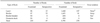
In serology, the unadjusted percentage of seroprevalence rates of layers and broilers was not significantly different (26% [25/96] and 23% [22/97], respectively). The seroprevalence rate of individual birds, however, was twice as high in the layers (13% [187/1440]) as in the broilers (6% [91/1455]). The AIV was not isolated from the seropositive flocks that showed no clinical signs when sampling. Some hemagglutinating agents were detected in the allantoic fluid inoculated with specimens of seropositive layers, but were not verified as an AIV with a test kit (Anigen, Korea). Thus, further testing for identification of the AIV was not performed (Table 1).
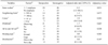
Distribution of HI titers
Table 2 presents the distribution of HI antibody titers against AIV among the flocks. Titers obtained from the layers ranged between 16 and 512 (mean = 89), and were higher than the broilers (mean = 27; p < 0.001). Of 181 seropositive layers, the number of birds with a HI titer of 64 (45 birds) was most frequent, followed by titers of 32 (43 birds), and 16 (40 birds).
Analysis of cross-sectional study
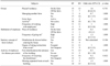
A multivariate analysis using the logistic regression model is shown in Table 3. Of the seven risk factors, only farms that hired one or more workers were found to have a significant association with the risk of being seropositive (POR = 11.5, p = 0.031); other characteristics were not significantly associated with seropositive layers.
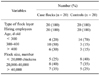
Table 4 shows the seroprevalence of layers by age in the GPK. There was a significant pattern, i.e., the older layers had a higher seroprevalence. The seroprevalence (40%) of the groups older than 400 d old was greater than twice that of the layer flocks younger than 300 d old. This demonstrated that the OR increased while the layers in the GPK were aging, with an adjusted OR of 4.9 (p = 0.017) for layers over 400 d old.
Analysis of retrospective data (Table 5) indicated that there was little significant difference (OR = 2.3, p = 0.082) in poultry producers with experience regarding clinical signs of AI between seropositive layers and seronegative layers. Having experience indicated that the poultry producers recognized at least one clinical sign, such as decreased egg production, respiratory syndromes, and increased mortality. Of 25 seropositive flock growers, 13 growers (52%) recognized at least one clinical sign, but 32% of the growers with seronegative layers recognized one clinical sign as well.
Analysis of the case-control study
There were 20 pairs of case and control flocks that were matched for type of flock, hired employees, flock age, and flock size. All cases and controls were layer flocks. Of 20 case-control pairs, 20 (100%) were successfully matched for hired employees. Case farms had a large number of flocks in comparison with control farms and were more likely to have older chickens than control farms. The details of the results of matching are shown in Table 6. As shown in Table 7, frequent cleansing with disinfectants resulted in a decreased risk of seropositivity (OR = 0.2, p = 0.022). Seropositivity had no association with the place of residence for the employees, frequency of going out, disinfection, and taking a shower when coming back to the farms after going out. Although there was little statistical association, usage of a foot disinfectant at the entrance of the building carried a decreased risk of seropositivity (OR = 0.3, p = 0.105).
Discussion
For determination of the minimal sample size per flocks, it was calculated that the minimum prevalence was 20% when the LPAI (H9N2) viruses were introduced into a flock. It was difficult to determine the precise seroprevalence of LPAI because of the sampling anomalies. However, a 20% attack rate was determined based on several studies [11,12,14]. In Pakistan, the seroprevalence of AI against subtype H9N2 was at least 54% (30/55 birds) [12]. In Iran, mortality in affected flocks with H9N2 was between 20 and 65% [14]. In addition, when the first outbreak of LPAI (H9N2) occurred in Korea, a 20-40% mortality rate was reported [11].
In this study, there was no virus isolation from seropositive flocks without clinical signs of infection. It could be inferred that for successful AIV isolation, specimens should be taken early after the onset of clinical symptoms, as described in other reports [4,6,12,14,20]. AIV can be isolated within 7-10 days infection [4,18], but antibodies are detected 7-10 days after infection; thus, it may be difficult to identify AIV from the birds that are seropositive. For instance, the AIV was not isolated from any samples for a long time after diagnosis with the disease, although many layers in a complex continued to be seropositive [22].
Thus, attempts for successful viral isolation must be performed within a few days of onset, but not after detecting antibodies. The WHO also recommends that specimens for AIV isolation should generally be taken during the first 3 days after the onset of clinical signs [19]. In a cross-sectional study, broiler chickens were not analyzed because of maternal antibody persisting for up to 4 weeks [15]. When the antibodies were detected from broilers, it was not easy to differentiate between maternal antibody and antibody arising due to infection. Thus, only the data of 96 layer flocks was analyzed.
In this case, farms with employees were a significant factor for seropositivity in layers in the GPK. The presence of farm workers means that a poultry farm owner hired one or more people who participated in the farm work. This may be related to an increased chance of introducing AIV into the flocks by increased personnel movement, as most studies concluded that the secondary spread of the AIV was principally by the movement of personnel and equipment between farms [1].
In addition, the present study is supported by other studies reporting that HPAI spread more rapidly on farms with employees [9,21]. Other characteristics, such as frequency of disinfection, were not significantly associated with seropositive layers. These results are similar to other reports. For example, a study [9] also suggested that various routine biosecurity and presence of wild birds on the premises were not significantly associated with infection of low pathogenicity H7N2 AI virus during an outbreak in West Virginia in 2002.
This study indicated that age was a significant risk factor for maintenance and introduction of LPAI. To compare the seropositivity by age, all of the tested layers were divided into 3 groups (< 300 d old, 300-400 d old, and > 400 d old) since the average age of the layers was 317 d. As shown in Table 4, the seroprevalence of older layers was over twice that of younger layers. This may have resulted from the increased susceptibility with age due to decreased immunity and an increased opportunity for virus exposure via personnel and transportation, which were the main source for the spread of the AIV [1].
The analysis of retrospective data showed that the growers with seropositive flocks might have experienced at least one sign of LPAI. Because the duration of the clinical period was short and the symptoms were mild, many poultry producers in Korea claimed that the clinical signs of LPAI were not easy to detect. Therefore, they did not report the occurrence of LPAI in their flocks. Thus, this study tried to evaluate if the poultry producers with seropositive layers recognized the clinical signs of LPAI when infected with the disease. As shown in Table 5, the poultry producers did recognize the clinical signs of LPAI because all farmers in this study examined the abnormality of their flocks daily. The present study suggested that more intensive education should be added for more effective LPAI control.
As the spread of AIV was usually associated with human involvement [2], a cross-sectional study indicated that having employee(s) was a major risk factor for seropositivity. To evaluate more specific risk factors in regard to farm workers, four categories were investigated. Frequent cleansing with disinfectants was a decreased risk factor and using foot disinfectants was a possible factor for decreased risk. Clearly, if the employees were active in the prevention of disease, the risk of seropositivity could be decreased. The risk could become even lower, for example, if the disinfectants were frequently used, as the Ministry of Agriculture and Forestry (Korea) recommended (i.e., disinfectants for boots and vehicles should be changed 2-3 times per week) [10]. This study strongly emphasized the needs for continued high levels of direction or supervision to control or prevent LPAI circulating in GPK.
This study had several potential limitations. In a cross-sectional study, some questions could be interpreted subjectively by the poultry producers. For example, the question regarding the observation of wild birds around farms may have been interpreted as on the premises in some cases, but as around (within 1 km) in other cases. The question defined disinfection as practicing entire places related to the farm, such as an entrance to the farm and nearby road, in and out of the poultry house, and entering traffic. Some growers may have interpreted this as on the premises, however, others may have interpreted it as any area around the farm. The questionnaire responses may have been affected by recall bias, especially with respect to the retrospective data. Some growers with seropositive flocks may not have stated their actual experiences because interviewers were public officers working at the VSCG.
In a case-control study, the number of cases and controls were small, limiting the power of the study to demonstrate significant associations.
In conclusion, this study indicated that LPAI (H9N2) has occurred in portions of layers and broilers in the GPK, but it has remained undetected or undiagnosed. It was also shown that many poultry producers did not notify the occurrence of LPAI in their flocks, even though they recognized the clinical signs. However, it was not easy to confirm the disease by viral isolation from the seropositive flocks because LPAI viruses were not detectable in a chicken within a few days after infection. Today, only the flocks with AIV isolation are under control programs, thus it is recommended that the current policy be modified for the effective control of LPAI in Korea. In addition, to reduce the risk of the introduction of the LPAI (H9N2) virus into farms, it is strongly suggested that farm employees should be more proactive in the prevention of disease.


 XML Download