

 PDF
PDF ePub
ePub Citation
 Print
Print


Feline leukemia virus (FeLV) is a retrovirus that causes a wide range of proliferative diseases in cats, including lymphoid and myeloid leukemia [6]. Acute myeloid leukemia may be misinterpreted as acute lymphoid leukemia if the blast cells are classified using only Romanowsky stained smears [10]. Cytochemical staining has been employed to aid in the differentiation of acute leukemias [2]. Assessment of cell ultrastructure by transmission (TEM) or scanning electron microscope (SEM) has also been used to enhance the magnitude of cell identification, especially with poorly differentiated cells [1]. Although FeLV is a common infectious disease in young cats, no clinical cases ofacute monoblastic leukemia in FeLV-infected cats in Thailand have been reported previously. In the present report, a case of acute monoblastic leukemia in a FeLV-positive cat is described.
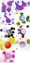
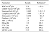
A 1.6-year-old male domestic short hair cat was brought to the Veterinary Teaching Hospital, Kasetsart University, with a history of anemia, depression, and weight loss. Physical examination revealed dyspnea, as well as cervical, axillary, and popliteal lymph node enlargement. The initial laboratory tests included a complete blood count (CBC) and serological test (Fast Test FeLV; MegaCor Diagnostic, Austria). The CBC is summarized in Table 1, and shows severe anemia, thrombocytopenia, and marked leukocytosis with undifferentiated blasts in more than 50 percent. Morphologically, these cells were round to ovoid in shape, with finely stippled nuclear chromatin and distinct nucleoli (Figs. 1A and B). Some presented prominent cytoplasmic tails (Fig. 1C). The serological test was positive for FeLV. Further examination using nested polymerase chain reaction, as described elsewhere [9], also confirmed the presence of FeLV nucleic acid in the blood. The initial treatment was started with 1 mg/kg dexamethasone IV and fluid therapy (5% dextrose in half-strength saline), with oxygen being given all day.
Although a bone marrow examination was recommended, the poor condition of the patient limited this procedure. To further classify undifferentiated blasts, selected cytochemical staining was performed, including peroxidase (PER), Sudan black B (SBB), α-naphthyl acetate esterase (ANAE), periodic-acid Schiff [4], and β-glucuronidase (β-GLU) [3]. Five hundred cells from each of the cytochemically-stained smears were counted following staining in which positive- and negative-stained cells were differentiated. For SEM and TEM, blood cells were processed as described elsewhere [11]. Identification of blasts by SEM and TEM was based on the relative number, size, shape, cytoplasmic complexity, and nuclear appearance.
Unfortunately, the owner denied the hospital from admitting the cat. Three days later, the cat died and his carcass was submitted to necropsy. The hallmark lesions showed splenomegaly, hepatomegaly, and enlargement of several lymph nodes. Histopathologically, massive neoplastic cells contained round, finely chromatic nuclei; amphophilic cytoplasm infiltrated these organs.
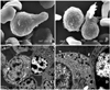
Detection using ANAE and β-GLU staining yielded 100% positive blasts (Figs. 1G and H), while PER, SBB, and PAS stains revealed only a few positive cells (Figs. 1D, F and I). The cytochemical profiles are summarized in Table 2. Using SEM, the blasts appeared round to ovoid in shape with a ruffled membrane and deep fissures, whereas pseudopodia were clearly observed (Figs. 2A and B). Ultrastructurally, a round to irregular nuclear shape with marginated nuclear chromatin and light cytoplasmic appearance with some electron-dense granules and organelles such as endoplasmic reticulum cisternae were shown (Figs. 2C and D). From these results, the patient was diagnosed acute monoblastic leukemia.
Definitive diagnosis of acute leukemia requires a panel of cytochemical and electron microscopic analysis. A panel of cytochemical stains of blood smears can be applied in order to determine the lineage of leukemic cells. In addition, SEM can be used to evaluate cell surfaces while TEM presents the ultrastructural images of organelles. The most useful cytochemical stain in monoblastic leukemia is the reaction for nonspecific esterase activities such as ANAE [5], while SBB- and PER-positive patterns support the myeloid lineage.
Though the most common form of leukemia in cats infected with FeLV is of the lymphoid lineage [12], a myeloid lineage was the affected progenitor subset found in the patient described in this study. This finding may be due to retrovirus-induced chromosomal translocation involving chromosome 11q 23, and rearrangement of a gene referred to as myeloid/lymphoid or mixed lineage leukemia at the translocation breakpoint [7,8].

 XML Download