

 PDF
PDF ePub
ePub Citation
 Print
Print


Introduction
The mammary gland of transgenic farm animals has been proposed as the best available bioreactor for the production of human pharmaceutical proteins [4,5,13,21]. Dairy goats have been used as a bioreactor because of their relatively short gestation period and low maintenance costs compared to cattle. Many studies have reported on the production of transgenic goats [6-8,11]. The possibility of large-scale production for industrial application has been demonstrated [9,10]. In Korea, transgenic goats have been used to produce human granulocyte colony stimulating factor (G-CSF) in their milk [15,18].
To date, laparotomy methods have generally been used for goat embryo transfer (ET). However, this method can cause adhesions in the reproductive tract following repeated surgical ET and requires relatively long intervals before the re-use of a recipient female [15,18]. To overcome the limitations of laparotomy, laparoscopic ET has been performed in various species including sheep [19,20], cows [12] and pigs [3]. The laparoscopic method has also been performed in goats [1,2,16,17,24]. However, in the above mentioned studies, the laparoscopic method was used only for oocyte recovery by ovum pick-up and embryo recovery but not for embryo transfer.
Estrus synchronization is essential for successful ET and corpus luteum (CL) formation is necessary for pregnancy maintenance. However, it is difficult to know the exact status of the ovaries if they are not observed directly by exploratory surgery or ultrasonography. Therefore, if the CL is not formed during ET, artificial formation of the CL by follicle puncture is necessary for ovulation and progesterone support is required to maintain the pregnancy or the ET must be postponed until CL formation.
In the present study, we performed laparoscopic ET to overcome the limitations of laparotomy in the production of transgenic goats. The pregnancy rates resulting from the two methods were compared in the Korean native goat (Capra hircus). In addition, the pregnancy rates were compared between ovulated and non-ovulated animals in the laparoscopic ET group.
Materials and Methods
Synchronization and embryo collection
In this study, Korean native goats with a body weight ranging from 15 to 25 kg were used as donors and recipients from September to April 2001-2002. All goats were fed alfalfa/grass hay and a commercial diet with free access to water and trace-minerals. The estrous periods of the donors were synchronized using an intravaginal progesterone devices such as a controlled internal drug-releasing insert (CIDR; Pharmacia & Upjohn, New Zealand) for 13 to 14 days irrespective of the natural estrous cycles (Table 1). Superovulation was induced following combined treatment with FSH (ICPbio, New Zealand), PMSG (Horizon Technology, Australia) and hCG (Sigma, USA). FSH (0.9 mg/goat) was administered to the goats over a 4-day period, at 12-h intervals, starting 2.5 days before CIDR removal and continuing within 1 day of CIDR removal. PMSG (150 IU) was administrated at the time of the first FSH administration and hCG (200 IU) was administrated at the time of the last FSH injection to induce ovulation. The donors demonstrated estrus within 24 h following the CIDR removal and were mated with fertile bucks. At 66 h after the CIDR removal, embryos were surgically recovered by flushing both oviducts. All donors were fasted 24 h prior to surgery. A low dose of xylazine hydrochloride (0.12 mg/kg BW; Bayer Korea, Korea) was injected (im) as a pre-anesthetic agent. After a subcutaneous injection of lidocaine (0.1 g/ animal, Kwangmyung Pharm, Korea) for local anesthesia, a midventral incision was made and the reproductive tract exteriorized. The ovaries were examined for fresh ovulation sites to provide an estimate of the number of embryos. The oviducts were flushed with sterile phosphate-buffered saline. The recipient goats were also synchronized with the donor doses for 13 d using the CIDR with a single injection of 400 IU PMSG two days before CIDR removal. As no FSH was administered to the recipient goats, the injection of hCG and removal of the CIDR were both performed one day before they were performed in the donor goats.
Embryo manipulation and microinjection
Immediately after flushing, the number of oocytes/embryos was evaluated for each donor under a stereomicroscope. Zygotes were microcentrifuged at 10,000 g for 7 min to improve pronuclei visualization and the injection of DNA. A 3.7 kb BaaH II/Kpn I fragment of pGbc-hGCSF, in which the hG-CSF (human granulocyte-colony stimulating factor) gene was fused as the promoter sequence with the goat β-casein gene, was injected into one of the pronuclei of the 1-cell embryos. Following microinjection, the embryos were placed into modified synthetic oviduct fluid (mSOF) supplemented with 10% FBS and cultured for 1 or 2 h in a humidified (38.5℃) 5% CO2 incubator until transfer [22].
Embryo transfer
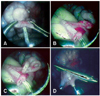
The equipment used for laparoscopic surgery included the following: a 5-mm laparoscope (MGB, Germany), a charge coupled device (IK-C43H 47; Toshiba, Japan), a flexible fiber-optic cable (Olympus, Japan), a camera control unit (IK-Cu43A; Toshiba, Japan), a light source (CLV-E; Olympus, Japan), a 5-mm trocar (MGB, Germany), a 5-mm laparoscopic assistant forceps for dissection, atraumatic grasping and allis forceps (MGB, Germany), and a 5-mm injection needle (MGB, Germany). Embryo transfer was performed 4 days after removal of the CIDR. The recipient goats were starved for 48 h prior to ET, and xylazine hydrochloride (0.7 mg/kg, IM) was administered as an anesthetic agent. The anesthetized goats were suspended head down on a laparotomy table at an angle of 45°. After disinfection of the surgical area, 2% lidocaine was infused for local anesthesia at the site of the proposed puncture. A Verres needle (Vomed, Germany) was inserted through the abdominal wall to create a pneumoperitoneum using a CO2 automatic insufflator. After obtaining a sufficient pneumoperitoneum, a 5-mm middle incision was made in the skin cranial to the mammary gland. The trocar was passed through the abdominal wall, the trocar sleeve was inserted and the laparoscope was inserted through the trocar sleeve. An injection needle was inserted cranial to the laparoscope and forceps were inserted lateral to the injection needle. After examination of the ovaries, oviducts and uterine horns, the embryos were transferred.
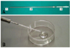
The stage and quality of the embryos were evaluated under a stereomicroscope, and the embryos were loaded into a polyethylene tube (SP65; Nastume, Japan) attached to the injection needle (Fig. 1). With the forceps, the infundibulum was grasped (Fig. 2A), the polyethylene tube was inserted into the oviduct via the infundibulum (Fig. 2B), and 2 to 3 embryos were then transferred (Fig. 2C). After transferring the embryos, the back flow of the medium, into the abdominal cavity, was prevented by grasping the infundibulum with the forceps (Fig. 2D). The polyethylene tube was washed with medium, then checked for any remaining embryos. Recipient goats were used up to three to four times if no pregnancy was established after the ET.
To compare the pregnancy rates, ET by laparotomy was performed as described previously [18]. Briefly, 2-3 embryos were surgically transferred into 1 oviduct ipsilateral to the ovulated ovary, using a syringe connected to a sterile polyethylene tube, which was inserted into the oviduct lumen via the fimbria. The pregnancies were diagnosed by transrectal ultrasound scanning (SonoVet 600; Medison, Korea) using a transrectal 5-MHz linear array probe on day 30 and 40 following ET in both groups.
Experimental design
The pregnancy rates following ET were compared between the laparoscopy and laparotomy groups in experiment 1. In experiment 2, the recipient goats were classified into two groups based on whether they had ovulated or non-ovulated ovaries (GF; ovary with Graafian follicle that was non-ovulated, CH; ovary with corpus hemorrhagicum after ovulation). The pregnancy rates were compared after the laparoscopic ET. In the GF group, the non-ovulated follicle was ruptured artificially by needle puncture prior to the ET. This experiment was performed to investigate the effects of artificial rupture, of non-ovulated Graafian follicles, on the efficiency of laparoscopic ET.
Results
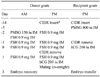
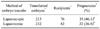
A comparison of the pregnancy rates between the laparoscopic ET and ET by laparotomy revealed that there was a significant difference between the two methods of ET (Table 2). Following the laparoscopic method, 213 transgenic embryos were transferred to 76 recipient goats and 35 recipients (46.1%) became pregnant. However, with the ET by laparotomy, only 22 out of the 82 recipients (26.8%) became pregnant. No significant difference was observed in the pregnancy rates between the ovulated (CH) and non-ovulated (GF) groups (Table 3).
Discussion
The results of this study showed a significantly higher pregnancy rate with laparoscopic ET compared to ET by laparotomy. Tittel et al. [23] noted that laparoscopic adhesiolysis resulted in a significantly reduced number of new adhesions compared to open surgery. The operation was performed only in transferable recipients after laparoscopic exploration of the ovary and uterus.
For efficient production of transgenic goats, by pronuclear microinjection, the pregnancy rate following ET is important. Relatively higher pregnancy rates have been recorded when transferring non-transgenic embryos by laparotomy ET [14], compared to the pregnancy rates after the transfer of transgenic embryos [18]. In previous studies using Korean native goats as recipients, the pregnancy rate following laparoscopic ET was lower than the rate observed in our study (25.7% and 36.8% vs. 46.1%).
The findings of our study suggest that by decreasing the disadvantages of ET by laparotomy, we achieved better results with laparoscopic ET. However, our pregnancy rate with ET by laparotomy was lower than reported in a previous study [11], suggesting that additional studies might lead to an improvement in pregnancy rates of ET by laparotomy.
We also compared the pregnancy rates between the GF and CH groups to evaluate the effects of artificial rupture on the non-ovulated Graafian follicles. When Graafian follicles were identified by laparoscopic ET, they were ruptured artificially for formation of the CL, essential for the maintenance of a pregnancy. We then investigated the effects of the artificially ruptured Graafian follicles by comparison of the pregnancy rates. The most appropriate period for transferring an embryo is within 24 h after ovulation. Although the recipient goats were synchronized with progesterone and PMSG for the ET, some of the recipient goats had not ovulated at the time of the ET. Out of 55 recipient goats, nine goats (16.4%) had not yet ovulated with the Graafian follicle and 46 goats (83.6%) were estimated to have passed beyond the 24 h after ovulation by observation of the corpus luteum (CL). In comparison of the pregnancy rates, there was no significant difference between the CH and GF groups (41.3% vs. 33.3%, respectively, p > 0.05). Although the pregnancy rates were lower than in the CH group, an acceptable pregnancy rate was achieved by artificial rupture in the GF group. Therefore, in cases with a non-ovulated Graafian follicle, artificial rupture was efficient for the formation of the CL, essential for pregnancy maintenance after embryo transfer. However, if artificial rupture was not performed in the Graafian follicle, medical induction of ovulation or additional embryo transfer after CL formation was needed.
The results of this study demonstrated that laparoscopic ET was a reliable and effective technique for efficient production of transgenic goats after pronuclear DNA microinjection. In addition, we found that artificial rupture of the Graafian follicle was an efficient method for the formation of the CL for pregnancy maintenance. More work is needed to better understand the factors involved in this process for further improvement of the pregnancy rate in caprine laparoscopic ET.

 XML Download