

 PDF
PDF ePub
ePub Citation
 Print
Print


Introduction
Photobacterium damselae (Ph. d.) subsp. piscicida, which was first isolated from fish by Snieszko et al. [31], is the etiological agent of pasteurellosis. This disease has been responsible for great economic losses in the marine aquaculture industry [7,13,15,19,27], and a means by which to control this disease is necessary in order for the industry to flourish. Many groups have attempted to develop a vaccine against pasteurellosis [6,7,16,20,25,32], with some examining the effect of culture conditions on the antigenicity of the bacterium [4,5,22,26].
To date, a number of antigens involved in the pathogenesis of Ph. d. subsp. piscicida have been identified [1,3,4,21-23,26,28]. The influence of culture conditions on the expression of virulence factors of Ph. d. subsp. piscicida have been examined [2,4,9,21,26]. For example, when glucose-rich medium (GRM) is used to culture the bacterium [9], a distinct capsule is produced by the bacterium, which is thought to be important to the virulence of the pathogen [10,21]. Iron restriction (IR-) has been used to mimic "near in vivo" culture conditions in vitro [2,11,22], whereby iron-regulated outer membrane proteins (IROMPs) are induced by this restriction. IROMPs help to transfer iron to the bacterium, and also play a role in the virulence of the pathogen by directly sequestering iron from the transferrin and lactoferrin of the host. The IROMPs of Aeromonas salmonicida were the first iron-regulated proteins to be identified as potentially protective antigens located on a fish pathogen [14].
A number of studies have been carried out to characterize the antigens of Ph. d. subsp. piscicida, which are recognized by the immune responses of fish [1,3-5,26]. In the present study, differences in the antigenicity of the bacterium were examined when the bacterium was cultured in vitro in tryptone soya broth (TSB) or under "near in vivo" conditions in TSB + IR-, GRM, or GRM+ IR-. Enzyme-linked immunosorbent assay (ELISA) and Western blot analysis were utilized to investigate these differences using sera collected from sea bass that were either infected with live Ph. d. subsp. piscicida bacteria or immunized with heat-killed bacteria.
Materials and Methods
Bacteria
The isolate of Ph. d. subsp. piscicida (isolate I752) used in this study came from the bacterial collection of the Dipartimento di Scienze della Produzione Animale, Universita di Udine, Italy, and was recovered from sea bream (Scophthalmus maximus) infected with pasteurellosis in 1996.
The bacteria were cultured in four different media: (1) TSB with 1.5% NaCl; (2) iron-restricted TSB with 1.5% NaCl and 0.175 mM 2,2'-dipyridyl (TSB + IR-); (3) GRM (D-glucose 20 g/l, special peptone (Oxoid) 10 g/l, yeast extract 5 g/l, KH2PO4 0.25 g/l, MgSO4 · 7H2O 0.01 g/l, CaCl2 0.6 mg/l, FeSO4 · 7H2O 2.4 mg/l, MnSO4 · 7H2O 0.45 mg/l, NaCl 10 g/l; pH 7.2); and (4) iron-limited GRM which had the same constituents as (3), excluding FeSO4 · 7H2O, but with the addition of 0.25 mM 2,2'-dipyridyl (GRM+IR-). The bacteria were first cultured in TSB at 22℃ for 18 h, and were then diluted 1 to 10 in the chosen culture medium. The bacteria were cultured in either TSB or GRM for 18 h or in TSB + IR- or GRM+ IR- for 36 h, respectively, without agitation. The bacteria were then transferred into fresh media (again using a 1 to 10 dilution) and cultured to log phase prior to use.
Bacteria were grown and harvested by centrifugation at 2,900 × g for 30 min at 4℃, and were washed twice with sterile phosphate-buffered saline (PBS: 0.02 M NaH2PO4 · 2H2O, 0.02 M Na2HPO4 · 2H2O, 0.15 M NaCl; pH 7.2). The concentration of the washed bacteria was adjusted to an absorbance of 1.0 at 610 nm with PBS, and colony-forming units (CFU/ml) of the suspension were determined retrospectively. A portion of the washed bacterial preparation was heat-killed at 60℃ for 60 min, and aliquots of the killed bacteria were stored at -70℃ and used for immunizing fish and screening sea bass antisera.
Production of anti-Ph. d. subsp. piscicida sea bass sera
Sea bass weighing 40 g or 350 g were bought from a commercial fish farm in Italy. The fish were maintained in a land-based flow-through seawater aquarium belonging to the Dipartimento di Scienze della Produzione Animale, Universita di Udine. The tanks were equipped with a system for sterile drainage water, and both the water and the fish were determined to be Ph. d. subsp. piscicida-free. The water temperature was maintained between 25 and 26℃, and the water salinity was 24 ‰.
Eight fish (350 g) were injected intraperitoneally (i.p.) with 5 × 10³ CFU/fish (1 ml) of live Ph. d. subsp. piscicida. The bacteria were grown in TSB at 22℃ for 18 h, and were washed twice with sterile PBS before adjusting the concentration of the suspension prior to injection. Four fish were injected with PBS as a negative control. The fish were bled 3 weeks later from their caudal vein, and the sera were used for Western blot analysis.
Sixty-three fish (40 g), which were also maintained as described above, were injected i.p. with a live preparation of Ph. d. subsp. piscicida. The bacteria were grown in TSB as described above, washed twice with PBS, and adjusted to a concentration of 1 × 10³ CFU/fish (0.2 ml). Another 44 fish were injected with 0.5 mg/fish of heat-killed bacteria in 0.2 ml PBS. The bacteria were grown in TSB, washed twice with PBS, and heat-killed prior to use as described above. Ten fish were injected with PBS (0.2 ml/fish) and used as a negative control. Fish were bled 3 and 4 weeks post-injection, and the antibody levels present in their sera were examined by ELISA.
ELISA
Antibody levels produced by the fish were measured using ELISA according to the method of Bakopoulos et al. [5], with slight modifications. ELISA plates (Immulon; Dynatech, USA) were coated with 0.01% poly-L-lysine in carbonate-bicarbonate buffer, pH 9.6 (100 ml/well), and were incubated for 1 h at 20℃. The plates were washed three times with low salt wash buffer (LSW: 0.02 M Trizma base, 0.38 M NaCl, 0.05% (v/v) Tween-20, pH 7.4), and 100 ml/well of bacterial suspension was added to the plates. Bacteria cultured in the four different media were adjusted to a concentration of 2 × 10⁸ bacteria/ml. The plates were incubated at 22℃ for 1.5 h, and bacteria were then fixed to the wells by the addition of 50 ml/well glutaraldehyde (0.05% v/v) diluted in PBS for 20 min. The plates were again washed by three LSW buffer washes. Non-specific binding sites were blocked with 100 ml/well of a 3% (v/v) solution of H2O2 for 1 h, and were then incubated with 250 ml/well 1% (w/v) gelatine solution in LSW. The ELISA plate was washed three times with LSW buffer. Fish sera, which were diluted 1 : 100 in PBS containing 0.01% (v/v) Tween-20, were added in duplicate (100 ml/well). Sera from fish injected with PBS were used as a negative control, along with a negative control of LSW. The plates were incubated at 20℃ for 1.5 h, washed 5 times with high salt wash buffer (0.02 M Tris, 0.5 M NaCl, 0.1% Tween-20, pH 7.8), and allowed to stand for 5 min during the last wash prior to the removal of the buffer. Anti-sea bass IgM monoclonal antibody (MAb) (Aquatic Diagnostics, UK) was added to the wells (100 ml/well) for 1 h at 20℃. The plates were again washed with the HSW as described above prior to incubation for 1 h with horseradish peroxidase conjugated to anti-mouse-IgG (Diagnostics Scotland, UK) diluted 1 : 1000 in LSW. Plates were washed with HSW as described above, and chromogen/substrate [120 ml of 43 mM tetramethylbenzidine dihydrochloride in 2M acetic acid added to 12 ml of substrate buffer (0.1 M citric acid, 0.1 M sodium acetate, pH 5.4, containing 0.33% v/v H2O2)] was added to each well (100 ml/well). The reaction was stopped after 7 min by the addition of 50 ml/well of 2M H2SO4, and was read spectrophotometrically at 450 nm using an ELISA reader (Dynatech, USA). Optical densities exceeding or equal to 3 times the mean background absorbance were considered positive.
Western blot analysis
Some of the bacteria used in Western blot analysis were freshly isolated from infected fish onto tryptone soya agar (TSA), and were then immediately sub-cultured in one of the four different culture media described above (Fig. 1a-e), while bacteria used in Fig. 1 (f-h) were subcultured several times (more than ten) on TSA prior to culturing in the four different culture media. Sodium dodecyl sulfate-polyacrylamide gel electrophoresis (SDS-PAGE) was performed on Ph. d. subsp. piscicida. In brief, a 12% acrylamide separating gel was prepared, onto which a 4% stacking gel was layered. Bacteria that had been grown in the four different media and washed in PBS as described above were then adjusted to an OD of 1.0 at 610 nm. The bacteria were prepared in sample buffer as described by Bollag et al. [8], with boiling for 5 min. Samples were placed on the gel (15 ml/well), and 180 V was applied to the gel for 45 min. Pre-stained molecular standard markers (Bio-Rad, USA) were used as standards for each gel. Gels containing whole bacteria were transferred onto nitrocellulose membranes by applying 60 V for 70 min using a Hoeffer transblotter. Non-specific binding sites on the nitrocellulose membrane were blocked with 1% w/v bovine serum albumin in Tris-buffered saline (TBS: 10 mM Tris, 0.5M NaCl, pH 7.5) for 1 h at 20℃. The membranes were then washed three times with TBS containing 0.1% (v/v) Tween-20 (TBST); each wash lasted 10min. The membranes were then incubated in anti-Ph. d. subsp. piscicida sea bass sera (diluted 1 to 10 with TBST) overnight at 4℃ with gentle agitation, after which they were washed as described above. Sera collected from fish injected with PBS were used as a negative control. The membranes were incubated with anti-sea bass IgM MAb for 3 h at 20℃. They were again washed three times with TBST, and were then incubated with anti-mouse IgG-HRP conjugate diluted 1 to 500 with TBST. The membranes were incubated with the conjugate for 1 h at 20℃. Blots were washed three times with TBST, 10 min per wash, and were then washed once with TBS and once with PBS. The reaction was then developed by the addition of 20% chromogen (4-chloro-naphthol, 3 mg/ml in methanol) in PBS and 0.01% H2O2 until bands appeared. The reaction was stopped with distilled H2O.
Results
Antibody response of sea bass against Ph. d. subsp. piscicida determined by ELISA
Sera from sea bass infected with live bacteria exhibited significant differences in their antibody responses at 3 weeks post-infection when screened against non-iron-restricted and iron-restricted bacteria using ELISA, with the latter producing greater antibody responses compared to those of non-iron limited bacteria (Table 1). When heat-killed bacteria were used to immunize sea bass, no significant differences were observed in the antibody responses of their sera against non-iron-limited and iron-limited bacteria, as indicated by screening with ELISA at 3 weeks post-infection (data not shown). However, significant differences were found in these sera between bacteria cultured in TSB and bacteria cultured under the other three culture conditions at 4 weeks post-immunization (Table 1). No antibodies were detected against Ph. d. subsp. piscicida in control fish injected with PBS.
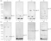
The response of anti-sea bass Ph. d. subsp. piscicida sera against bacteria grown under different culture conditions, compared using Western blot analysis.
Each gel represents serum collected from an individual fish at 3 weeks after injection with Ph. d. subsp. piscicida. The patterns of staining in the blots presented in Fig. 1 (A-E), where the bacteria had been freshly isolated from an infected fish, showed differences between bacteria cultured under iron-restriction (TSB + IR-, GRM+ IR-) and those grown under non-restricted conditions (TSB, GRM). However, when bacteria had been passaged several times on TSA prior to culturing in the four different media (Fig. 1F-H), a similar pattern of staining was obtained with the antisera against bacteria grown under all four culture conditions. This is clearly shown in Fig. 1A and 1F, where the same serum was used but the staining pattern changed between the freshly-isolated bacteria (Fig. 1A) and bacteria passaged several times on TSA (Fig. 1F).
The reaction of sea bass sera to the freshly-isolated bacteria (Fig. 1A-E) resulted in staining of a 22 kDa band in the TSB + IR- and GRM+ IR- bacterial profiles. The sea bass sera, however, recognized a band at 24 kDa in bacteria cultured in TSB and GRM, which was approximately 2 kDa higher than that observed with TSB + IR-- and GRM+ IR--cultured bacteria. Two further bands were detected at 47 and/or 57 kDa in some of the profiles of TSB- and GRM-cultured bacteria using freshly-isolated bacteria (Fig. 1A-D). When bacteria had been passaged several times in vitro, two main bands were identified at 24 and 47 kDa; the 22 kDa band was only observed in two repeatedly passaged samples grown under iron-restriction (Fig. 1F & G). Generally, when freshly-isolated bacteria were used, less staining of low molecular weight material was evident with the TSB + IR- and GRM+ IR- bacterial samples compared with TSB and GRM bacteria. However, a similar level of staining of low molecular weight material was observed with the bacteria subcultured several times, as shown in Fig. 1F-H.
Discussion
It is important to understand the antigenic characteristics of a pathogen in order to develop effective detection methods and vaccines against the pathogen. Insight into the nature of the antibody response elicited against the pathogen and the establishment of the means by which alterations of culture conditions can affect the antigenicity of the pathogen will aid in this understanding.
Agglutination [18,30] and ELISA [1,4,5,24,26] have both been used to measure the levels of antibodies produced by fish against various preparations of Ph. d. subsp. piscicida. Mazzolini et al. [24] immunized sea bass with formalin-inactivated Ph. d. subsp. piscicida, and examined their antibody responses by both agglutination and ELISA. The sea bass produced agglutinating antibodies 2 to 3 weeks after vaccination, which persisted for 3 to 5 months when measured by agglutination, and for 7 months when measured by ELISA. The sea bass also responded very soon after immunization in the study performed by Bakopoulos et al. [5]. These investigators found that fish injected with live bacteria responded more quickly to produce specific antibodies, while fish injected with dead cells took longer to respond, reflecting the lower ELISA results obtained in the present study with sea bass sera produced against heat-killed Ph. d. subsp. piscicida. In a later study, Bakopoulos et al. [6] examined the antibody response of 20 g sea bass against whole cell preparations in adjuvant (AW) and whole cell preparations containing extracellular products (ECP) and crude capsular polysaccharide (cCPS) in adjuvant (AWEC). Fish showed a significant increase in antibody response at 3 weeks post-immunization with the AW and AWEC preparations compared to the PBS-injected control fish, and this difference was still significant at 9 weeks post-immunization. When the antibody response [ECPs, outer (OM) and cytoplasmic membrane (CM), LPS, O-antigen (Ag-O), and extracellular material (EM)] was assessed in 100 g gilt-head seabream (Sparus aurata) immunized with formalin-killed Ph. d. subsp. piscicida, a significant increase in antibody titers was obtained four weeks later, with the greatest responses shown against OM, CM, Ag-O, and then ECPs, respectively. However, when fish received a booster immunization, the order of this response changed such that the highest antibody titer was obtained against the bacterin, ECPs, OM, and then LPS, respectively [1].
In the present study, the antigenicity of Ph. d. subsp. piscicida was found to differ between bacteria grown in TSB, TSB + IR-, GRM, and GRM+ IR- when examined by ELISA and Western blot analysis using anti-Ph. d. subsp. piscicida sera from sea bass infected with either live bacteria or immunized with heat-killed bacteria. Significant differences were found in ELISA between non-iron-limited and iron-limited bacteria with sera sampled from fish infected with live bacteria at 3 weeks post-infection, the latter resulting in a higher antibody reaction compared to non-iron-limited bacteria. However, no significant differences were found between TSB-cultured bacteria and bacteria cultured under the other three culture conditions until 4 weeks post-immunization with sera raised against dead bacteria.
Many reports have focused on producing a high antibody response in immunized fish by altering the immunogens through culture conditions or by adding antigen-related substances, but the antibody response against the immunogen may not be related to protection [12,17,26,29]. Two points are important when examining this correlation. One such point is the determination of which antigens should be present in the test used to screen the antibody response and the other is the establishment of which antigens are responsible for triggering a protective immune response. Antibodies produced during infection may differ from those produced against bacteria cultured in an artificial medium in vitro. Therefore, if antibody levels from infected fish are screened with bacteria grown in vitro, their levels may be represented incorrectly. It has been shown in the present study and the study by Arijo et al. [1] that level of antibodies measured in sera from infected fish depend on the antigens present on the bacteria used to screen the antibody response.
The bacteria used to examine the reactivity of the sea bass antibodies had either been freshly isolated from fish infected with pasteurellosis or had been subcultured several times on TSB. Thick bands were detected at 22 kDa on bacteria cultured in either TSB + IR- or GRM+ IR- using sera raised against live bacteria, whereas a band was detected at 24 kDa on bacteria cultured on TSB and GRM, together with a band at 47 kDa. However, bands were found at 24 kDa rather than 22 kDa when cultured in either TSB + IR- or GRM+ IR-, when the bacteria had been passaged several times in artificial medium prior to culturing in the four different conditions.
Using sera from sea bass infected with Ph. d. subsp. piscicida, Bakopoulos et al. [5] detected a band at 15.5 kDa on bacteria grown in TSB with 2% NaCl and a band at 12 kDa on bacteria cultured in TSB + IR-. These investigators later identified antigens at approximately 21 and 14 kDa in bacteria cultured in modified yeast extracted peptone medium [4]. Nitzan et al. [26], who carried out similar experiments, observed antigenic bands at 36 and 22 kDa on bacteria cultured in the presence of 0.5% NaCl, while only the 36 kDa was observed on bacteria cultured in 2.5 and 3.5% NaCl. The 21 and 22 kDa bands may represent the same molecule as the 22 kDa band observed in bacteria cultured in TSB + IR- or GRM+ IR- in the present study.
LPS and/or lipoprotein antigens were detected at the running front of gel lanes containing bacteria cultured in TSB and TSB + IR- [2], bacteria cultured in a variety of media [2], and bacteria cultured with 0.5% NaCl [26]. In the present study, a similar reaction was observed at the running front of lanes containing bacteria that was freshly isolated and cultured in TSB + IR- or GRM+ IR-, and also in bacteria passaged several times on TSA.
The changes that occurred in the antigenic profiles of bacteria cultured in this study appear to be related to the culture conditions used and the fresh isolation of the bacterium from fish. The ability to make these changes may be part of the bacterial strategy for coping with variations in environmental conditions. The 36 kDa band recognized by Nitzan et al. [26] has been identified as a Na+/H+ antiporter that may play a vital role in maintaining the homeostasis of the bacterium in extreme environments. The epitopes on the 22 and 24 kDa antigens appear to be conserved, with the immune response of the fish still capable of recognizing them after the shift in molecular weight. This was shown earlier in Fig. 1A and F, where the same serum was used but the staining pattern changed between the freshly-isolated bacteria and bacteria passaged several times on TSA.
Ph. d. subsp. piscicida seemed to be very adaptive to its surrounding environmental conditions, showed by repeated passaging on TSA and culturing in the various media used in this and other studies. The antibody response of sea bass against Ph. d. subsp. piscicida differs depending on the antigens used to screen it. However, sea bass sera from infected fish are still able to detect epitopes on antigens after changes in their molecular weight. It remains to be determined how these changes were related to the antigens present on the bacterium during infection, what the extent of relatedness between the 22 kDa and 24 kDa bands may be, and whether the changes that were observed can confer protection against infection.

 XML Download