

 PDF
PDF ePub
ePub Citation
 Print
Print


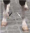
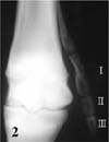
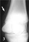
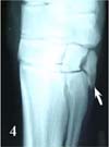
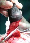
A 7-month-old Berber colt was referred to the National Veterinary School of Maisons-Alfort, France, for evaluation of right sided polydactyly. According to the owner, the abnormal foot was not present at birth. The owner noticed the polydactyly during the first weeks of the foal's life. On admission, the foal appeared to be in good health. Clinical examination was normal and the colt showed no signs of lameness when trotted on soft or hard surfaces. Inspection and palpation of both forelimbs revealed an additional supplementary mobile foot at the site of MC II on the right side (Fig. 1). Palpation of the left forelimb revealed a mobile distal MC II and a 0.5 × 0.3 cm cornified region at the medial aspect of the left fetlock joint. Radiographic examination of the right metacarpal and carpal region revealed a supernumerary digit composed of proximal, middle and distal phalanges at the distal aspect of metacarpus (MC) II (Fig. 2) as well as the development of the first carpal bone. Radiographic evaluation of the left metacarpal and carpal region showed a supernumerary digit composed of one phalanx at the distal aspect of MC II (Fig. 3) and a well-developed first carpal bone (Fig. 4).
Bilateral amputation of the supernumerary digits including the corresponding distal MC II was performed at the owner's request. The results of the foal's pre-anaesthetic examination were normal including auscultation, blood analysis, and biochemistry values. One hour prior to induction, the colt received acepromazine maleate (0.01 mg/kg, IV; CEVA, France). Procain penicillin (22,000 IU/kg, IM; Intervet, France), gentamycin (6.6 mg/kg, IV; Virbac, France) and phenylbutazone (2.2 mg/kg, IV; Vétoquinol S.A., France) was administered 30 min prior to general anaesthesia. The colt received romifidine (0.05 mg/kg, IV; Boehringer Ingelheim, France) and 1% morphine HCl (0.1 mg/kg, IV; Cooper, France) 5 min prior to induction. General anaesthesia was induced with ketamine (2.4 mg/kg, IV; Merial, France) and diazepam (0.05 mg/kg, IV; Roche, France). The colt was intubated with an oro-tracheal tube and was placed in right lateral recumbency. Anaesthesia was maintained with isoflurane (Abott, France) vaporized in oxygen, by using a large animal anaesthesia machine as well as intermittent positive pressure ventilation (Respirator GT; Stephan, Germany). The patient monitoring included: clinical assessment, ECG, direct blood pressure, spirometry, respiratory gas analysis (Datex AS3; Datex Ohmeda, Finland) and arterial blood gas analysis (ABL 77; Radiometer Medical, Denmark).
The right forelimb was prepared aseptically from the carpus to the hoof. A straight incision was performed over the medial aspect of the right MC II. The incision started about 15 cm distal to the carpus and continued distally up to the supplementary hoof, which was incised about 7 mm proximal to the coronary band (Fig. 5). The subcutaneous tissue was bluntly dissected. The supernumerary digit was similar to the normal digit and had an extensor tendon, flexor tendons, a suspensory ligament and sesamoid bones as well as nerves and vessels. The vessels were ligated and subsequently cut as far proximal as possible. An osteotomy of the MC II was performed in its middle part with the use of an osteotome. The distal MC II and the supernumerary digit were removed. The proximal and remaining middle part of MC II was sufficiently attached to MC III. Fascia and subcutaneous tissue were sutured with 3-metric polyglactin 910 (Vétoquinol S.A., France) using simple continuous suture pattern. The skin was closed with a simple interrupted and interrupted horizontal mattress suture pattern using 3-metric monofilament polypropylene. A sterile, modified Robert-Jones bandage was applied to the limb for recovery from anaesthesia.
The site of surgery was the same on the left forelimb. The surgical procedure consisted of an amputation of the supernumerary digit and the distal MC II by osteotomy. The cornified region at the medial aspect of left fetlock did not adhere to the supernumerary digit and was therefore not excised. The cosmetic aspect of both surgical sites was good and the colt recovered from general anaesthesia uneventfully.
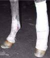
Postoperatively the colt was given procain penicillin (22,000 IU/kg, IM, BID) for 5 days and phenylbutazone (4.4 mg/kg, IV, BID) for one day then 2.2 mg/kg BID for 5 days and 1.1 mg/kg for 3 days. Compressive bandages were changed every 3 days for 2 weeks (Fig. 6). The colt was subsequently bandaged with cotton bandages for 5 weeks. The foal was confined to a box for 4 weeks, followed by inhand walking exercise for 2 weeks, paddock rest for 4 weeks and subsequent pasture exercise. Follow-up information obtained 18 months after surgery revealed a completely sound horse. The cosmetic appearance of both forelimbs was, according to the owner, good.
Polydactyly is a congenital anomaly characterised by the presence of one or more supernumerary digits. This anomaly has been described in man and domestic animals (e.g. cats [12], dogs [8], horses [2,3,16] cattle [1,9,14]). This malformation can occur as an isolated anomaly or together with rare developmental or inherited anomalies [4,15]. In man, polydactyly can occur sporadically but it can also be inherited with in many cases an autosomal dominant pattern of inheritance. Many genes have been associated with limb malformations in man [6]. In rare cases, polydactyly may be caused by external factors like exposure to toxins [5].
The occurrence of polydactyly is rare in horses; however, this anomaly constitutes the most frequently described congenital phalangeal malformation in the horse [13]. About 80% of polydactyl cases show a supernumerary digit at the medial aspect of the forelimb. In one study of 100 horses, 60% of the animals had one supernumerary digit, 24% bilateral involvement and 15% had this anomaly in all four limbs [10]. The etiology of polydactyly remains unknown in horses; various classifications of polydactyly have been described. One of the most common is the three-group classification: In the teratogenic form, the supernumerary digits are due to a teratogenic splitting of the basipodial elements, i.e. schistodactyly. Most cases have all phalangeal bones affected whereas the splint bones are often normal. In the developmental or atavistic form, the digit usually derives from the distal end of the splint bone. The bilateral symmetric-inherited form is a hereditary condition in poultry and dogs [7]. However, the above classification is controversial and characteristics of more than one form might be present in the same individual [13]. In the above case, no cytogenetic analysis was performed as described by Giofré et al. [5]. Neither the dam nor sire had any signs of polydactyly. Therefore, the etiology of the polydactyly in the colt described here is unknown.
The diagnosis of polydactyly is normally based on clinical examination. A complete radiographic evaluation of the metacarpal and metatarsal region and adjacent joints, e.g. the carpus, is necessary to determine possible osseous abnormalities [2] (e.g. the presence of a supernumerary MC I [3]). Surgical removal of the supernumerary digits is provided to improve cosmetic appearance and to prevent injury to the digit [11], but should not destabilize the involved articulation. The incomplete removal of supernumerary digits might cause an insufficient cosmetic outcome and/or lameness in horses [11]. In the above described case, the removal of the distal digit and the adherence of MC II to MC III decreased the risk of postoperative lameness and resulted in a good cosmetic outcome. However, the well-developed first carpal bone might constitute a risk for future degenerative joint disease.
 XML Download