

 PDF
PDF ePub
ePub Citation
 Print
Print


Japanese encephalitis (JE) is a mosquito-borne viral zoonosis that is becoming increasingly important in terms of public health. Japanese encephalitis virus (JEV), a member of the genus Flavivirus in Flaviviridae, is an emerging virus that is spreading to new areas. Several species of mosquito, including Culex tritaeniorhynchus in Asia, are thought to be vectors for JEV. Domestic and wild animals, including pigs, horses, cattle, sheep, goats, pigeons, chickens, gray herons, and reptiles, are susceptible to the virus. Adult swine, horses, cattle, and sheep usually do not manifest clinical symptoms of the disease, but they may serve as viral amplifiers [3]. Seroepidemiological surveys of JEV in pig populations have been conducted as part of the preventative measures against JE in several countries, including Korea [1,9-11]. Since the hemagglutination inhibition (HI) antibody to JEV infection in pigs is long lasting, and pigs serve as viral-amplifying hosts, serological sampling of pigs may not show the exact prevalence of JE in a given period. In this study, we investigated the seroprevalence of JEV in domestic goats to determine a more exact JE infection rate and improve our understanding of its transmission in the period from October to March. The seroprevalence survey consisted of a total of 804 goat serum samples from seven provinces from 144 farms in Korea between May 2005 and May 2006 [Gyeonggi (n = 59), Gangwon (n = 31), Chungbuk (n = 103), Jeonnam (n = 186), Jeonbuk (n = 224), Gyeongbuk (n = 91), and Gyeongnam (n = 110)]. Most of the samples were collected between October 2005 and March 2006. In order to estimate the JEV antibody status of the goat sera, the HI test was performed in 96-well microtiter plates using the standard method [2]. Viral antigens were prepared from suckling mice brains infected with the Nakayama strain using the sucrose-acetone extraction method [2]. Briefly, the infected suckling-mouse brain was homogenized with 5 volumes of 8.5% sucrose solution and the homogenate was added to 20 volumes of chilled acetone. After shaking vigorously, the milky supernatant was discarded and an equal volume of acetone was added to the bottle. This preparation was incubated for 1 h at 4℃ to dehydrate the sediment. The supernatant was discarded and the sediment was dried using a vacuum pump. The dried antigen was suspended in saline. After centrifugation for 10 min at 10,000 rpm, the supernatant was used as the HI test antigen. The serum specimen was pretreated with kaolin to remove any non-specific inhibitors and then adsorbed with washed goose red blood cells to eliminate natural agglutinin. An HI titer of 1 : 20 or greater was considered positive.
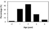
The incidence of JEV-positive cases was 12.1% (97 out of 804 sera), and 45 of the 144 farms tested had positive cases. The regional distribution of positive JEV cases was 20.0 (22/110), 16.1 (30/186), 15.6 (35/224), 5.8 (6/103), 5.1 (3/59), 1.1% (1/91), and 0% (0/31) in Gyeongnam, Jeonnam, Jeonbuk, Chungbuk, Gyeonggi, Gyeongbuk, and Gangwon province, respectively (Table 1). In addition, while there were no positive reactions from British Saanen goats imported from Australia and raised in Gangwon province (0/31), the incidence in Korean native black goats was 12.5% (97/773), with titers ranging from 1 : 20 to 1 : 320 (Table 2). The highest prevalence of seropositive animals was observed in 2-year-old goats, of which 60 out of 97 had positive sera (Fig. 1). Since the HI antibody titer to JEV is not persistent in goats and lasts only about 4 weeks, it is thought that the age of the goats is not important when determining the antibody positive rate and titer, and that only the abundance and distribution of the mosquito vector are thought to be important for the JEV infection of goats during the experimental period. Of the 520,000 goats currently being raised in Korea, most are Korean native black goats that are used for meat production, as well as some British Saanen that are used for milking. Although several diseases associated with goats, such as rotavirus and bovine viral diarrhea infections [4,5] have been reported, no nationwide seroepidemiological survey of arboviral infection in goats has been reported. A serological survey of JEV infection in domestic animals, including sheep and goats, was carried out in 1956 in Korea, but the ovine serum samples were collected only in Kyongju city and the positive rate for JEV was 21.7% (26/120) in goats [6]. In this study, three districts of Korea, primarily in southern regions, showed a relatively high seroprevalence of JEV. The results suggest that JEV is actively transmitted in the southern regions of Korea from October to March (winter). Epidemiologically, goats older than one year were more likely to be exposed to JEV than younger goats. Rajendran et al. [8] reported that HI titers against JEV in goats were low, and these levels declined to undetectable levels by approximately 4 weeks after seroconversion. In addition, Peiris et al. [7] reported that the JE seroprevalence in cattle and goats was a better predictor of the human infection risk than the porcine seroprevalence. Therefore, we believe that goats would serve as a good sentinel animal for serological monitoring of JEV infection in domestic animals because they are currently not vaccinated against JEV in Korea. A sero-monitoring system for goats and swine should provide a clear picture of the epidemiological characteristics of JEV transmission in the natural environment. Further work is required to determine which species of mosquito serve as vectors of JEV transmission in goats.


 XML Download