

 PDF
PDF ePub
ePub Citation
 Print
Print


Bovine spongiform encephalopathy (BSE) in cattle is a neurodegenerative disorder belonging to the transmissible spongiform encepahlopathies (TSE), a group of diseases that include sheep scrapie and human Creutzfeldt-Jacob disease (CJD) [8]. The accumulation of abnormal isoforms of the host encoded cellular prion protein (PrP-C), as a protease-resistant prion protein (PrP-Sc) in the nervous tissue, has been proposed as the cause of the TSEs [6]. TSE's are characterized by long asymptomatic incubation periods [9]. The pathognomonic lesion is a combination of both spongiform change in gray matter neuropil and neuronal vacuolation of certain brain stem nuclei [4,10].
In March 1996 in England, the announcement of a new and variant form of CJD in humans that could be linked to BSE increased awareness of the possible danger of BSE to human health [3]. BSE has been a notifiable disease in Turkey since 1997, with the prohibition of the importation of live cattle and products of bovine origin, such as meat and bone meal (MBM), gradually instated between 1996 and 2001 [7]. The Turkish Ministry of Agriculture and Rural Affairs has been responsible for the passive and active surveillance of BSE since 2001. The Geographical BSE risk (GBR) assessment of Turkey by the EU Scientific Steering Committee (SSC) was released in 2002, which evaluated as Level III, i.e. "it is likely but not confirmed" that one or several domestic cattle are (clinically or pre-clinically) infected with the BSE-agent [7]. In the decision making of the GBR report of Turkey, the importation of cattle and MBM, or feed stuffs containing MBM, from the UK and other BSE risk countries, was considered an external challenge to the animal stock of Turkey. A break down of the imported live animal and MBM into 5-year periods, as well as their affect upon the animal population as an external challenge risk, was evaluated as being very high between 1986-1990 and 1991-1995 and high between 1980-1985 and 1996-2000 [7]. The purpose of this presented study was to establish a targeted surveillance, on a regional scale, within a population of animals at risk, with respect to age and period, according to the GBR report of Turkey.
Between 2004 and 2005, a total of 30,847 cattle were slaughtered for human consumption in Bursa slaughterhouses. Samples were collected from the animals that might be considered to be in the age and period risk groups for BSE; therefore, animals at an age of 30 months and older originating from intensive feeding farms were the target population. Among the 30,847 animals, 8,385 were within this range, with a total of 420 obex cerebri and medulla oblongata [1,4] samples collected from these animals. Tissues were immediately placed in 10% neutral buffered formalin and fixed at least 48 h [4]. The remaining brain stem samples were kept at -20℃ for further Western Blot analysis in case a BSE positive or suspected result was obtained. Samples of the obex cerebri were treated with 96% formic acid for 60 min to enhance antigen retrieval and decontamination [4]. Sections from the obex cerebri were processed for hematoxylin-eosin (H&E) and immunohistochemical (IHC) staining. As a BSE positive control tissue, obex samples provided by Dr. Anne Buschmann, the Friedrich Loeffler Institute, Germany, to Dr. Yavuz Ulusoy, Central Veterinary Control and Research Institute, Etlik, Turkey, were used, with written permission, in this study.
Conventional methods were used in the preparation of tissues for H&E staining. For the IHC, a commercially available monoclonal antibody F99/97.6.1 (Pullman TSE-IHC/99; VMRD, USA) was utilized, with the tissues prepared according to the manufacturer's instructions. Briefly, the tissues were decontaminated in 95-98% formic acid for 60 min, and then autoclaved for 20 min at 121℃ in citrate buffer. For the proteinase K treatment, 100 µl of the enzyme was applied for exactly 1.5 min. The slides were then incubated with 100 µl MAb F99/97.6.1 (freshly diluted 1 : 100) for 10min. Following the application of the secondary antibody, and consecutive applications of streptavidin-HRP and amino-ethyl-carbazole (AEC), the slides were mounted with immune mount. Sections prepared from the obex cerebri of BSE positive and healthy cattle, as negative control, were included in each cycle of the staining procedure.
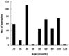
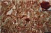
The age distribution of the 420 animals is shown in Fig. 1. Of these animals, 56.4% were at an age of 50-months and older. Except for limited mononuclear infiltrations in the meninges of two cattle, no other pathological lesions were observed in the sections of the brain tissue stained with H&E. While no positive immunostaining for the PrP-Sc antigen accumulation was observed in any of the 420 cattle brain tissue samples, sections from the BSE positive cattle brain showed positive brown to red PrP-Sc deposits, in the form of clusters and plaques, within the perikaryon (Figs. 2 & 3).
It is estimated that only 7.2% of BSE infected cattle have an incubation period of less than 36 months and rapid BSE tests do not generally have the ability to detect early BSE cases [1,4]. In this presented study, all samples were collected from animals older than 30 months. The use of IHC, which has been considered as a golden standard for the detection of PrP-Sc in formalin fixed paraffin embedded material, has been applied successfully in both active and passive surveillance [2,4,5]. Using this technique, the detection of BSE cases in the last month of incubation, prior to the occurrence of vacuolar changes, seems possible [4]. All of the obex cerebri samples in this study were treated with IHC, but no positive reaction to the F99/97.6.1 monoclonal antibody was observed. The effectiveness of the monoclonal antibody, F99/97.6.1, in the detection of BSE has been documented [2,5], and reacts with the epitope conserved in most ruminant species [5].
The GBR report of Turkey stated that the system had been exposed to very high external challenges due to live cattle and MBM imports from BSE risk countries; however, the uses of MBM in cattle is not economical, and since plant proteins are more affordable, these have been used as the protein source of cattle feed in Turkey [7]. Again; within the same period, 76% of the 1,141,476 live cattle imported into Turkey were at and age of either 18 months, for fattening, or 24 months, for immediate slaughtering. However, for the purpose of risk assessment, the SSC report assumed that a small fraction of these cattle might have entered the national cattle herd and increased the external challenge of Turkey [7]. Thus, in the recommendations section of the same report, it was stated that improvement of the passive surveillance and expanding the active surveillance system should be made by increasing the sampling of asymptomatic animals from the at-risk cattle populations [7]. The size of the sample population used in this study was not sufficient for an accurate assessment of such risks; however, the tissue samples were from animals that were either born during the risk period or were the offspring of risk period animals. In conclusion, while closely monitoring animal movement and products of bovine origin, a larger scale of active surveillance, similar to that presented in this work, should be performed without interruption throughout the nation.

 XML Download