

 PDF
PDF ePub
ePub Citation
 Print
Print


Introduction
Several methods of anesthesia for flank laparotomy in standing cattle have been described [2,11]. These methods include infiltration (line block and inverted L-block), paravertebral (proximal and distal), epidural and subarachnoid (with or without a catheter) anesthesia. Each method has some advantages and disadvantages for clinical use. Infiltration anesthesia, in particular, has been widely used in clinics because it is easy to perform without special knowledge and skill, although a higher volume of anesthetic solution, between 50 and 100 ml, is needed to desensitize at least three vertebral nerves [the last thoracic (T13), the first (L1) and second lumbar (L2) spinal nerves] for flank surgery. On the other hand, segmental dorsolumbar epidural anesthesia is considered to be a difficult and challenging technique to perform; however, it may be the most effective method of anesthesia delivery when performed by an expert [2,11]. In addition to the wide individual variation among the animals, the fact that a needle needs to be inserted into the epidural space, which is just above the spinal cord, may make one fearful and hesitant to use segmental dorsolumbar epidural as a first choice for flank anesthesia in cattle [6].
After identifying several influencing factors on dorsolumbar epidural anesthetic effect [3,10], the method was modified to minimize the effects of epidural pressure and epidural fat [7,8]. The modified dorsolumbar epidural anesthesia method using a combination of 0.025mg/kg of xylazine hydrochloride and 0.1 mg/kg of lidocaine hydrochloride induced analgesia that was suitable for laparotomy in standing conscious cattle [5]. However, it is impractical to measure the bodyweight of each cow in a bovine clinic or field before surgery. In a previous study, the clinical usefulness of a fixed volume of anesthetic with this modified method was examined in clinical cases at a veterinary medical teaching hospital [4]. The anesthetic combination composed of 1 ml of xylazine and 3 or 4 ml of lidocaine was sufficient for anesthesia in dairy cattle undergoing flank surgery [4]. The degree of difficulty with which beginners learn and use modified dorsolumbar epidural anesthesia in cattle was also examined in the same hospital [6]. In this study, the introduction of the modified dorsolumbar epidural anesthesia method with a fixed volume of anesthetic was tested in a bovine referral center for 10 months.
Materials and Methods
One hundred and thirty, 2- to 12-year-old (5.1 ± 2.0 years) female Holstein cattle were included in this study. The cattle were transported from several farms to the Hamanaka branch of the Kushiro District Agricultural Mutual Aid Association and were scheduled to undergo flank surgery in a standing position between June 2004 and March 2005. Each cow was positioned in a chute and its body condition score (BCS) was determined on a 5-point scale (1, emaciated; 2, thin; 3, average; 4, fat; 5, obese) with increments of 0.25 for scores between 2.25 and 4.00 [1].
The skin surrounding the first interlumbar (L1-L2) space was shaved and aseptically prepared. A 16 G, 120 mm Tuohy needle (Hakko Medical, Japan) was inserted using a dorsal midline approach. When the needle tip reached the ligamentum flavum, the stylet was removed and saline was added to the hub of the needle. The needle was then slowly inserted into the epidural space. If the L1-L2 epidural space could not be reached, the space between the last thoracic and first lumbar (T13-L1) vertebra was used. The entrance to the epidural space was identified using the hanging drop technique [11], and air was then allowed to freely enter the space for about 1 min in order to decrease the effect of negative epidural pressure [5]. The epidural needle was then slowly inserted about 1 cm deeper in order to penetrate the epidural fat [8]. The insertion of the needle was stopped if the cow showed any signs of discomfort, such as a sudden movement or 'dipping' of the back [4,5,8]. After confirming that there was no blood or cerebrospinal fluid present in the aspirate, 90 cattle (group 1; age, 5.2 ± 2.1 years; BCS, 2.86 ± 0.36) received a 4 ml mixed anesthetic solution containing 1 ml of 2% xylazine hydrochloride (xylazine; Daiichi, Japan) and 3 ml of 2% lidocaine hydrochloride (xylocaine; AstraZeneca, Japan) according to the method described in the previous study [4]. The dose of xylazine was reduced in cases where the cattle showed signs of weakness, dehydration and/or lameness. These 18 cattle (group 2; 5.0 ± 1.8 years; 2.90 ± 0.42) received a mixed anesthetic containing 0.5 ml of xylazine and 3 ml of lidocaine. The solution was administered at a rate of 0.5 ml/s with the needle bevel directed cranially, and the needle was then removed. The distance from the skin to the epidural space and the appropriate depth of injection were determined by measuring and subtracting the distance between the skin and needle hub from the length of the needle (distance between the needle hub and end of the needle) [9]. Infiltration anesthesia (line block) was performed in 22 cattle (group 3; 5.8 ± 2.4 years; 3.06 ± 0.46) because the L1-L2 and T13-L1 space could not be reached. After preparation of the surgical area, including shaving and disinfection, surgery was performed on the right side of the flank for left displacement of abomasum (LDA), right displacement of abomasum (RDA) and intestinal volvulus, and on the left side for cesarean section.
The existence (score 0) or non-existence (score 1) of light sedation was defined in terms of the drooping of the animal's upper eyelids, the position of the head relative to the shoulders, and the reduction in the animal's awareness of its surroundings. The analgesic effect was assessed and scored by the animal's response to skin, muscular and peritoneal incisions (0, no responses; 1, movement with no kicking; 2, movement with a little kicking; 3, struggling with repeated kicking). When the cattle showed purposeful movement, indicated by a score of 3, additional infiltration anesthesia (line block) with lidocaine was performed before any further incisions were made. However, when the animal showed non-purposeful movement, indicated by a score of 2, the surgery was performed without additional anesthesia. The degree of ataxia was assessed and scored by observing the posture of the animal (0, standing with 2 limbs; 1, swaying or standing with 1 limb; 2, leaning against the chute; 3, sternal recumbency). After surgery, the cattle came out of the chute and were transported back to each farm by truck.
Descriptive statistics (mean ± SD), one-way ANOVA and Scheffe tests were utilized to compare the characteristics of the cattle in the three groups. The Mann-Whitney's U test was used to compare the sedative, analgesic and ataxic effects after administration of mixed anesthetics. A value of p < 0.05 was considered significant.
Results
The entrance to the epidural space was successful in 108 of 130 cattle (83%). A mixed anesthetic solution was injected into the L1-L2 space of 94 cattle and into the T13-L1 space of 14 cattle by several veterinarians. There was no significant difference in age or BCS between the cattle in groups 1 and 2. Although the 22 cattle in group 3 were generally older and had higher BCS, there were no significant differences between these cattle and those in the two groups in which the epidural space could be reached. The mean distance from the skin to the epidural space and the depth of anesthesia in group 1 (81 ± 7 and 90 ± 6 mm) were similar to those in group 2 (81 ± 4 and 90 ± 5 mm). The surgeries in groups 1 and 2 began about 12 min after the epidural administration of a mixed anesthetic solution (11.6 ± 3.5 vs. 12.2 ± 3.6 min) and lasted for about 36 min (36.0 ± 18.8 vs. 35.6 ± 23.6 min).
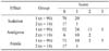
The sedative, analgesic and ataxic effects after epidural administration of mixed anesthetics in 108 cattle are summarized in Table 1. Twenty cattle from group 1 and one cow from group 2 showed light sedation after epidural administration of a mixed anesthetic. Analgesia was sufficient for flank surgery in almost all of the cattle except for one cow in group 1 in which infiltration anesthesia (line block with lidocaine 50 ml) had to be performed. Although 13 cattle in group 1 and two cattle in group 2 showed some movement with a little kicking, the surgery was performed successfully without struggling or need for additional infiltration. Five cattle in group 1 showed sternal recumbency during and/or after surgery. Three of these cattle were in poor health condition, and the other two had problems with their feet. Four of them stood up within 1 h after the surgery, and the other one stood up the next day.
Seventy-two of the animals underwent omentopexy for LDA, 29 underwent omentopexy for RDA, six underwent cesarean section and one underwent surgical correction of intestinal volvulus. Of the 22 cattle whose epidural space could not be reached, 15 underwent omentopexy for LDA and seven underwent omentopexy for RDA after infiltration anesthesia (line block).
Discussion
In this referral center, right paramedian abomasopexy on a hydraulic surgical table was the primary method used to treat left and/or right displacement of abomasum for several years. However, the number of surgeries to be performed has been on the rise in recent years, making it necessary to effectively use time and manpower in a limited space in order to reduce the cost of each surgery. The method for performing right flank omentopexy and/or abomasopexy in a standing position substituted the use of two chutes for a hydraulic surgical table. Under these circumstances, the first author became a part of the bovine referral center after graduation from veterinary school and introduced modified dorsolumbar epidural anesthesia to her co-workers. Although dorsolumbar epidural anesthesia has been considered a difficult technique to perform in cattle [2,11], veterinarians at this referral center easily mastered and used the modified method in their surgical practices.
Insertion of the epidural needle into the epidural space was unsuccessful in 22 of 130 cattle, and the unsuccess rate of 17% was slightly higher than the rate estimated in a previous study [4]. The primary cause of difficulty in reaching the epidural space with the epidural needle in the T13-L1 or L1-L2 intervertebral space may be attributed to the ossification of this space due to aging [11]. However, there were no significant differences in age and BCS between the 22 unsuccessful and 108 successful insertion cases, even though age and BCS tended to be higher in the unsuccessful cases. The higher rate of unsuccessful insertions may be a result of the introduction of this new method because all of the veterinarians were new to this method except for one. With time and experience, there will be an increase in the success rate to that which was estimated in a previous study [6].
The mean distance from the skin to the epidural space (about 80 mm) and the mean depth of injection (about 90 mm) in this study were consistent with those in previous studies [4-6,8], and these depths are very important indexes with regard to the feeling of the resistance in the three ligaments during epidural insertion of needle [6]. The supraspinous ligament and ligamentum flavum are very resistant to needle penetration, but the interspinous ligament exhibits little resistance. Although there are individual variations in size, body weight and BCS among cattle, the combination of these two pieces of information will lead to more successful and safer insertions of the epidural needle in conscious cattle.
The use of 1 ml of xylazine in healthy adult animals was recommended in a previous study [4]; however, a reduction in the dose of xylazine was also recommended in order to prevent severe sedation and recumbency in weak animals. In this study, the recumbency of cattle was reduced by decreasing the xylazine dosage from 1 ml to 0.5 ml. This dose was about 0.017 mg/kg in cattle with a body weight of 600 kg. Before performing this epidural anesthesia method, veterinarians should decide the appropriate xylazine dosage based on patient examination.
Unfortunately, in this study, detailed information regarding the surgeries in the 22 cattle whose epidural space could not be reached was not recorded. However, routine flank laparotomy with infiltration anesthesia (line block) can be easily imagined. During flank surgery in cattle in a bovine practice, kicking and violent movements have been considered natural or common occurrences. This may be due to the cattle experiencing sensations of pain and fear; therefore, proper sedation and analgesia should be performed during the standing surgery [4]. All of the veterinarians at this referral center were satisfied with the better analgesia afforded by modified dorsolumbar epidural anesthesia in comparison to that of the infiltration anesthesia (line block) with procaine and/or lidocaine, which they had been using for several years.
In this referral center, shaving, scrubbing, disinfecting and draping of the surgical site were systematically performed after epidural administration of a mixed anesthetic. Thus, the surgery began soon after the epidural was administered and lasted for about 36 min. This allowed the veterinarians to effectively use their time and manpower to continue the surgeries in a limited space. Simultaneously performing two surgeries with two chutes was more effective than doing one surgery with a hydraulic surgical table. Some time and effort was required to manipulate the table and to restrain the cattle on the table when performing the other type of surgery. The modified method of anesthesia was a good fit for this situation.
In this study, it was clearly shown that the modified dorsolumbar epidural anesthesia method could be easily introduced and effectively used in a bovine referral center. Furthermore, it was not difficult for the veterinarians in the clinic to learn and use this modified method. This modified method has several advantages. Analgesia of both flanks could be obtained by a single injection of a small volume of anesthetic with minimal desensitization of the hindlimbs. This would allow veterinarians to save time and effort, and consequently lower the cost of each surgery while securing the welfare of the cattle through painless surgery and preventing injury to the veterinarian.
 XML Download