

 PDF
PDF ePub
ePub Citation
 Print
Print


Introduction
Babesia gibsoni (B. gibsoni) is a well-known causative pathogen of canine babesiosis and causes severe hemolytic anemia in infected dogs [1,4,5]. Our previous study [6] demonstrated that the serum from dogs infected with B. gibsoni retarded the maturation of canine reticulocytes and decreased the activity of erythrocyte 5'-nucleotidase in a dose dependent manner. The in vitro replication of B. gibsoni also induced a significant decrease in enzyme activity. During the final stages of maturation of reticulocytes, to their mature state, erythrocytes undergo a series of biochemical and physiological transformations. The maturation of reticulocytes is known to be heavily dependent upon the function of erythrocyte 5'-nucleotidase. This enzyme mediates the hydrolysis of ribose or deoxyribose in various 5'(3')-nucleotides, producing inorganic phosphate and the corresponding nucleoside. The substrate of this enzyme appears to be derived from the degradation of reticulocyte RNA in ribosomes during the maturation of reticulocytes. Therefore, this enzyme aids in the removal of useless ribosomal RNA which is involved in the morphological maturation of reticulocytes by contributing to the degradation of reticulocyte RNA in ribosomes [8,21].
We previously showed that B. gibsoni parasites preferentially invade and multiply in reticulocytes rather than in mature erythrocytes when cultured in vitro [14,23]. In general, the reticulocytes contain ribosomes, polyribosomes and mitochondria, and have a higher concentration of adenosine triphosphate, reduced glutathione, amino acids, and nucleic acids compared to mature erythrocytes [13,22]. Severe anemia often occurs in dogs infected with B. gibsoni in spite of a markedly low percentage of parasitized erythrocytes in their peripheral blood [2,10]. Anemic dogs infected with B. gibsoni have many young erythrocytes, including polychromasia, reticulocytosis and occasionally increased numbers of nucleated erythrocytes in vivo [4,5]. During in vitro incubation of canine reticulocytes, with serum from dogs infected with B. gibsoni, the inhibitory effect of serum in infected dogs on the maturation of reticulocytes may contribute in part to reticulocytosis in vivo in canine babesiosis. However, severe hemolytic anemia often occurs in dogs infected with this parasite in spite of a very low percentage of parasitized erythrocytes in their peripheral blood [2,10]. Furthermore, the infected serum has been shown to inhibit the activity of erythrocyte 5'-nucleotidase measured with cytidine 5'-monophosphate (5'-CMP) and inosine 5'-monophosphate (5'-IMP) in vitro [6].
In the course of a prior study, we found that the serum from dogs infected with B. gibsoni had a suppressive effect. Therefore, we considered that it might contain certain factor(s) that retard the maturation of canine reticulocytes in vitro. The serum might change the properties of reticulocytes in the dogs infected with B. gibsoni. In vitro culture supernatant of Plasmodium falciparum was used for the study of immunogenicity and isolation of antigents for serology [17,20]. The supernatants of in vitro cultures of B. divergens from human erythrocytes were obtained from glycoproteins excreted and/ or released from merozoites [3]. Confirmation of these possibilities requires further elucidation. The purpose of the present study was to investigate the serum from dogs infected with B. gibsoni, and to determine the effect of the culture supernatant of erythrocytes infected with B. gibsoni on the maturation of canine reticulocytes.
Materials and Methods
Reagents
We obtained the necessary reagents from the following sources: cellulose microcrystalline from Merck (Germany); Percoll gradient solution from Amersham Pharmacia Biotech (Sweden); alpha-modification of Eagle medium (α-MEM) from Life Technologies (USA); and potassium benzylpenicillin (penicillin G; Meiji, Japan) and streptomycin sulphate (Meiji, Japan). All other chemicals were purchased from Wako Pure Chemicals (Japan).
Preparation of canine serum
Sera were collected from four clinically healthy dogs and three dogs chronically infected with B. gibsoni. The reticulocyte percentage was 0.6 ± 0.3% (mean ± SD, range 0.1-1.0%) in the clinically healthy dogs and 5.5 ± 3.7% (range 3.2-9.8%) in the infected dogs. The strain of B. gibsoni used in this study was originally obtained from a dog infected naturally with B. gibsoni in the city Nagasaki in 1973; this strain has been maintained in dogs at Hokkaido University since then. All experimental procedures were in accordance with the guidelines for animal use at the Graduate School of Veterinary Medicine, Hokkaido University, Japan.
Preparation of canine reticulocytes
Canine reticulocytes were prepared by the method of Maede and Inaba [12], with some modifications. Four clinically healthy dogs weighing about 10-12 kg were used. About 200-240 ml blood was taken from the cervical vein of each dog once daily for three days. On the third day after bleeding, 130 ml of whole blood was collected into heparinized syringes. At that time, the reticulocyte count in the peripheral blood from each dog was 6.0-8.5%, as determined by microscopic examination of a blood smear stained with new methylene blue staining solution [9]. The collected blood was washed twice with 10 mM phosphate-buffered saline (PBS, pH 7.4) and resuspended in PBS with a packed cell volume (PCV) of about 25-30%. The reticulocytes were separated from the washed cell suspension by Percoll discontinuous gradient centrifugation. 45% (v/v) and 64.5% (v/v) Percoll solutions containing 150 mM NaCl, 0.1% (w/v) bovine serum albumin, and 20 mM HEPES/Tris buffer (pH 7.5) were then used for preparation of the discontinuous Percoll gradients. The solutions had specific densities of 1.070 and 1.096 g/ml, respectively. The erythrocyte suspension was carefully layered over the Percoll gradient and centrifuged at 1,800 × g for 15 min at room temperature. The reticulocyte-rich (reticulocyte count 70-95%) portion was located at the interface of the two Percoll solutions. The separated reticulocytes were washed twice with PBS and then three times with α-MEM supplemented with sodium pyruvate (0.11mg/ml), potassium benzylpenicillin (100 units), and streptomycin sulphate (100 µg/ml). The reticulocytes with a final PCV of 2% were incubated at 37℃ in a humidified atmosphere containing 5% CO2 and 95% air in an incubator (Model 6100-100; Napco, USA). The following was added to the incubation media to examine the effects on the maturation of canine reticulocytes: culture supernatant of erythrocytes infected with B. gibsoni, incubated erythrocytes supernatant, serum free culture supernatant of erythrocytes infected with B. gibsoni and serum free incubated erythrocytes supernatant were obtained. In addition, serum from dogs who were chronically infected with B. gibsoni, serum from normal dogs, fractions of serum from dogs chronically infected with B. gibsoni (>150 kDa, 150-70 kDa and <70 kDa), and fractions of culture supernatant of erythrocytes infected with B. gibsoni (>150 kDa, 150-70 kDa and <70 KDa). The final concentration of each serum sample, supernatant, fraction and the time of incubation are shown in each figure legend.
Preparation of fractionation of serum infected with B. gibsoni and culture supernatant of erythrocytes
B. gibsoni was cultured according to the method reported by Hossain et al. [7]. Venous blood was collected from clinically healthy dogs using ethylenediaminetetraacetic acid as an anticoagulant. The Buffy coat was removed after centrifugation, and the erythrocytes were washed twice with PBS and then washed three times with α-MEM containing the supplements described above. The washed erythrocytes were resuspended in a culture medium consisting of 80% α-MEM and 20% serum from normal dogs. For cultivation of the parasites, B. gibsoni-infected erythrocytes, with a high parasitemia in subculture, were added to the prepared erythrocyte suspension to yield a parasitemia of 2% and a PCV of 3%. The erythrocytes were separated using the method described above, were washed three times with PBS, and then these erythrocytes were added to the media with serum from normal dogs or the serum free media for a suspension at a PCV of 3%. All of the suspensions were placed in each well of a 96-well flat-bottomed microculture plate and incubated at 37℃ for 6 days in a humidified atmosphere containing 5% CO2, 5% O2 and 90% N2, using an incubator (APMW-36; Astec, Japan). Every 24 h, 60% of the culture supernatant and the incubated supernatant of erythrocytes were removed without disturbing the sediment of erythrocytes; they were replaced with an equal volume of either fresh culture medium or serum free media. The culture supernatant of erythrocytes were collected after 6 days of cultivation of the erythrocytes infected with B. gibsoni, and the incubated supernatant of erythrocytes without infection, were deep frozen at -70℃ and then used for fractionation and incubation. Serum from dogs chronically infected with B. gibsoni and the culture supernatant of erythrocytes infected with B. gibsoni were fractionated using high-performance centrifugal concentrators (Orbital Bioscience, USA) to fractions of >150 kDa, 150-70 kDa and <70 kDa.
SDS-polyacrylamide gel electrophoresis (SDS-PAGE)
Proteins from the serum of dogs infected with B. gibsoni and the infected culture supernatant of erythrocytes were separated by SDS-PAGE according to the method of Laemmli [11] using a 7.5-10.0% gradient gel (Atto, Japan). Samples were dissolved in Laemmli sample buffer (Bio-Rad, USA) and electrophoresis was carried out using a current of 20 mAmp for 1.5 h. The gels were stained with Coomassie brilliant blue (Bio-Rad, USA) or Stains-All (Sigma, USA). The apparent molecular weights of the protein bands were estimated by comparison with a SeeBlue Plus2 Pre-Stained Standard (Invitrogen, USA).
Results
SDS-PAGE analysis on infected serum and culture supernatant of erythrocytes
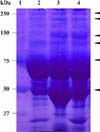
SDS-PAGE patterns showed the results of the serum from normal dogs in comparison with the serum from dogs chronically infected with B. gibsoni and the infected culture supernatant of erythrocytes. The infected serum bands were stronger and broader bands; the arrows (Fig. 1) indicate the protein composition in comparison with the serum from normal dogs. The infected culture supernatant of erythrocytes bands were prominently broader bands, similar to the serum from dogs chronically infected with B. gibsoni.
Effect of the culture supernatant of erythrocytes infected with B. gibsoni on the maturation of canine reticulocytes
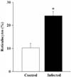
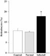
Canine reticulocytes collected by gradient centrifugation rapidly lost their cytoplasmic reticulum, and the percentage of reticulocytes in the supernatant of cultured erythrocytes infected with B. gibsoni decreased during the 24 h incubation period in vitro (Fig. 2). The rate of the disappearance of reticulum was significant (*p < 0.005); it was slowed by the addition of culture supernatant of erythrocytes infected with B. gibsoni. However, the incubated supernatant of erythrocytes had no effect on the morphological maturation of the reticulocytes. Furthermore, the percentage of reticulocytes decreased rapidly with the addition of 20% serum from healthy dogs; this represents normal and serum free media that served as a control for the incubated erythrocytes supernatant. The rate of reticulocyte maturation was significantly (*p < 0.005) retarded by the addition of the serum free culture supernatant of erythrocytes infected with B. gibsoni (Fig. 3).
Effect of fractionated serum and culture supernatant of erythrocytes infected with B. gibsoni on the maturation of canine reticulocytes
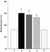
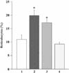
The effects of fractionated serum and culture supernatant of erythrocytes infected with B. gibsoni on the morphological maturation of canine reticulocytes are shown in Fig. 4 and 5. The canine reticulocytes incubated with >150 kDa and 150-70 kDa fractions of serum infected with B. gibsoni were significantly (*p < 0.005 and **p < 0.001) retarded in comparison with cells incubated with normal dog serum and <70 kDa fraction of infected dogs serum (Fig. 4). In addition, the percentage of reticulocytes was significantly higher when fractions >150 kDa and 150-70 kDa of culture supernatant of erythrocytes infected with B. gibsoni were added to the incubation medium, compared to the serum of normal dogs and <70 kDa fraction of culture supernatant of erythrocytes infected with B. gibsoni (Fig. 5).
Discussion
The results of prior studies indicate that serum from dogs infected with B. gibsoni and probably the parasite itself or the metabolites produced by replication of the parasites, might inhibit the morphological maturation of canine reticulocytes. In the present study, SDS-PAGE analysis demonstrated that infected culture supernatant of erythrocytes and serum from dogs infected with B. gibsoni had bands identified that indicated greater molecular mass and protein composition. We found that SDS-PAGE analysis of immune mediated hemolytic anemia in dogs (IMHA Acute and recovery), experimentally induced anemic dog serum and 25-30% erythrocyte hemolysates, did not exhibit a broader band compared to the serum from normal dogs. In addition, samples incubated with canine reticulocytes had no apparent effect on the morphological maturation of reticulocytes (data not shown). In the present study, in vitro incubation of canine reticulocytes with culture supernatant of erythrocytes infected with B. gibsoni resulted in delayed maturation of the cells in comparison with the incubated erythrocyte supernatant. In addition, serum free culture supernatant of erythrocytes infected with B. gibsoni strongly suppressed the maturation of canine reticulocytes. However, there were no significant changes in the maturation of cells observed in response to the addition of either serum from normal dogs or serum free incubated erythrocytes supernatant.
Our previous study showed that serum from dogs infected with B. gibsoni decreased the activity of canine pyrimidine 5'-nucleotidase subclass I (P5N-I) and purine-specific 5'-nucleotidase [6]. Serum biochemically modified by B. gibsoni infection may specifically inhibit canine P5N-I and purine-specific 5'-nucleotidase. This may lead to the maturation of reticulocytes due to the delayed removal of intracellular ribosomal RNA. The results of our study suggest that B. gibsoni itself and/or its metabolites released into the culture supernatant of erythrocyte and serum from infected dogs might cause the delayed maturation of reticulocytes.
At the same time, we examined fractions obtained from the dog serum infected with B. gibsoni and fractions from the culture supernatant of erythrocytes infected with B. gibsoni to evaluate the morphological maturation of canine reticulocytes, and compared the serum from the normal dogs with the serum from the dogs infected with B. gibsoni. We found that >150 kDa and 150-70 kDa fractions of both infected serum and culture supernatant of erythrocytes significantly retarded the maturation of canine reticulocytes. These results suggest that the infection might have decreased the activity of erythrocyte 5'-nucleotidase and simultaneously delayed maturation of reticulocytes. The delayed maturation of reticulocytes in culture appears to favor replication of the parasites, because the B. gibsoni parasite preferentially replicates in reticulocytes rather than in mature erythrocytes when cultured in vitro [14,23].
The SDS-PAGE analysis demonstrated that the broader protein bands, i.e., >150 kDa and 150-70 kDa fractions of both serum and culture supernatant of erythrocytes infected with B. gibsoni, significantly retarded the maturation of canine reticulocytes. Consequently, the data from the present and previous studies show that infected culture supernatant of erythrocytes and the serum from dogs infected with B. gibsoni contain >150 kDa and 150-70 kDa fractions of proteins that might be responsible for the delayed maturation of canine reticulocytes; these proteins might be released by B. gibsoni. In vitro cultivation of B. gibsoni and infected serum shows decreased activity of P5N-I and purine-specific 5'-nucleotidase; subsequently, accumulation of nucleotides might also contribute in part to low parasitemia in vivo [7]. Serum from dogs infected with B. gibsoni might exhibit any number of immunological and biochemical responses, for example, the protective effect of parasite-specific antibodies, toxicity of free radicals [16,18,19], and/or increased phagocytic activity of macrophages [15]. Therefore, it is difficult to identify any particular factor currently.
In conclusion, the results of the present study demonstrate that culture supernatant of erythrocytes infected with B. gibsoni had a suppressive effect and might contain certain factors that retard the maturation of canine reticulocytes in vitro.
In summary, the results of this study show that reticulocyte maturation was retarded by >150 kDa and 150-70 kDa fractions of infected culture supernatant of erythrocytes and serum from dogs infected with B. gibsoni. B. gibsoni itself and/or its metabolites might be released into the culture supernatant of erythrocytes and serum from the infected dogs and as a result suppresses the maturation of canine reticulocytes. The determination of the precise role of infected culture supernatant of erythrocytes and the serum from infected dogs in the delayed maturation of reticulocytes requires further study.
 XML Download