Laminitis, which results into severe lameness, has been reported commonly in equine, bovine, caprine, and porcine [2,4]. There isn't any report on laminitis in dromedary. This paper reports on the case of laminitis in a male camel, which was confined in stall and fed daily with pearl millet grains (Pennisetum typhoideus).
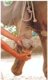
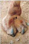
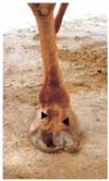
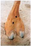
About six year old medium sized male camel was presented at the hospital for treatment of severe lameness. On clinical examination enormous onyx enlargement was observed in both forelimbs (Fig. 1, 2 and 3), and left hind limb (Fig. 4) but onyx of right hindlimb was normal. The digital veins of affected limbs were engorged (Fig. 2, 3 and 4). On progression animal was walking with short strides and putting weight on heels with leaning on left side.
The onyx of left forelimb turned from anterior border of toe towards ventro-caudal direction and occupied 1/4th of cranial solar surface (Fig. 1 and 2); in right forelimb it turned from cranial border toward ventro-medial direction (Fig. 3), whereas, in left hindlimb the onyx growth from cranial border was turning towards ventral direction (Fig. 4). Anamnesis revealed that due to scarcity of roughage during the famine period, the animal was kept under confinement and fed with pearl millet grains without the roughage as daily ration for more than five months.
The animal was sedated with xylazine (0.1 mg/kg, IM) and restrained in lateral recumbancy. The onyxes were trimmed at the level of cranial border of the toe. The pain was not resolved even at 24 hrs. after the nail trimming. Non steroid anti inflammatory drug, phenylbutazone sodium (5 mg/kg, IM) was administered for 7 days and feeding schedule was changed to incorporate the roughage in daily ration of the animal. Though continuous improvement in gait was observed on consecutive days but the draught capability of the animal was re-attained after about ten days.
Onyx enlargement is a common feature in canine and felines that warrants trimming at regular interval. Enormous hoof enlargement in bovine, caprine, equine and porcine species has been reported due to laminitis [1,3,4]. Present findings resembles the grain founders in bovine and equine where eating of considerable quantities of grains as a daily ration alters the bacterial balance resulting in increased lactic acid producing bacteria, primarily lactobacillus and streptococcus. The lactic acidosis even in mild form may be a major factor in the development of acute laminitis [6]. The increased lactic acid and decreased pH causes lyses of cell wall of Gram-negative bacteria with resultant release of endotoxins, these events ultimately contribute to the onset of laminitis [4]. In present case enlarged onyxes and engorged digital veins were similar to elongated hooves, engorgement of digital veins and walking with short strides as reported in the early stages of bovine and equine laminitis [2,4]. Nonsteroid anti-inflammatory drugs administered at early stage of laminitis have been reported to resolve the condition [4], similar results were observed in present case. Rotation of distal phalanx associated with chronic laminitis in bovine [2,5] and equine [4] was not found in present case probably due to earlier detection and therapeutic measures taken for resolving the lameness.


 PDF
PDF ePub
ePub Citation
 Print
Print


 XML Download