

 PDF
PDF ePub
ePub Citation
 Print
Print


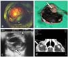
A mongrel male dog of three years old was presented with a pain and enlargement on the right eye. Ophthalmic findings of the right eye included uveitis, glaucoma, and hyphema characterized by a buphthalmic globe, corneal edema, episcleral injection, severe hyperemic conjunctiva, corneal neovascularization, aqueous flare, and blood clot in the anterior chamber. The intraocular pressure in the affected eye was 44 mmHg compared to 14 mmHg in the normal eye. The right eye was blind with no direct or consensual pupillary light response. Additionally, protruded mass in episcleral region and white solitary mass in the distorted iris of right eye were noted (Fig. 1-a). No other ocular abnormalities were detected and physical findings were normal. The values of complete blood count and serum chemistry were within normal limits. The thoracic and abdominal radiographies were completed and revealed no abnormalities. B-mode ocular ultrasonography confirmed complete retinal detachment, posterior lens subluxation and a solid mass of tissue from the iris to the choroid. In addition, an echo-dense material in the anterior chamber indicative of hyphema was noted (Fig. 1-c). Computed tomography (CT) was performed to evaluate the extraocular extension of the mass and the involvement of the adjacent structure was not founded (Fig. 1-d).
As the eye was inflamed, painful, and blind, it was enucleated. Gross examination of the enucleated globe revealed a heavily pigmented mass extended from the dorsolateral aspect of the iris to the choroid, and into the vitreous chamber. The anterior portion of iridal mass was white (Fig. 1-b). The globe was embedded in paraffin, sectioned at 5 µm, and stained with hematoxylin and eosin for light microscopic examination.
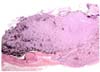
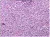
Histological examination revealed that intraocular mass consisted of compact sheets of pleomorphic cells that infiltrating into adjacent anterior uveal tract and sclera (Fig. 2). The neoplastic cells were spindle to polygonal in shape with variably distinct cell borders and had moderate amounts of eosinophilic cytoplasm (Fig. 3). The neoplastic cells often contained abundant amount of dark brown granular pigment and had a round nuclei with a stippled chromatin and single prominent nucleolus. The mitotic rate was 0 to 6 in ×400 fields. The neoplastic cells were expanding to the iris and choroid with scant fibrovascular stroma, and tumor emboli were found in the sclera and uveal tract. Moderate numbers of lymphocytes and melanophages were noted throughout the neoplasm.
Uveal melanomas have been the most common primary intraocular neoplasm in dogs [1,2,3,4], and classified as benign or malignant based on the morphologic findings of the neoplastic cells [2,4]. The majority of both benign and malignant melanoma arises in the anterior uvea [1,2,4]. The neoplasm was involved in both the anterior uvea and the choroid in this case. The melanomas usually arise in the anterior uvea and involvement of choroid is an extension from the anterior uvea [7], but the site of origin was ambiguous in this case. Iridal mass was partially amelanotic. The melanomas are commonly heavily pigmented, but amelanotic melanoma can occur [1,4].
The clinical characteristics of this dog were similar to other previous reports [2,5,8,9,11]. There were secondary changes associated with tumor such as hyphema, uveitis, glaucoma, and blindness. The primary canine uveal melanoma has been thought to have a low risk of distant metastasis [3,4,11]. The evidence of metastatic disease has been rarely reported, even though in malignant form of ocular melanoma [4,6,8,10,11]. There was no evidence of systemic metastatic disease, but a long-term follow-up evaluation would be needed because of the malignancy of the tumor.
 XML Download