

 PDF
PDF ePub
ePub Citation
 Print
Print


Introduction
New types of vaccines such as recombinant vaccines or DNA vaccines in some diseases have largely supplanted the traditional methods of vaccination, which utilize live or dead whole microorganisms [9,30,38]. Although DNA vaccines have many advantages over conventional vaccines, some new generations of vaccines have the drawback that DNA vaccines require repeated doses or mixing with strong adjuvants to achieve an effective immune response [13,25,28]. Therefore, recent vaccine studies have focused on discovering efficient adjuvants.
Adjuvants with DNA vaccines are essential for achieving sufficient levels of protection and long-term immunity. Suitable adjuvants must be able to increase the immune response without having any undesirable side effects. Conventional oil-based adjuvants deteriorate the meat quality at the injection sites. Hence, either naturally occurring adjuvants such as cytokines or other types of compatible adjuvants are needed [25].
Interferon-γ (IFN-γ) is a cytokine produced by stimulated T cells and has important effects in immunomodulation [8]. Its properties as an adjuvant have been examined in a large number of animal studies [1,5,11,25,40,46]. Dimethyl dioctadecyl ammonium bromide (DDA) has been recognized as an adjuvant for enhancing both humoral and cell-mediated immune responses [18]. Unmethylated cytosine-phosphate-guanosine (CpG) dinucleotides are sequences that are found in the bacterial DNA that have adjuvant properties in the mammalian immune system by activating antigen-presenting cells (APCs) [20,24]. In addition, CpG-containing oligodeoxynucleotides (CpG-ODNs) induce the secretion of various cytokines, including interleukin-6 (IL-6) and type II interferon (IFN-γ) [47,48].
Infectious bursal disease (IBD) or Gumboro disease, which is caused by the infectious bursal disease virus (IBDV), is an acute and highly contagious disease in chickens 3 weeks of age and older. The disease has an important economic impact on the poultry industry worldwide because it is associated with a high mortality and immunosuppression in recovered chickens, which leads both to a variety of secondary infections and a a decreased response to vaccinations [26]. After the emergence of highly virulent IBDV strains that cause high mortality in Europe and the antigenic variants of IBDV in USA, these IBDVs have spread rapidly despite conventional vaccination programs to protect chickens against IBDV infections [1,4,34].
This study examined the adjuvant effects of a plasmid encoding chicken IFN-γ, of DDA, and of CpG-ODN on a plasmid DNA vaccine (pcDNA-VP243) against the highly virulent IBDV SH/92 strain, which was developed in our laboratory [17], in chickens. The results suggest that CpG-ODN and IFN-γ failed to enhance the DNA vaccine efficacy, and that DDA actually had an adverse effect on the level of protection against the IBDV.
Materials and Methods
Chickens
The fertilized eggs of specific pathogen free (SPF) White Leghorn were obtained (Sunrise Farm, USA) and hatched. The hatched chicks were placed into plastic isolators operated under a positive air pressure and provided with food and water ad libitum during the experimental period.
Construction and preparation of plasmids
The DNA vaccine, pcDNA-VP243, encoding the VP2, VP4, and VP3 proteins of the highly virulent IBDV SH/92 strain, is described elsewhere [17]. The plasmid DNA was purified from the transformed Escherichia coli using the Endofree Plasmid Giga Kit (Qiagen, USA).
For cloning of the IFN-γ gene, the spleens were obtained aseptically from 8-week old SPF chickens. After the spleens had been passed through a plastic cell strainer (Becton Dickinson Labware, USA), the spleen lymphocytes were separated by Histopaque-1077 (Sigma, USA). The prepared splenocytes were rinsed three times in Hanks balanced salt solution (HBSS) and incubated at 1 × 10⁷cells/mL for 6 h in RPMI-1640 medium containing 10% fetal bovine serum (FBS) supplemented with 12.5 µg/mL Concanavalin A (ConA, Sigma, USA), at 40℃ and 5% CO2. The total RNA was isolated and purified from harvested splenocytes using the TRIzol reagent (Invitrogen, USA) according to the manufacturer's recommendation, and the cDNA was synthesized using random primers (Invitrogen, USA). The PCR fragments were synthesized from cDNA using the primers, CIG-F (5' GCCGCCGCCATGACTTGCCAGACTTACAAC 3') and CIG-R (5' TTAGCAATTGCATCTCCTCTG 3'), which were synthesized according to the published sequence of chicken IFN-γ [6]. PCR was performed with 35 cycles of denaturation at 95℃ for 1 min, annealing at 55℃ for 1 min, and extension at 72℃ for 2 min. The final extension step was performed at 72℃ for 10 min. The PCR products were analyzed on a 1.0% agarose gel.
The PCR products were purified utilizing a GENECLEAN Turbo kit (Q-biogene, USA) according to the manufacturer's instructions. The purified PCR products were cloned into the pcDNA 3.1/V5/His-TOPO vector (Invitrogen, USA) and transformed into competent Escherichia coli (TOP 10) cells (Invitrogen, USA). The plasmid DNA was isolated using the E.N.Z.A plasmid miniprep kit I (Omega Bio-tech, USA). The nucleotide sequence and the orientation of the plasmid construct were confirmed by DNA sequencing. The verified plasmid construct was named pcDNA-ChIFN-ã, and large quantities of the plasmid were prepared using a Endofree Plasmid Giga Kit (Qiagen, USA).
Synthetic CpG-ODN and preparation of DDA solution
The CpG-ODN (2007) sequence [32] is TCGTCGTTGTCGTTTTGTCGTT (underlining indicates CpG dinucleotides), which was produced with a phosphorothioate backbone (Bioneer, Korea). Synthetic CpG-ODN (10 µg/bird) along with the DNA vaccine was injected. A DDA (Sigma, USA) solution (2 mg/bird) was prepared as described previously [16] and also injected along with the DNA vaccine.
In vitro transcription and translation
The in vitro expression of pcDNA-ChIFN-γ was performed using the TNT Quick Coupled Transcription/Translation System (Promega, USA) and visualized using the Transcend Colorimetric Translation Detection System (Promega, USA) according to the manufacturer's recommendations. The samples were electrophoresed on a 12% discontinuous SDS-PAGE gel and transferred onto nitrocellulose membranes for visualization. The membranes were washed with Trisbuffered saline (TBS) and incubated in a blocking buffer (TBS containing 0.5% Tween 20). For visualization, streptavidin alkaline phosphatase was added to the membranes, which were rocked gently for 60 min and visualized by adding Western Blue Stabilized Substrate (Promega, USA).
Immunization protocols
Two-week-old SPF chickens were randomly divided into 6 experimental groups (9 birds/group), which included the normal control (without vaccination and challenge), challenge control (without vaccine but with challenge), DNA vaccine alone (with pcDNA-VP243 DNA vaccine and challenge), DNA vaccine plus DDA (with pcDNA-VP243 DNA vaccine plus DDA and challenge), DNA vaccine plus CpG-ODN (with pcDNA-VP243 DNA vaccine plus CpG-ODN and challenge), DNA vaccine plus ChINF-γ (with pcDNA-VP243 DNA vaccine plus ChINF-γ plasmid and challenge). Twice at a 2-week interval, 2-week-old chickens were injected intramuscularly (100 µg) and intraperitoneally (100 µg) with the DNA vaccines. For the DNA vaccine plus ChINF-γ group, 125 µg of the pcDNA-ChIFN-γ plasmid was injected intramuscularly into separate sites.
Two weeks later, all the groups except for the normal control group were re-injected with the same dose and by the same route as used for the primary immunization. Two weeks after the second immunization, all the groups except for the normal control were challenged orally with a 1 × 104.8 50% egg lethal dose (ELD50) of the highly virulent IBDV SH/92 strain [21]. The chickens were then monitored daily for any clinical signs over a 10 day period. At 10 days postchallenge, all the remaining chickens in each group were euthanized, and the spleens and the bursa of Fabricius were collected aseptically. The bursa/body weight (B/B) ratios were calculated by (bursa weight)/(body weight) ×1,000.
Peripheral blood and splenic lymphocyte proliferation assay
The lymphocyte proliferation assay was performed as described previously [17]. Briefly, the whole blood and spleens were collected aseptically from the chickens before the challenge as well as at 10 days after the challenge, and the cell suspensions were obtained. The lymphocytes were separated using Histopaque-1077 (Sigma, USA), washed three times, and then resuspended in RPMI-1640 medium supplemented with 10% FBS. The cells (PBL at 1.25 × 10⁶ cells/well and spleen at 2.5 × 10⁶ cells/well) were placed in 96-well flat-bottomed tissue culture plates. ConA (12.5 µg/mL) was added to each well except for the negative control well. The plates were incubated for 48 h at 40℃ in a 5% CO2 atmosphere. The lymphocyte proliferation activity was measured using a WST-8 working solution [29]. The optical density (OD) was determined at 450 nm and the stimulation index (SI) was calculated using the following formula: SI = mean OD of ConA-stimulated cells/mean OD of non-stimulated cells.
Antibody assay by enzyme-linked immunosorbent assay
Blood samples were collected from the birds in each experimental group both before the challenge and at 10 days after the challenge. The serum antibody titers of the experimental groups were determined using an infectious bursal disease antibody test kit (IDEXX, USA) as described elsewhere [17]. Titers >396 were considered positive.
Statistical analysis
All the analyses were performed using the statistical package SAS 8.01 (SAS Institute, USA). The non-parametric Kruskal-Wallis rank test with a pairwise multiple comparison, which used the Dunn method for post-hoc analysis, was used to evaluate the differences in the B/B ratios between the groups [49]. One-way ANOVA was used to assess the individual differences in the serum antibody titers and lymphocyte proliferation assays. The Levene's test for homogeneity of the data was used to determine the equality of the variances among the groups [12]. A P-value <0.05 was considered significant.
Results
In vitro transcription and translation
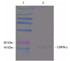
The protein expressed by pcDNA-ChIFN-γ was detected using an in vitro transcription/ translation and detection system (Fig. 1), and a band with a molecular weight of 17-18 kDa was observed. According to the observations reported by Song et al. [35], the molecular weight of the recombinant ChIFN-γ expressed in E. coli was 17-18 kDa, which corresponds to the nonglycosylated ChIFN-γ form.
Evaluation of adjuvant in chickens
The protective efficacy against IBDV in the chickens immunized with the DNA vaccine mixed with selected adjuvants, DDA, CpG-ODN and ChIFN-γ was examined by challenging the chickens with the IBDV SH/92 strain two weeks after the second immunization and observing them clinically for 10 days. The efficacy of the adjuvants in these chickens was evaluated by the mortality, B/B ratios, serum antibody titers, and peripheral blood and splenic lymphocyte proliferation assays (Table 1, Fig. 2 & 3).
The clinical signs (anorexia, depression, and ruffled feathers) began to appear on three days after the challenge, and the mortality peaked over a period of 4-5 days after the challenge. The groups that received the DNA vaccine plus DDA or CpG-ODN had significantly lower survival rates than the group given the DNA vaccine alone (p < 0.05). However, the survival rates of the groups given the DNA vaccine alone and DNA vaccine plus ChIFN-γ were similar (p > 0.05). In particular, the DNA vaccine plus DDA group showed a similar mortality to the challenge control group.
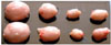
Bursal atrophy in the chickens receiving the DNA vaccine plus CpG-ODN or ChIFN-γ was severe compared with that observed with the DNA vaccine alone and with the normal control group (Fig. 2). The bursae of the challenge control and the DNA vaccine plus DDA groups were not evaluated because all the chickens in this group died within the postchallenge period. The B/B ratio for the DNA vaccine plus CpG-ODN group was not significantly lower than that observed with the DNA vaccine alone (p > 0.05) (Table 1). The B/B ratio for the DNA vaccine plus ChIFN-ã was lower than for the DNA vaccine alone or normal control but the difference was not significant (p > 0.05).
None of the chickens in all groups had any detectable antibodies to the IBDV before the challenge. However, all the surviving chickens of each group except for the normal control group showed detectable antibodies to the IBDV at day 10 after the challenge (Table 1). Similar ELISA antibody titers were detected in all groups except for the normal control group (p > 0.05).
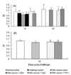
The kinetic changes in ConA-induced peripheral blood and splenic lymphocyte proliferation in the chickens in each group were measured using the WST-8 assay before and after the challenge with the IBDV SH/92 strain (Fig. 3). The SI of the peripheral blood in all groups was similar immediately before the challenge except for the normal control group (p > 0.05). The SI at day 10 post-challenge in the DNA vaccine plus CpG-ODN or ChIFN-γ groups was almost identical to those of the group receiving the DNA vaccine alone. The SI for the splenic lymphocytes on day 10 after the challenge were higher in the DNA vaccine plus CpG-ODN and ChIFN-ã groups than in the DNA vaccine control group but the difference was not significant (p > 0.05).
Discussion
Various compounds have been examined and recommended as adjuvants for stimulating the immune response. However, the commercially available adjuvants are still mostly limited to the conventional adjuvants that contain oil emulsions and insoluble salts of aluminum [13]. Although mineral oil emulsions are used in a wide range of food animals, their undesirable side effects, such as deterioration in the meat quality at the injection sites and the risk of mineral oil residues in the meat from these animals, highlights the need for new adjuvants [13]. This study evaluated DDA, CpG-ODN and ChIFN-γ, which have been reported to be immunomodulators against various infectious diseases in many animal models [7,14,16,22,42], as adjuvants for pcDNA-VP243 vaccine against IBDV [17].
The pre-challenge serum antibody titers of the chickens, whether vaccinated or not, were all negative as tested by ELISA, but all the post-challenge serum samples revealed high antibody titers except for the normal control group. The commercial IBDV antibody test kit used in these experiments was specifically designed to evaluate the immune status of the chickens for the IBDV, and only those serum samples with antibody titers >369 were considered positive. Although it was reported that protection against IBDV correlates with the levels of neutralizing antibodies in chickens [26], the antibody titers for the DNA vaccine alone and for our adjuvant groups were not in proportion to their mortality or B/B ratios. However, there were no significant differences between the groups receiving the DNA vaccine alone and those receiving the DNA vaccine plus CpG-ODN or ChIFN-γ. This is despite the fact that the adjuvant groups had higher mortality rates than the group given the DNA vaccine alone. It appears that other protective mechanisms might have been induced in the chickens from the vaccine-only group. The chickens were partially or fully protected against the IBDV challenge, even though the VP2-VP4-VP3-expressing plasmid, which was constructed with the classical IBDV strain STC, induced only low or undetectable levels of antibodies in the chickens before the challenge [3]. In this study, the high post-challenge serum antibody titers appeared to have been induced by the challenge virus.
The lymphocyte proliferation assay is widely used to evaluate the cell-mediated immune responses in chickens in the normal and disease states [27,29]. The DNA vaccine alone and DNA vaccine plus CpG-ODN or ChIFN-γ produced in higher levels of peripheral lymphocyte proliferation in response to ConA at 10 days post-challenge than the prechallenge. However, the differences between these groups were not statistically significant. In addition, the splenic lymphocyte proliferation assay at day 10 post-challenge showed a similar of mitogenic response between the 3 groups. In a previous study with the DNA vaccine [17], the level of peripheral blood lymphocyte proliferation in the challenge control group decreased after the challenge.
DDA has been identified as an effective enhancer of the immune response to bacteria [15] and viruses [18,19] in mammalian models. In chickens, DDA enhanced the antibody response after a secondary boost with the Newcastle disease virus vaccine [33] and augmented the humoral and T-cell immunity to coccidial antigens [23]. DDA improved the pseudorabies virus-specific humoral immune and cell-mediated responses after DNA vaccination against the psedorabies virus [41]. However, DDA had an adverse effect on the efficacy of the pcDNA-vp243 vaccine based on the mortality and B/B ratio when co-administered with the DNA vaccine. The amount or preparation method of DDA might have to be modified in future studies because several researchers have used various DDA formulations in their experiments.
The efficacy of CpG-ODNs as immunomodulators has been reported in many studies [20,24,36,37,47,48]. According to these studies, CpG-ODNs activate and induce the maturation of several cells such as B cells, macrophages, NK cells and dendritic cells which play important roles in the immune system. However, the CpG-ODNs used in this study worsened the DNA vaccine effect. Although the co-administration of DNA vaccine with CpG-ODNs (ODN2007) in chickens surviving after challenge induced an almost identical antibody titer and a similar stimulation index score in the lymphocyte proliferation assay compared to those obtained with DNA vaccine alone, co-administration also increased the mortality rate and induced bursal atrophy. This suggests that the CpG-ODN was not an effective immunomodulator for the pcDNA-VP243 DNA vaccine. Another CpG-ODN (ODN2135) previously showed a high stimulation index in a proliferation assay of PBMC in the peripheral blood from chickens [32] in another experiment. However, there was no significant difference between the pcDNA-VP243 alone and pcDNA-VP243 plus ODN2135 (data not shown). Conflicting results obtained with the co-administration of DNA vaccines and CpG-ODNs were reported [43,44]. The co-administration of CpG oligodeoxynucleotide with the DNA vaccines against IBDV carrying VP2 genes enhanced the protective immune response of the DNA vaccine in chickens [43]. However, the DNA vaccine, the CpG oligodeoxynucleotide, and the formulations used were different from these experiment. An ODN dose-dependent reduction in gene expression from a plasmid was reported in mice [44]. Different formulations (different times or sites) of the CpG-ODN and DNA vaccine also did not augment the antibody responses, possibly due to competition between the CpG-ODN and DNA vaccine for entry into the cells in addition to the lower nuclear resistance and poor stability of CpG-ODN in vivo. Several types of CpG-ODNs have been studied in vitro and in vivo [32]. The other CpG-ODNs might augment immune response to the pcDNA-VP243 DNA vaccine.
The effects of IFN-γ on the immune response appear to be dependent upon the animal species and age, the types of combined antigen, the nature and dose of IFN-γ, and the particular promoter/enhancer of the plasmid expressing the foreign gene. The co-administration of ChIFN-γ expressed using a baculovirus system with the inactivated Salmonella enteritidis antigen enhanced the level of protection against Salmonella enteritidis challenge without increasing the antibody production [39]. The co-administration of the recombinant ChIFN-γ with sheep red blood cell antigens resulted in stronger antibody responses and allowed a lower dose of the antigen to be used more effectively than in the chickens that received the antigen alone [25]. Harms et al. [10] reported a down-regulatory effect of IFN-γ on both the SV40 and CMV promoter/enhancer-driven transgenes in most cell lines tested, and an up-regulatory effect of IFN-γ on the MHC I promoter/enhancer-driven transgene in all the cell lines tested. In particular, the addition of IFN-γ in myoblasts drastically reduced the SV40 and CMV promoter-driven expression in myoblasts.
The effect of IFN-γ depended upon the promoter driving expression of the viral antigen. The immune responses to the antigen-expressing vectors carrying a viral promoter such as the SV40 were lower in presence of IFN- γ. However, IFN-γ had no effect on a vector expressing the antigen under the control of the MHC class II promoter [46]. In this experiment, the ChIFN-γ-expressing plasmid under the control of the CMV promoter given at separate site did not increase the level of protection against the vvIBDV by the IBDV DNA vaccine. In another experiment, the ChIFN-γ-expressing plasmid co-injected with the IBDV DNA vaccine at the same site had an adverse effect on the level of protection against the vvIBDV in chickens (data not shown). The inhibitory effect of IFN-γ was stronger with the co-injected DNA vaccine than with the separately injected DNA vaccine, which suggests that the effect of IFN-γ might occur locally. These results agree with the results of most other researchers using the CMV or SV40 promoter/enhancer-driven transgenes, although the antigens expressed in the plasmids were different.
These studies showed that CpG-ODN and ChIFN-γ had no significant adjuvant effect and DDA had a negative effect on the DNA vaccine of pcDNA-VP243. Further studies will be needed to identify the optimal adjuvants for the DNA vaccine against the IBDV.

 XML Download