

 PDF
PDF ePub
ePub Citation
 Print
Print


Introduction
Paratuberculosis (PTB), Johne's disease, is a chronic progressive disease of ruminants caused by infection with Mycobacterium avium subsp. paratuberculosis (MAP). Although infection usually occurs in the first few months of life [35], the first sign of clinical disease may not appear until 6 months to 15 years post infection [5,23]. This long latent period has been attributed to control difficulties because subclinically infected cows become transmitters of PTB, and shed causative bacteria in feces before progressing to the terminal disease stage.
PTB causes substantial economic loss to the beef and dairy industries [4,19,29]. Therefore, an appropriate control program should be implemented to reduce the negative impact of PTB. Moreover, the initial step required for the successful development of a control program is the determination of the regional distribution of infected herds. Although cultivation of MAP from fecal or tissue samples is considered the reference test for PTB, it is a cumbersome and expensive method for detecting infected animals, especially when the numbers involved are large. Moreover, culture requires up to six months, and the method is not sufficiently sensitive to detect animals early in the course of infection. ELISA provides an alternative; it is faster (results take two to three days), provides increased sensitivity, and importantly is less expensive and can be used to test large numbers of animals [6]. For this reason, the authors developed an 'in house' absorbed ELISA method (P-ELISA) as a screening test, and compared this with a commercial ELISA (C-ELISA) using field samples from all provinces in Korea, excepting Jeju-do. P-ELISA yielded results similar to those obtained using C-ELISA. This study provides first data on the prevalence of MAP in Korea, information that will prove invaluable for the development of a national strategy to control the disease.
Materials and Methods
Test samples
Sera were randomly collected by the National Veterinary Research and Quarantine Service as part of an annual investigation of bovine infectious diseases. A total of 2,161 bovine sera samples from 1,056 beef cattle in 448 farms and 1,105 dairy cattle in 219 farms, were collected from eight provinces (Gyeonggi, Gangwon, Chungbuk, Chungnam, Jeonbuk, Jeonnam, Gyeongbuk and Gyeongnam) in Korea from September to November in 2002.
C-ELISA
All sera were tested using a commercial ELISA kit (Parachek; CSL, Australia) according to the manufacturer's instructions. Briefly, samples were diluted 1 : 20 in green diluent containing M. phlei, and then transferred to 96 well plates in duplicate. Positive and negative control solutions supplied by manufacturer were also tested in duplicate in each plate to validate test results. After washing, secondary antibody was diluted 1:100 in blue diluent. TMB was used as substrate. Optical density (O.D.) values were measured using an ELISA reader (Tecan, Australia) at 450 nm. The cutoff value for positive sera was defined as the mean of negative controls plus 0.100.
P-ELISA
MAP ATCC 19698 was grown in Watson-Reid medium [36] at 37℃ for 12 wks. Bacterial cells were washed twice in phosphate buffered saline (PBS, pH 7.4) and resuspended in PBS. Cells were then sonicated twice on ice for 30 min, and centrifuged at 20,000 × g (Beckman, UK) at 4℃ for 30 min. Supernatant was then harvested and filtered using a 0.2 µm pore size filter. This filtrate was used as a capture antigen after measuring its protein concentration by spectrophotometry (Eppendorf, Germany). M. phlei was cultured in Dorset-Henley medium at 37℃ for 8-10 wks, and then prepared as described above for use as an absorption antigen.
Polystyrene ELISA plates (Maxisorp; Nalgen Nunc International, USA) were coated with 0.4 µg of capture antigen in 100 µl of 50 mM carbonate buffer (pH 9.6), and incubated overnight at 4℃. Coated plates were washed once with 100 µl of PBS (pH 7.4) containing 0.05% Tween20 (PBST), and incubated with 300 µl of 1% bovine serum albumin (BSA) in PBST for 2 h to block non-specific binding. Test sera were absorbed at a dilution of 1 : 20 in absorbent diluent (150 µg/ml of M. phlei, 5% fetal bovine serum, 2% BSA in PBST) and incubated for 30 min. After blocking, the plates were washed with PBST and incubated with absorbed sera (100 µl/well, in duplicate) for 30 min. Positive and negative controls were included in each plate. After washing, 100 µl of a 1 : 1500 dilution of horse radish peroxidase-labeled goat anti-bovine IgG (H + L) (Kirkegaard & Perry Laboratories, USA) was added to each well. Plates were then incubated for 30 min, washed 3 times, and 100 µl of peroxidase substrate (ABTS; Kirkegaard & Perry Laboratories, USA) was added. This reaction was stopped using 50 µl of 1M HCl, and plates were read in an ELISA reader (Tecan, Australia) at 405 nm.
Serum from a seropositive and fecal culture positive cow was used as a positive control, a serum pool from four seronegative animals from different herds, which had been seronegative and culture negative for more than 2 years, was used as a negative control. All steps were conducted at room temperature except the antigen coating step.
Statistical analysis
The receiver operating characteristic (ROC) analysis [3], kappa statistics [2], and percent agreement were used to compare P-ELISA with C-ELISA. Percent agreement (P) was defined as according to Eq. 1 [33],
P = (a + d) / n × 100,
where 'a' is the number of positive reactions, 'd' is the number of negative reactions, and 'n' is the number of total samples tested.
The test prevalence of PTB was calculated by dividing the number of positive sera by the number sera tested. This value was then adjusted to calculate the estimated test-positive prevalence (etp) at a nationwide level. The calculation takes into account bias due to different sample sizes and populations in the different provinces, as detailed by Eq. 2 [17],

where 'B1' is the number of total beef cows, 'D1' is the number of total dairy cows, 'p1' is the proportion of positive beef cattle, and 'q1' is the proportion of positive dairy cows in province 1. To calculate the estimated true prevalence (ETP) in Korea, etp was adjusted to compensate for the lack of sensitivity (Se) and specificity (Sp) of C-ELISA using Eq. 3 [24],
ETP = (etp + Sp - 1) / (Se + Sp - 1)
National population data were obtained from the Ministry of Agriculture and Forestry in Korea (28). All statistical analyses were carried out using commercially available software (Analyse-it, UK)
Results
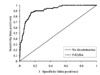
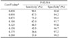
Based on C-ELISA results, ROC analysis was performed to analyze the efficacy of P-ELISA as a screening test and to determine a suitable cutoff value. Area under the curve (AUC) and the standard error of AUC were 0.913 [95% confidence interval (CI), 0.883 to 0.943] and 0.015, respectively (Fig. 1). Generally, the determination of an optimal cutoff value for the differentiation of a positive and negative reaction is difficult since the O.D. values of samples are not clearly divided into two groups. For this reason, after measuring Se and Sp of P-ELISA at various cutoff values (Table 1), a cutoff point of 0.100 was arbitrary chosen for further analysis, because this point gave relatively high Se and Sp values for P-ELISA, and this cutoff was then used to differentiate positive and negative sera in preliminary experiments (data not shown). This cutoff value was two times higher than that of the negative controls, and higher than the mean of negative controls plus standard variation (data not shown). Based on P-ELISA results using a 0.100 cutoff point, the kappa value and percent agreement between P-ELISA and C-ELISA were 0.322 and 92.5%, respectively (Table 2).
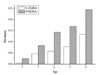
We also obtained information on the ages of 1,650 of the 2,161 cattle tested. Although P-ELISA detected about twice as many seropositive cows than C-ELISA, both ELISAs showed that a significant correlation existed between age and a positive response (Fig. 2).
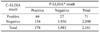
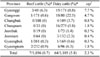
C-ELISA test results revealed that 71 of 2,161 cows (3.3%, 95% CI, 2.6% to 4.1%) were seropositive (Table 3). Thus, based on the recently reported Se (28.4%) and Sp (99.7%) of C-ELISA [9] and the known population size, the national ETP was estimated to be 7.1% (Eqs. 2 and 3). Moreover, the proportion of seropositive dairy cattle was significantly greater than that of beef cattle (p < 0.01), i.e., 7 of 1,056 beef cattle (0.7%, 95% CI, 0.3% to 1.4%) versus 64 of 1,105 dairy cattle (5.8%, 95% CI, 4.5% to 7.3%). In terms of comparisons between provinces, Gyeonggi showed the highest proportion of seropositivity in total and beef cattle. Gangwon had a higher proportion of seropositive dairy cattle than other provinces. For beef cattle, all provinces, except Gyeonggi (6.3%), showed less than 1% seropositivity (Table 3).
Discussion
To evaluate the efficacy of P-ELISA, it should be compared with other reference methods, such as bacterial cultures or other antibody based tests. However, since it was not possible to obtain fecal or tissue samples from the cows tested, C-ELISA, which has been evaluated for detecting infected cows in several studies [8,10,27], was used as the reference method in this study. For ELISA tests, the general problem encountered is the degree of signal overlap of samples from diseased and non-diseased animals, especially for PTB. Moreover, the Se and Sp values of ELISAs have been found to vary depending on cutoff point. Therefore, the cutoff of 0.100 for P-ELISA was chosen for further analysis for the reasons mentioned above in Results.
Using a cutoff of 0.100 for P-ELISA, the kappa value for the two ELISAs showed a low level of agreement, which has been reported for ELISAs previously in terms of detecting antibody to MAP [9,12,34]. This low agreement may have been caused by the presence or absence of certain antigenic components that react with some specific or crossreactive antibodies. Moreover, specific test antigens are the most important component of sensitive and specific ELISA tests. However, MAP is known to have antigenic components in common with other species of mycobacteria, and with related organism such as Corynebacterium spp., Norcardia spp., Actinomyces spp., and Eschericia coli [5,16,37]. In addition, different Sps of absorbed ELISAs for PTB were found in serum samples from different regions, which may reflect regional cross-reactive antibodies [31]. Our findings indicate that some cross-reactive antibodies remained after absorbing sera with M. phlei, and that this affected the Sps of the two ELISAs. In addition, although the capture antigens were prepared from the same organism, the antigenic composition of MAP preparations can be different depending on the method of preparation [10,15]. For these reasons, each of the ELISAs used in the present study might have only detected a subset of specific or cross-reactive antibodies. Nevertheless, the high AUC and percent agreement [33], and the similar age distribution patterns observed demonstrated that P-ELISA can be used as a herd screening test and as a pre-screening test for individual and followed with other identification tests, such as PCR or bacterial culture.
We tested cows up to six years of age, and both ELISAs revealed a significant correlation between animal age and a positive result. PTB is characterized by its long latent period, and thus, seroconversion is more readily detected in older animals. Thus, although cows are likely to be infected with MAP whilst young, most infections go undetected. Other studies have yielded similar results, although these studies also found that seropositivity is reduced in animals over six years of age [13,14], which may be due to the culling of symptomatic cows.
To date, few studies have been performed on PTB in Korea, and these have been limited in scope [20-22]. The present study is the first seroprevalence study conducted on PTB at a nationwide level. Only C-ELISA results were used to estimate of prevalence because this test has been used worldwide and well evaluated. In the present study, although the overall seroprevalence of PTB in Korea was found to be low to moderate compared with those of other countries [1,11,14,18,26,30,32], some provinces showed much higher seroprevalences. Many factors probably contribute to differences in prevalences between provinces, such as herd characteristics, climate, and environment effects. For example, Gangwon has been known by veterinarians to be an endemic region for PTB, and number of overpopulated herds in Gyeonggi may have contributed to this high seropositivity. In terms of dairy and beef cattle, our data reveal that the seropositive rate of beef cattle is significantly lower than that of dairy cattle (p < 0.01), as was previously reported by Kim et al. [21], which suggested a low overall prevalence in beef cattle in Korea. Similar results have also been reported in other countries [11,25]. This finding may be due to restricted transmission opportunities among beef cattle because they are culled earlier than dairy cattle.
Taken together, our data provide a general indication of the true state of PTB in Korea, and suggest that a national control program should be considered to control the disease. Our findings also suggest that different control systems might be needed in different provinces depending on the prevalence of PTB with consideration of the economic models of Johne's disease [7], and that programs should focus on limiting the spread of PTB among provinces. As a first step in any control program, large numbers of cattle should be tested, and due to its low cost and accuracy, we suggest that P-ELISA can be used for this purpose as a screening test for infected herd or for individual animals followed by other methods to verify PTB infection.
 XML Download