

 PDF
PDF ePub
ePub Citation
 Print
Print


Multilobular tumor of bone is an uncommon, slow growing, locally invasive, and malignant tumor of the flat bones of the canine skull, although such tumors have been reported to occur in non-cranial sites such as the mandible, maxilla, zygomatic arch, and tympanic bulla [1,3,6]. A few cases have been reported in humans, cats, a horse, and a ferret [5,7]. This tumor often recurs locally after surgical excision, and it has been found metastasize to the lungs [2,4]. There is no report on breed or sex predilection to this tumor. This tumor primarily occurs in older and medium- to large-breed dogs and occasionally in young and small dogs [1,6]. In humans, it often affects children and young adults and involves the wrist and the sole.
Depending on the location of its growth, compression of the adjacent structures by the tumor can manifest in various clinical signs in the affected animals; these include difficulty in mastication, sinus obstruction, neurological signs, exophthalmia, and disfiguration of the face and head due to protruding tumor mass [1,6].
Other synonyms, such as chondroma rodens, multilobular osteoma, multilobular chondroma, calcifying aponeurotic fibroma, juvenile aponeurotic fibroma, cartilage analogue of fibromatosis, multilobular osteoma and chondroma (MLO/C), and multilobular osteochondrosarcoma (MLO), have been used by various authors to describe this tumor. Currently, to avoid the confusion caused by using various synonyms, the term multilobular tumor of bone is preferred [6].
In this article, we aim to describe a multilobular tumor of bone in the mandible of a dog with secondary salivary gland mucocele.
A 2.5-year-old male Pekingese dog was brought to a local veterinary clinic with a firm mass at the cervical region near the junction between jaw and neck. On fine needle aspiration, there was low cellularity with scattered mononuclear macrophage type cells with highly vacuolated cytoplasm, nondegenerate neutrophils, rare hemotoidin crystals, and interspersed eosinophilic homogenous materials, and the case was diagnosed as mucocele and referred for surgical excision. No radiographic examination was performed. One year after the surgery, the owner brought the dog in again. At this time, the mass had increased to almost 5 times its previous size, and it extended up to the mandible. The practitioner carried out surgical excision of the mass and submitted it to the Department of Veterinary Pathology, College of Veterinary Medicine, Seoul National University, for diagnosis. The specimen was routinely processed, embedded in paraffin, stained with hematoxylin and eosin (H&E), and examined under light microscope.
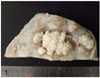
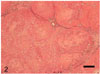
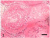
Grossly, on the cut surface, the mass consisted of multiple, variably sized, gritty, grayish-white to yellow nodules separated by thick collagenous septa (Fig. 1). Histologically, these nodules contained multiple lobules of irregularly shaped and sized islands, or nests, of well-differentiated osteoid and cartilage, separated by anastomosing fibrovascular septa (Fig. 2). Within the cartilagenous areas, chondrocytes are within irregular lacunae, and in more osseous islands, osteocytes are within lacunae in a bony matrix (Fig. 3). The island margins are delineated by thin layers of flattened cells that blend with the septa. The islands are surrounded by a variably thick zone of pale eosinophilic mesenchymal stroma containing spindled cells with indistinct cell borders, moderate amounts of eosinophilic fibrillar cytoplasm, and elongated, finely stippled to vesiculate nuclei with one to two prominent nucleoli. Mitotic figures were not evident. A diagnosis of canine multilobular tumor of bone was made on the basis of histologic features.
Multilobular tumor of bone most often occurs in the skull of dogs and, rarely, in non-cranial sites of the canine skeleton such as maxilla, mandible, orbit, tympanic bulla, and base of the zygomatic process [1,6]. It is documented to occur most commonly in older dogs and occasionally in younger dogs. However, in our case, we observe this tumor emerging in a non-cranial site in a 2.5-year-old dog. Our histological observations, which include multiple lobules of irregularly shaped and sized islands of well-differentiated osteoid and cartilage within a connective tissue matrix surrounded by fibrovascular septa, are in line with the observations made by earlier workers [5,6,7].
To date, the exact histogenetic origin of this tumor is still under speculation. In most of the available reports, these tumors have been found to originate in the bones of the chondrocranium and viscerocranium, which develop from the common source of embryonic cartilage ossification.
Since the tumor mass is not likely to be completely removed due to its anatomical location and strong adhesion to the mandible, local recurrence is highly expected. Median time of local recurrence is about 797 days, with a range of 30 to 1,332 days [1]. Furthermore, chondrosarcoma that developed from a previous multilobular tumor of bone site has been reported in a cat [5]. Considering all of this information together, the patient should be carefully monitored.
 XML Download