

 PDF
PDF ePub
ePub Citation
 Print
Print


Introduction
Thyroid hormones are iodine-containing amino acids synthesized and secreted in the thyroid gland by changes in the circulating concentration of pituitary thyrotropin. All circulating thyroxine (T4) and 20% of 3,5,3'-triiodothyronine (T3) are derived from the thyroid gland [7,8,11]. In the blood, more than 99% of T4 and T3 is bound to plasma proteins, with T4 more highly bound than the other [15]. Dogs have lower thyroid hormone binding to serum proteins than humans, resulting in lower total serum concentrations of T4, T3, higher free hormone concentrations, and more rapid clearance rates [2].
Hypothyroidism is the most common endocrinopathy of the dog. Unfortunately veterinarians today face choosing from a wide variety of diagnostic tests of thyroid function, none of which is optimal in all clinical cases. Determination of baseline serum concentrations of thyroxine (tT4), free thyroxine (fT4), 3,5,3'-triiodothyronine (T3), and provocative tests of thyroid secretory reserve (e.g., thyroid stimulating hormone [TSH] response test) have been the most common diagnostics for the assessment of thyroid gland function in dogs [1,13]. Also, a relatively new assay is available to measure canine TSH (cTSH) but a sole measurement of endogenous TSH concentration should not be used to diagnose hypothyroidism [19].
tT4 can be best used to rule out the hypothyroidism. If the tT4 is normal, the dog is unlikely hypothyroid. If the tT4 is less than normal, the dog may or may not be hypothyroid. Numerous non-thyroidal factors such as medications [19] and chronic illness can suppress T4 concentration to less than the normal range, so called 'euthyroid sick syndrome' [15].
The concentration of tT4, history of previous medication, signalment, clinical signs, complete blood count, and biochemistry panel may support a diagnosis of hypothyroidism or rule out other diseases. It means that the evaluation of the daily fluctuation of thyroid hormone may be important to reach the accurate diagnosis of hypothyroidism in practice.
The purpose of this study was to determine the daily fluctuation of serum tT4, fT4, and T3 concentrations in healthy dogs during a day by using the enzyme chemiluminescent immunoassay (ECLIA).
Materials and Methods
Experimental animals
Eleven healthy adult dogs of 9 male and 3 female dogs, weighing from 10 to 20 kg, were used in the study. The dogs were healthy, dewormed and vaccinated one month before the experiment. They were housed individually and fed commercial dry food (Woosung Feed, Korea). All dogs were bright, alert and responsive. They were not being given any medications for the last 6 months and during the period of the study. Food was withheld for 12 hours before and throughout the experiment.
Thyroid stimulating hormone (TSH) response test
For TSH response test, blood samples were collected by the jugular venipuncture into glass tubes without anticoagulant before and 6 hours after administration of thyroid stimulating hormone (TSH). The thyroid sitmulating hormone from bovine pituitary (Sigma-Alderich, USA) was injected intravenously at the dose of 0.1 IU/kg, up to 1 unit for an individual dog.
Thyroid hormones were measured by using Access Immunosystem (Sanofi Diagnostics Pasteur, USA).
Sample collection and processing
Blood samples from all dogs for the measurement of tT4, fT4, and T3 concentrations were obtained five times at 3 hour intervals from 8 : 00 to 20 : 00 by the jugular venipuncture into glass tubes without anticoaglulant.
Blood samples were stored at 4℃ and the serum was obtained after centrifugation at 1,000 × g for 20 minutes. tT4, fT4 and T3 concentrations were measured by the ECLIA method [20].
Statistical analysis
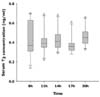
Statistical analysis was performed with ANOVA. All data were expressed as mean ± standard deviation. Results were displayed graphically as box plots. For each box plot, the T-bars represent the measured data, which in most instances are in the normal range. The horizontal bar in the body represents the median. For all statistical analysis, values of p < 0.05 and p < 0.001 were considered significant.
Results
Thyroid stimulating hormone (TSH) response test
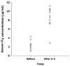
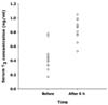
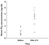
Mean tT4 concentration before TSH injection was 2.15 ± 1.12 µg/dl and that 6 hours after TSH administration was 7.43 ± 2.95 µg/dl (Fig. 1). Mean T3 concentration before TSH injection was 0.37 ± 0.11 ng/ml and that 6 hours after TSH administration was 0.79 ± 0.21 ng/ml (Fig. 2). Mean fT4 concentration before TSH injection was 0.81 ± 0.43 ng/dl and that 6 hours after TSH administration was 2.86 ± 1.24 ng/dl (Fig. 3).
Thyroxine (tT4) concentrations
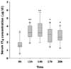
Mean tT4 concentrations for the 12-hour sample collection period in healthy dogs were within the reference range (Table 1). Mean tT4 concentration was 1.75 ± 0.75 µg/dl (0.53 to 3.32) at 8 : 00, 3.28 ± 0.86 µg/dl (1.88 to 4.46) at 11 : 00, 3.54 ± 1.15 µg/dl (1.96 to 5.73) at 14 : 00, 2.90 ± 1.03 µg/dl (1.39 to 4.46) at 17 : 00, 2.7 ± 0.90 µg/dl (1.24 to 4.09) at 20 : 00. tT4 concentrations at 11 : 00 and 14 : 00 were significantly different compared to serum tT4 concentrations at 8 : 00 (p < 0.001) and those at 17 : 00 and 20 : 00 were significantly different compared to serum tT4 concentration at 8 : 00 (p < 0.05) (Fig. 4).
3,5,3'-triiodothyronine (T3) concentrations
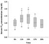
Mean T3 concentrations for the 12-hour sample collection period in healthy dogs were from 0.14 to 0.70 ng/ml (Tabel 1). Mean T3 concentration was 0.43 ± 0.18 ng/ml (0.14 to 0.70) at 8 : 00, 0.42 ± 0.13 ng/ml (0.22 to 0.69) at 11 : 00, 0.44 ± 0.12 ng/ml (0.29 to 0.68) at 14 : 00, 0.37 ± 0.09 ng/ml (0.28 to 0.61) at 17 : 00 and 0.46 ± 0.10 ng/ml (0.33 to 0.66) at 20 : 00. There were no significant differences (Fig. 5).
Free thyroxine (fT4) concentrations
Mean fT4 concentrations for the 12-hour sample collection period in healthy dogs were within the normal range except at 8 : 00 and 20 : 00 (Table 1). Mean fT4 concentrations was 0.967 ± 0.245 ng/dl (0.6 to 1.27) at 8 : 00, 1.30 ± 0.37 ng/dl (0.73 to 1.9) at 11 : 00, 1.35 ± 0.12 ng/dl (0.97 to 1.97) at 14 : 00, 1.05 ± 0.34 ng/dl (0.65 to 1.64) at 17 : 00 and 0.94 ± 0.32 ng/dl (0.59 to 1.4) at 20 : 00. Mean fT4 concentration at 11 : 00 and 14 : 00 were significantly different compared to serum fT4 concentration at 8 : 00 (p < 0.05) (Fig. 6).
Discussion
Thyrotropin is produced and secreted by the pituitary gland and stimulates the thyroid gland to produce and secrete thyroid hormones. The principal secretory product of the thyroid gland is T4. T3 is secreted in smaller amount and is mainly a result of deiodination of T4 in peripheral tissues [6,9]. Most of the circulating T4 and T3 are bound to protein, and the remainder is unbound or "free". Only the free portion of thyroid hormones is able to penetrate cell and accomplish their function [6]. Therefore, determination free thyroid hormone concentrations is thought to reflect thyroid gland function of animals more accurately than that of total thyroid hormone concentrations, which involves both bound and free hormone concentrations [6,19].
Two methods are used to measure fT4: radioimmunoassay (RIA) and equilibrium dialysis. RIA is less expensive but is not reliable in dogs with euthyroid sick syndrome, providing no additional diagnostic value over measurement of tT4 [13]. Unfortunately the equilibrium dialysis method has not been widely used in veterinary clinics. Therefore, most veterinary clinicians may rule out the hypothyroidism based on interpretation of T4 concentrations obtained from the RIA, clinical signs, hemogram and biochemistry panel.
The ECLIA was one of methods measuring thyroid hormone and was performed in this study. One report showed that the calibration curve slope of the radioimmunoassay versus ECLIA curve was close to unity [20] and the results of TSH response test that was measured by the ECLIA method were produced correctly.
Several laboratories have established reference values for serum and plasma tT4, fT4, and T3 concentrations in clinically normal dogs. However, the ranges were sometimes quite broad and the random fluctuation in the serum tT4 and T3 concentrations throughout the day, with the occasional low value, which could result in a misdiagnosis of hypothyroidism [12]. But in that report, there were some problems that lack of the number of experimental animals (only four animals) and the different circumstances in each animal.
Based on the results of this study, hypothyroidism may be easily ruled out through the measurement of tT4 concentration at 11 : 00 and 14 : 00, because tT4 concentrations at these times were constantly higher than other times as opposed to fluctuation of fT4 concentrations. The fT4 concentrations at 11:00 and 14:00 were higher than other times. It seems to be caused by increasing tT4 concentrations. It is likely that this meant that tT4 concentration were higher than other times. If tT4 concentration at that time is in the normal range, the case is unlikely to be hypothyroidism. However, if tT4 concentration at that time had lower base line, diagnosis of hypothyroidism became difficult.
It was known that the thyroid hormone concentration could be affected by the factors involving season [3], time of day [10], breed [4], body size [16,18], age [14,16], and the reproductive status of bitch [17,21]. In the survey of relation between signalment and thyroid hormone, small-breed dogs have higher serum concentration of tT4 than larger breed and there are no apparent differences between males and females not selected for specific reproductive states; nursing pups have considerably higher tT4 concentration; and dogs >6 years old have lower serum tT4 concentration than do dogs <1 year old [16]. In report of the relationship between the season and thyroid hormone, the basal tT4 level in January was the lowest, and was significantly lower than in December, February, March, April, June, August, September, October, and November, and basal tT4 levels in March, August, and September were significantly higher than in December, January, February, April, May, June and July. Basal fT4 levels in January and November were significantly higher than in December, February, April, May, June, July, August and October. No significant variation was found in serum cTSH levels among the twelve months [3]. However, it was uncertain that these results were produced all the year round and all breeds because they were performed in the fall season and with only 11 mongrel dogs.
It is thought that T3 concentrations were not affected by the sample collection time. The serum carries only 5% to 10% of the body's tissues such as muscle and skin that exchange T3 with serum only very slowly [6]. That result might have something in common with reason that T3 concentration was not recommended in the evaluation of hypothyroidism in dogs.
Thyroid hormone is affected by many factors including drugs, other endocrinopathy, stress and pregnancy which decrease the thyroid hormone levels at that time of thyroid function test [1,4]. Therefore it is important to confirm the euthyroid state for the diagnosis of canine hypothyroidism. Most clinicians have used the measurement of serum total thyroxine and free thyroxine for the screening of hypothyroidism any time of their routine work day. But daily fluctuations of thyroid hormone could affect the test results. In this study, it was shown that canine serum tT4 concentrations from 11 : 00 to 14 : 00 were significantly higher than other times and all the dogs had the similar fluctuation of tT4 concentration. Therefore, if blood sample for the diagnosis of hypothyroidism was collected at those times, hypothyroidism might be ruled out easily. It was thought that T3 concentration were unlikely affected by sample collection time.
Further studies of thyroid hormone fluctuation in healthy dogs, dogs with hypothyroidism and euthyroid dogs are required to support the results of this study and the experiments in the relation to thyroid hormone and photoperiod, region, environment and/or other hormones may be needed.

 XML Download