

 PDF
PDF ePub
ePub Citation
 Print
Print


Introduction
Staphylococcus aureus is a major cause of contagious bovine intramammary infection (IMI). This infection is often subclinical or chronic and results in significant economic losses in addition to being a potential human health threat. Staphylococcal IMI can be refractory to therapy, suggesting the influence of immunosuppression or a suboptimal immune response to this pathogen [1]. S. aureus can produce over 30 extracellular proteins with enzymatic, immunomodulatory, and/or toxic properties [15]. The virulence of bovine S. aureus strains has been correlated with constitutive and inducible factors that promote adhesion to the epithelium, formation of a capsule or pseudocapsule, and secretion of toxins [28]. However, a complete understanding of the virulence factors necessary for causing mastitis or other diseases has not been achieved.
Many bovine strains of S. aureus associated with mastitis produce staphylococcal enterotoxins (SEs) including staphylococcal enterotoxin C (SEC) [17]. The SEs and toxic shock syndrome toxin-1 belong to a family of pyrogenic toxins is known as superantigen (SAg) [4]. The molecular interactions of SAgs with the T cell receptor and major histocompatibility complex (MHC) class II molecules lead to oligoclonal activation of large numbers of T cells [31], resulting in proliferation [8], anergy [16], and apoptosis [5,7]. SAg may disproportionately affect different subpopulations of T cells [16] and reduce the CD4+:CD8+ T cell ratio by inducing CD8+ T-cell-mediated suppression of proliferation of CD4+ T cell [23].
We recently demonstrated that, SEC induces aberrant activation of a CD8+ T cell subpopulation expressing CD26 and a corresponding inversion of the CD4+:CD8+ T cell ratio [11,18]. In addition, staphylococcal infections were shown previously to induce immunosuppressive CD8+ T cells in vivo [9,25], although it is unclear whether SAg moderated the effect in those prior studies. To further characterize the responses of bovine peripheral blood mononuclear cell (PBMC) stimulated by SAgs, this present study examined bovine T cell proliferation, apoptosis, and cytokine profiles, associated with inversion of the CD4+:CD8+ T cell ratio.
Materials and Methods
SEC toxin and monoclonal antibodies (mAbs)
SEC was purified from cultures of S. aureus RN4220, harboring the recombinant sec structural gene from a bovine mastitis S. aureus isolate RN3170 [20]. Cultures were grown in medium containing beef heart broth and erythromycin (50 µg/ml). SEC was purified by ethanol precipitation from the bacterial cultures, followed by preparative isoelectric focusing with broad (PI 3-10) and narrow (PI 6-8) ranges of ampholytes in succession as described previously [10].
The mAbs used in this study were obtained from the Washington State University Monoclonal Antibody Center (USA) and are specific for CD4 (mAbs CACT138 and IL-A11A) or CD8 (mAbs 7C2B and CACT80A).
PBMC preparation
Bovine PBMCs were obtained from three purebred adult, mid-lactated healthy Holstein-Frisian cows housed at Washington State University Dairy Center (USA). Milk samples were collected, screened for S. aureus using standard culture methods, and confirmed to be culture-negative. Human PBMCs were isolated from venous blood obtained by venipuncture from healthy human donors. Routine gradient centrifugation methods described previously [11,14] were used to obtain enriched PBMCs from both sources.
Proliferation and apoptosis assays
3[H]thymidine incorporation was used as an indicator to monitor nucleic acid synthesis in PBMC cultures exposed to SEC [26]. Bovine or human PBMCs were plated in triplicate in 96-well plates. Cultures were supplemented with SEC (0.1 µg/ml) or concanavalin A (Con A; 5.0 µg/ml; Sigma, USA). After incubating for various periods of time, 3[H]thymidine (1.0 µCi/well) was added and the cultures were allowed to incubate for an additional 18 to 20 h before harvesting.
In some experiments, cell proliferation and apoptosis levels in PBMC cultures were assessed simultaneously using propidium iodide (PI) staining. Bovine PBMC suspensions were adjusted to 2.0 × 10⁶ cells per ml in full Dulbecco's Modified Eagle Medium (Gibco, USA) supplemented with SEC (0.1 µg/ml) or Con A (5.0 µg/ml) and incubated in 6-well plastic culture plates (5 ml/well). The cultures were then incubated at 37℃ in 5% CO2 for up to 4 days with no change of medium. Cultures maintained for longer than 4 days were supplemented with 4 ml of fresh medium on day 4. Cells were harvested at varying time points, washed in phosphate buffered saline (PBS), stained for surface markers using anti-CD4 or -CD8 mAbs as described previously [11], fixed with ice-cold 70% ethanol, and stored at -20℃ until final processing. After washing in PBS, the cells were incubated in phosphate citrate buffer (192 ml of 0.2M Na2HPO4, 8 ml of 0.1M citric acid, pH 7.8) at room temperature for 5 min, washed again, and placed in a solution of PI (20 µg/ml) and RNase A (100 µg/ml; Sigma, USA) for 30 min. Cells were then analyzed using a FACSCalibur flow cytometer operated with CellQuest software (BD Biosciences, USA). T cells were considered apoptotic if their PI fluorescence intensity was below baseline levels (<2n). Proliferating T cells exhibited elevated (>2n) PI fluorescence [3]. Specific subpopulations of cells were enumerated using fixed attractor regions, with a cut-off channel at 1.1 log. Activated cells, identified by their large size, were detectable on plots of forward/right angle light scatter. A cut off value of linear forward light scatter (typically >550 channels), was set to differentiate small cells (resting cells and cells in the initial stages of activation) from larger blast cells (cells in the later stages of activation and proliferation).
Analysis of cytokine gene expression
PBMC cultures were stimulated with SEC (0.1 µg/ml) as described above for the proliferation assays. Cells were harvested at 24 h intervals for 7 days and analyzed for cytokine expression. Reverse transcription-PCR (RT-PCR) amplification of interferon (IFN)-γ, interleukin (IL)-2, IL-4, IL-12, IL-13 and glyceraldehydes-3-phosphate dehydrogenase (GAPDH) mRNA was performed as previously described [12,13]. Amplified RT-PCR products were resolved on 4% NuSieve 3 : 1 agarose gels (FMC BioProducts, USA) containing ethidium bromide. mRNA quantities in each sample were determined by densitometric image analysis (IS-1000 Digital Imaging System and Alpha-EASE 3.21 software; Alpha Innotech, USA). The normalized expression index was calculated by dividing the quantity of cytokine mRNA by the quantity of GAPDH mRNA.
Results
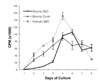
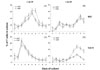
Con A-induced stimulation of bovine PBMCs and SEC-induced stimulation of human PBMCs resulted in a constant and nearly linear increase in PBMC nucleic acid synthesis in cultures for the first 4-5 days (Fig. 1). Coinciding with nucleic acid synthesis, a high percentage of bovine CD4+ and CD8+ T cells with >2n levels of cellular nucleic acid (52% and 65%, respectively) were observed within 2 days in cultures stimulated by Con A (Fig. 2C). The response in SEC-stimulated bovine PBMC cultures was delayed and less dramatic; no increase in PI fluorescence intensity was evident until day 3 (Fig. 2A). This response was associated with only a slight increase in a total nucleic acid synthesis within the first 4 days (Fig. 1). These differences suggest that mitotic activity of bovine CD4+ and CD8+ T cells in SEC-stimulated culture was low and possibly suppressed.
In Con A stimulated cultures, apoptosis (PI fluorescence intensity <2n) did not increase above background level until day 6 (Fig. 2D), which is 4 days after the day 2 peak in percentages of cells containing >2n amounts of nucleic acid (Fig. 2C). However, in cultures stimulated with SEC, a moderate level of apoptosis was initiated early (especially in CD4+ T cells) (Fig. 2B). It was sustained at a near constant level until after day 6, when a surge of apoptosis began. The increase of apoptosis in SEC stimulated bovine cells (Fig. 2B) preceded both an increase in percentages of cells with >2n amounts of DNA (Fig. 2A), and also an increase in nucleic acid synthesis (Fig. 1). These results suggest that some apoptosis in bovine PBMC cultures stimulated by SEC occurs prior to proliferation of these cells. These combined results suggest that an early wave of SEC-related apoptosis resulted from stimulation of PBMC by the toxin.
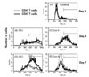
The numbers and sizes of bovine CD4+ and CD8+ T cells were assessed in cultures stimulated with 0.1 µg/ml SEC (Fig. 3). Stimulation with SEC consistently resulted in an inversion of the CD4+:CD8+ T cell ratio, from an initial value of 1.96 ± 0.60 to 0.35 ± 0.07 (mean ± SE, n = 6) by day 7. The observed changes in CD4+ and CD8+ T cell numbers and ratios correlated with the changes in cell sizes within each subpopulation (Fig. 3). Con A-induced enlargement of T cells occurred by day 4. However, SEC-induced cell enlargement was minimal at day 4; a high level of blast-sized cells was not observed until day 7 (Fig. 3). By day 7, Con A-stimulated cells had already substantially decreased in size (Fig. 3). This suggests that these cells were reverting to a quiescent state or were dying, as they also had relatively low side scatter values (data not shown). In contrast to cultures stimulated with Con A, the blast cell population in SEC-stimulated cultures contained more CD8+ T cells compared to CD4+ T cells consistent with the low CD4+:CD8+ T cell ratio.
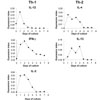
Both Th-1 (IL-12, IFN-γ, and IL-2) and Th-2 (IL-4 and IL-13) cytokine mRNAs were induced early, by 24 h. mRNA levels for IL-4 and IL-13 were sustained over a period of 4 days, and exhibited secondary peaks on day 3 (IL-4) or day 4 (IL-13) (Fig. 4). The mRNA for a key Th-1 cytokine, IL-12, dropped to background level on day 2 and declined to even further levels afterwards. No secondary peaks were observed in mRNA levels for IL-12, IFN-γ, or IL-2. These results indicate that stimulation of bovine PBMC cultures with SEC creates a Th-2-biased microenvironment, and SEC-induced proliferation of CD4+ and CD8+ T cells takes place in an IL-4- and IL-13-rich milieu.
Discussion
SAgs produced by S. aureus and other organisms are causative agents of human toxic shock syndrome and can induce shock-like illnesses in other animals [4]. Interestingly, toxic shock syndrome is not a described illness in dairy animals, despite the common colonization and occurrence of bovine infections caused by SAg-producing S. aureus [17,30]. This suggests that the bovine immune system responds differently to SAg stimulation. In this study, we showed that the increase in cell proliferation in bovine PBMC cultures stimulated with SEC is delayed compared to human PBMCs or to bovine PBMC exposed to Con A. The bovine T cell response in vitro is characterized by a slow proliferation and relatively early induction of apoptosis.
Mercado et al. [21] demonstrated that disease severity and progression of antigen-specific T cell responses are determined by events very early bacterial infections. Our results support the possibility that early events following exposure to SAgs induce the expansion of bovine CD8+ T cells which could influence the pathogenesis of staphylococcal infection. Previously, we also showed that SEC induces an aberrant increase in bovine CD8+ T cell populations and that most CD8+ T cells express CD26 (ACT3) [11,12,18]. In addition, the mRNA expression of Th-2 cytokines such as IL-4 and IL-10 was examined in SEC-stimulated cultures [12]. The results of this study were consistent with our previous report [12]. Analysis of cytokine mRNA expression was extended in this study with the addition of IL-13, another Th-2 cytokine, plus IL-12, a Th-1 cytokine. The results of our combined work demonstrate that SEC induces a prolonged Th-2 cytokine expression although Th-1 cytokines are expressed in early cultures after exposure to the toxin.
This present study confirms and extends our previous finding that a delayed SEC-induced proliferation of bovine T cells involves an increase in CD8+ T cell numbers after 7 days of exposure. The SEC-induced differentiation of T cells into Th-2 cells may be the result of early programming events and is consistent with Th-1 and Th-2 cell differentiation process reported previously [27]. These activated T cells may have an immunoregulatory role in the bovine mammary gland [29]. Since S. aureus is capable of entering bovine epithelial cells [2], induction of non-cytotoxic, CD8+-derived regulatory cells may reduce the capacity of effector cells to control and clear an infection. Thus, our results suppose that SEC-induced expansion of bovine CD8+ T cells may be involved in pathogenesis of S. aureus.
A reversal of a CD4+:CD8+ T cell ratio, due to a relative increase in CD8+ T cell numbers, is often associated with chronic disease states and an inability to mount a protective immune response [24,25]. For example, a subset of CD8+ T cells predominate in lepromatous lesions in leprosy patients [22], whereas CD4+ T cells are predominant in tuberculoid (healing) lesions [32]. Importantly, CD8+ T cells from lepromatous lesions, but not from tuberculoid lesions, could be activated by Mycobacterium leprae antigens to suppress proliferation of CD4+ T cells [22,32]. The SEC-induced reversal of CD4+:CD8+ T cell ratio is consistent with the results obtained with other SAgs in vivo [16,19], and it is likely to contribute to an inability of the immune system to generate a protective response to staphylococcal mastitis in cows.
In conclusion, we present evidence that the SEC-induced proliferation of bovine T cells is preceded by a period of a non-proliferative immunoregulation during which they are exposed to both Th-1 and Th-2 cytokines. Continued exposure leads to a Th-2 bias, inversion of the CD4+:CD8+ T cell ratio and induction of CD8+ T cells with immunoregulatory activities [6]. These responses, early and late, could promote intracellular survival of S. aureus and influence the pathogenesis of staphylococcal infection.
 XML Download