

 PDF
PDF ePub
ePub Citation
 Print
Print


Introduction
Echocardiography is an ultrasound technique that is an important diagnostic tool in cardiology [21], which has recently been introduced to veterinary medicine as a non-invasive method for evaluating the anatomy and function of the heart [12].
Conventional echocardiographic modalities include two-dimensional, M-mode, and Doppler modes [12]. An ultrasound examination of the heart and large vessels represents a significant technological advance in veterinary medicine. Echocardiography allows an evaluation of the space relationship between structures, cardiac movement and the blood flow features, the precise and non-invasive diagnostic of cardiac alterations, as well as a follow-up of therapy and to determine the prognosis through direct vision of the cardiac chambers. It is important that an echocardiographic examination be considered as part of a thorough cardiovascular evaluation including other examinations, such as clinical, radiographic, and electrocardiographic examinations [13].
The echocardiographic indices show significant breed variations and it is important to know the normal echocardiographic values for each dog breed considering the influence of body weight on the established echocardiographic values [3,6,23,26].
The aim of this study was to determine the echocardiographic indices of clinically healthy German shepherd dogs, and correlate them with the body weight, gender and age. It is expected that the data obtained will be valuable for the progress of studies on small animal cardiology.
Materials and Methods
This study examined 60 clinically health German shepherd dogs, 31 males and 29 females, ranging in age from 1 and 5 years. The animals were selected through a physical and electrocardiographic examination, those showing any clinical sign of cardiopathy were excluded. The dogs were examined by two-dimensional, M-mode, and Doppler echocardiography, and all data was recorded in the proper files containing the name, number, gender, age, body weight and echocardiographic measurements.
An ultrasound unit (UF5800; Fukuda Denshi, Brazil), 3.5 MHz transducer was used for the echocardiographic examination. The animals were positioned on the right and left lateral recumbency using a manual restraint. A U-shape table was used at the thoracic region. A thick acoustic gel layer was placed between the transducer and the site of examination with no hair removal. Three individual measurements of each variable were performed and the average data was obtained. All examinations were printed on thermosensitive paper and recorded on videotape for further evaluations.
For the two-dimensional mode images, the right and left parasternal positions, from the third to the fifth intercostal spaces, were used according to Thomas et al. [26]. In this mode, the left atrial apex-base and mediolateral diameters, area and volume were measured. The aorta diameter was also measured at the level of the aortic valve annulus.
For M-mode, the following measurements were made at the end-diastole according Lombard and Ackerman [19]: aorta (AO), right ventricular internal dimension (RVIDD), septal thickness (ST), left ventricular internal dimension (LVIDD) and left ventricular wall thickness (LVWT). The same measurements were made at the end-systole, including the left atrium (LA).
The following calculations were made: the left atrial to aortic root ratio (LA/AO), left ventricular fractional shortening (%FS), left ventricular mass (LVM) and the mitral valve Epoint to the ventricular septal separation (EPSS) (the distance between the ventricular septum and the maximal initial opening of the mitral valve E-point). The LVM was calculated according to the formula reported by Devereux and Reichek [10]: [1.04 × (LVIDD + STD + LVWTD)3 - LVIDD3 - 13.6].
Doppler mode transducer positioning was performed according to the recommendations by Darke et al. [7]. With each examination site using pulsed-waved Doppler, the transducer angle was guided by audio and by the waveforms with a greater velocity. The mitral valve flows were obtained in order to determine the velocity peaks, the ratios of the E and A waves and the mitral E wave deceleration time. Regarding the tricuspid valve, the velocity peaks and ratios of the E and A waves were examined. The velocity peaks for pulmonary and aortic valves were also obtained. On the left ventricular outflow tract flow, the time velocity integral (TVI) and aortic cross-sectional area were used to calculate the stroke volume. The stroke volume across the aortic valve was calculated by multiplying the cross-sectional area at the level of the aortic annulus by the time-velocity integral of flow across that valve. The area was calculated from the peak diameter at the insertion of the aortic leaflets in the parasternal long-axis view, assuming a circular configuration. The outflow velocities were obtained by pulsed Doppler echocardiography from the apex with the sample volume at the level of the measured diameter and scanned radially for optimal alignment with flow. The time-velocity integral was calculated by tracing the modal velocity (the darkest portion of the velocity spectrum representing the largest scatter) and averaging over three to five beats. The stroke volume was determined from the product of area and time-velocity integral . The cardiac output (CO) was then calculated by multiplying the stroke volume by the heart rate [28].
Statistical analysis involved correlating all the studied variables with the body weight and age. In relationship to gender, the Student t test was used to compare the mean values between males and females, and to set the significance level (%). The sample distribution was normal (Gaussian). Hence, the data is described as the average and standard deviation.
Results
The mean age of the dogs was 2.9 ± 1.21 years ranging from 1 to 5 years. The mean body weight was 30.2 ± 3.98 kg ranging from 22 to 37.2 kg. 48.3% were females and 51.7% were males.
Two-dimensional mode
The left atrial apex-base and mediolateral diameter was 3.73 ± 0.238 cm and 3.32 ± 0.186 cm, respectively. The volume and area of the left atrium was 10.00 ± 1.235 cm2 and 25.12 ± 4.210 ml, respectively. The diameter of the aorta at the level of the aortic valve annulus was 2.52 ± 0.16 cm. All these variables correlated with the body weight (correlation coefficient r = 0.38 to 0.58, p < 0.05), with no association with age or gender (p < 0.001).
M-mode
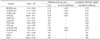
Table 1 shows the average and standard deviation of all the variables examined in this mode. A statistically significant correlation with body weight was observed for the left atrial, aortic diameter, left ventricular, septal and posterior wall dimensions (correlation coefficient r = 0.30 to 0.87, p < 0.05), with no age correlation (p < 0.001). A statistically significant gender difference was found for the right and left ventricular and posterior wall dimensions (p < 0.01).
Doppler mode
Mitral valve: During the diastole, the mitral valve flow was positive (over baseline), laminar, and divided in two main phases of a triangle aspect, E and A waves. The E wave peak of the velocity was 0.74 ± 0.084 m/s. The A wave, which is generally lower than the E value, was the result of atrium contraction, with velocity peaks of 0.44 ± 0.074 m/s. The relationship between the E and A waves (E/A) was always greater than 1.0. The E and A wave peaks were not associated with age or gender. The mitral E wave deceleration time was 132.6 ± 16.44 ms.
Tricuspid valve: a similar diastolic flow to the mitral valve was observed. The E wave velocity peak was 0.67 ± 0.117 m/s and the A wave velocity peak was 0.41 ± 0.094 m/s. The E and A velocity peaks showed no correlation with the body weight, age or gender.
Pulmonary valve: The pulmonary valve flow was observed as a negative, laminar and during systole. The velocity peak was 0.92 ± 0.129 m/s.
Aortic valve: Negative and laminar flows were observed during the systole, with velocity peaks of 1.02 ± 0.143 m/s.
Left ventricular outflow tract: This flow was observed during the systole, was negative and quite similar to the aortic flow, but with lower velocity peaks (0.92 ± 0.164 m/s). After its measurement, the stroke volume was calculated using the time velocity integral (average: 17.93 ± 2.277 cm) and the aortic valve annulus (2.52 ± 0.16 cm). The cardiac output was calculated by multiplying the stroke volume (average: 52.93 ± 7.843 ml) by the heart rate (average 63.45 ± 8.45 beats/min). The CO value was 3.28 ± 0.797 l/min. the pulmonary and aortic flows showed no significant correlation with the body weight, age or gender.
Discussion
Good two-dimensional echocardiographic images of 60 studied dogs were obtained, which were qualitatively similar to those described in other breeds [8,12,20,22,23,29]. On two-dimensional mode, the left atrium apex-base and mediolateral diameters, area and volume were examined. All these results were similar to those described by O'Grady et al. [22]. The left atrial size in the dog demonstrated a strong correlation between the changes in the left ventricular end-diastolic pressure and the left atrial pressure, with the left atrial size being a good indicator of left ventricle abnormalities, particularly cardiac failure. The same authors have also observed a significant correlation between the variables and the body weight but with higher correlation coefficients (r > 0.77). This might be explained by the breed, age and body weight homogeneity of our study sample. Crippa et al. [6] observed similar results in Beagles.
Morceff [21] and O'Leary et al. [23] examined the aortic diameter at the level of the aortic valve annulus and reported similar results to this study. They reported that the main preference for the measure in this place was its possible reproducibility, which is better than in other sites. O'Leary et al. [23] demonstrated that the high correlation for the aortic diameter measurement at the level of the aortic valve annulus was the result of the greater ease in defining the region of the sinuses, as opposed to the more difficult definition of the aortic annulus.
For M-mode, the left ventricular and septal wall thickness or left ventricular internal cavity dimensions measurements fall within the range of values considered to be normal for dogs of this body size, as reported elsewhere [8,19,26]. Echocardiography can help evaluate many cardiac diseases such as mitral regurgitation, cardiomyopathy, congenital defects or hypertrophic patterns. Knowledge of the normal measurements for a specific body weight can help to indicate the degree and direction of the change from normal. Adult German shepherd dogs were shown to have larger left ventricular dimensions and wall thicknesses than other small breeds of dogs. These differences suggest that the breed and body conformation in addition to the heart rate may need to be considered when interpreting echocardiographic studies in dogs. Caution should be taken in extending these generic reference echocardiographic values to an evaluation of echo studies. Similar values were obtained when comparing this data with other large breeds of dogs, such as Greyhounds [7,24]. One explanation for this might be the morphologic adaptations made during training as well as other factors such as breed and body conformation.
Brown et al. [5] proposed an indexing system for quantitative echocardiographic interpretations based on a calculation of the ratio indices in which each raw M-mode measurement is divided by the aortic root dimension. This system appears to be valid over a range of species and is independent of the size of the animal. German shepherd dogs have a high prevalence of cardiac diseases. Such cardiac disorders can lead to cardiac heart failure and sudden death, making early detection important. Early detection is also important in breeding animals in order to facilitate effective disease control, because undiagnosed or mild cardiac defects can be transmitted to their offspring before the parent develops any clinical signs. Another difference from the study of Brown et al. [5] is that a global echocardiography assessment of the heart was performed, including two-dimensional and color flow Doppler modes instead of just a M-mode study.
There were gender differences in the right and left ventricular dimensions in German shepherd dogs. It is believed that these differences are the result of work hypertrophy and the higher body weight in males than females. A further factor could be related to inherited characteristics, as reported by Page et al. [24] for Greyhounds.
The left atrial to aortic root ratio in our dogs was approximately 1.0, which is similar to that reported elsewhere [3,8,19]. The LA/AO ratio is a useful tool for recognizing relative left atrial enlargement. The LA enlarges most often with chronic, acquired mitral regurgitation and dilated cardiomyopathy.
The %FS is the most commonly used clinical measurement of the LV systolic function. The dogs in this study had a %FS within the range reported for dogs. The %FS is a measure of function even though it is generally considered to be an indicator of ventricular compliance and contractility. The three conditions that most affect the fractional shortening are preload, afterload and contractility. Each one of these may act individually or together to affect the FS [2,8,11,19,23].
The left ventricular mass also remained within the range reported for dogs [11]. Cardiac hypertrophy usually involves an increase in the left ventricular wall thickness and is not always apparent because there also may be wall thinning as the heart dilates. The degree of hypertrophy parallels the severity of overload, and detection of extreme hypertrophy may indicate a poor prognosis [9]. This increase in mass can be quantified using M-mode echocardiography. Devereux and Reichek [9] demonstrated that the M-mode echocardiography method plays a valuable role examining disorders characterized by LV hypertrophy in humans because it is an accurate, relatively easy, safe and reproducible method. The two-dimensional method of determining the mass is superior to the M-mode measurement because the entire geometry of the heart is considered. The M-mode method relies on accurate measurements of the wall and the septal thicknesses at specified locations. However, the two-dimensional method is an involved process that begins with a determination of the two-dimensional volume, which is not routinely performed during a clinical examination.
The E-point to ventricular septal separation (EPSS) values fell within the range of values considered normal for dogs. This is a useful, practical and easily reproducible clinical index of the left ventricular inflow blood and therefore the left ventricular function. Kirberger [15] reported that the major virtue of EPSS as a clinical measurement is its simplicity. However, EPSS is only a qualitative indication of the left ventricular function. It should be noted that a normal EPSS value might also occur in the presence of severe cardiac disease. It is a simple measurement, which if altered, should alert the examiner to the possibility of cardiac disease.
Doppler mode was performed over the left parasternal location according to Darke et al. [7]. The mitral, tricuspid, pulmonary and aortic valves flows and the left ventricular outflow tract were examined, and showed similar results to those reported by other authors [4,7,8,23,27].
Pulsed-waved Doppler (PW) was used on this study according to Bonagura [1], Henik [12] and Kirberger [14] because it allows the selection of a special area, and is sufficient to collect normal laminar flow as it occurs in healthy dogs.
Some authors [7,16,30] suggested that the apical long-axis (four-chamber) view of the left atrium and left ventricle was good for the transmitral flow. The normal values found in the E and A waves are similar to those reported elsewhere [16,30]. The relationship between the E/A waves was greater than 1.0, which was also related by Morcerf [21], who reported the importance of this index in cases of cardiac disease, which alter the diastolic function e.g. restrictive cardiomyopathy, making the E wave much greater then the A wave. The same authors did not find any significant correlation between the E and A waves velocity peaks and the body weight, gender and age, which is similar to our results. The deceleration times are also measured from the mitral inflow profiles. The values in German shepherds were found to be 132.6 ± 16.44 ms. The absence of clinical and ECG evidence of abnormalities supports the echocardiographic measurements of the deceleration time as being normal for this breed. This is one of the echocardiographic assessments of the diastolic function. Boon [2] suggested that the diastolic function of the heart is complex and involves many interactive components, which include myocardial relaxation, atrial contraction, rapid and slow filling phases and the loading condition. However, this parameter has not been reported in animals. Therefore, no comparison with other data could be made.
The tricuspid valve flow was measured as recommended in the literature [7,16]. The transducer was always moved by one intercostal space cranially from a four-chamber view, with moderate dorsal and often slight caudal angulation. The flow found was similar to the mitral valve, and similar to those reported elsewhere [16,30]. Kierberger et al. [16] reported that there was no significant correlation with body weight and gender, as observed in this work.
The pulmonary valve flow was measured according to recommendations reported in the literature [7,27,30]. The normal values for the velocity peaks were similar to those reported by Kirberger et al. [16]. There was no significant correlation between the pulmonary valve flow and body weight, age or gender. A possible explanation can be the homogeneity of the breed and size of this studied sample.
The five-chamber view was used for the aortic valve flow [7,16,30]. Kierberger et al. [16] reported a non-symmetric flow because it has a very rapid initial velocity, which is a feature of flows under systemic resistance, as observed in this study According to the same authors [16,30] there was no correlation between the velocity peak and body weight, gender or age, which was also observed in this work.
The left ventricular outflow tract flow was measured according to Kirberger et al. [16] and O'Leary et al. [23]. The values obtained for the aortic flow envelope and the velocity peaks were also similar to the left ventricular outflow tract flow. Using this flow, the TVI was calculated, and its values were similar to those reported by Brown et al. [4]. Lehmkuhl and Bonagura [18] reported higher TVI values in dogs with a subaortic stenosis in relationship to the normal values (70 cm), which shows how important it is to know the reference values when making a diagnosis. The stroke volume value was similar to the values reported elsewhere [4]. The aim was determine the stroke volume in order to calculate the cardiac output (CO), which reported to be was 3.28 ± 0.797 L/min in German Shepherd dogs, as reported by the same author [4]. The cardiac output is another measure of the global cardiac function. The CO can be measured for different diagnostic modalities. The echocardiographic method is a noninvasive technique for examining the cardiac performance. It has the advantage of being inexpensive with low risk to the patient risk. However, CO is a very insensitive indicator of the cardiac performance because many compensatory mechanisms act to maintain a normal CO, such as the heart rate and stroke volume. The factors causing errors in this technique are associated with the methods used to determine the aortic luminal area and requires a well-trained person to operate the machine for consistent echocardiograms. The CO value obtained using the thermodilution method was simple and harmless to the patient, and allowed repeated evaluations to verify a single reading. However, this method has the inherent disadvantage of high equipment cost and the need to insert pulmonary artery and right atrial catheters [10]. The Fick method has been used in most animal species. A major problem with this method is that the Fick determination requires a time delay for calculation, which can mask problems and eliminate the opportunity to carry out multiple measurements when technical difficulties develop [10]. Boon et al. [2] and Kittleson and Kienle [17] demonstrated that the CO calculated using Doppler echocardiography showed the highest correlation (r = 0.93) compared with the thermodilution derived cardiac output. Although technically demanding and with the potential for significant errors, the use of Doppler echocardiography in dogs showed a good correlation with traditional methods. This technique was found to be highly useful as a method of clinical examination because of its non-invasiveness. It was also reported that serial determination of CO is useful for determining the hemodynamic response to acute changes in the LV performance. Furthermore, this method might be useful in individual dogs for following certain trends because it accurately reflects the directional change in normal dogs.
There was no significant correlation between the measured variables through the left ventricle outflow tract flow and body weight, gender or age, which is different to the results reported elsewhere [4,16]. This observation can be explained by the following: less homogenous samples, different breeds and a lower number of animals.
 XML Download