

 PDF
PDF ePub
ePub Citation
 Print
Print


Introduction
The Mycobacterium tuberculosis complex group includes: M. tuberculosis, M. bovis, M. africanum, M. microti [19] and a newly described species M. canetti [24]. Mycobacterium tuberculosis is the primary causative agent of human tuberculosis, but may also infect animals in contact with infected humans [15]. Tuberculosis has re-emerged as one of the leading causes of death worldwide, causing nearly three million deaths annually [2]. In India alone, half a million people die of TB every year, i.e. more than 1000 people every day, and one patient every minute [29]. Both M. tuberculosis and M. bovis have been isolated from humans and animals in India [26]. However, the origin and transmission of infection between animals and humans have not been investigated. Therefore, in view of the global prevalence of tuberculosis, there is an urgent need to develop techniques that not only identify and characterize tubercular bacilli, but also facilitate epidemiological studies to trace the source of infection thereby facilitating formulation of effective control strategies.
Rarely does the antibiotic susceptibility patterns including: serotyping [10], biotyping and bacteriophage typing [11,18] allow for strain differentiation. DNA based techniques are now available for molecular characterization of M. tuberculosis. Restriction fragment length polymorphisms (RFLP) using probes for insertion sequences IS986, IS1081 and IS6110 that have been extensively used to differentiate strains of M.tuberculosis [12,21]. However, the relatively complex nature of the standard methods, as well as the lack of utility of some of the probes (such as IS6110) for some of the Indian strains indicates the need for alternate rapid procedures. Random amplification of polymorphic DNA (RAPD) is a multiplex PCR-based molecular system [27,28]. This method uses short oligonucleotide primers of an arbitrary sequence, and low-stringency PCR, to amplify discrete DNA fragments that can be used as molecular markers. RAPD analysis is rapid, inexpensive, easy to perform and can be used for determination of genetic heterogeneity based on DNA sequence diversity [3,4,27,28].
This method, which requires no previous genetic knowledge of the target organism, relies on the presence of low-stringency priming sites, for a single arbitrary primer on both strands of the DNA molecule, close enough to permit PCR amplification. This DNA fingerprinting has been successfully used to type M. tuberculosis [8,13,17,22] and other bacteria including: E. coli [5], P. multocida [7] and Staphylococcus aureus [9]. The present study reports on the use of RAPD analysis of M. tuberculosis strains to identify the heterogeneity in these strains.
Materials and Methods
Mycobacterial strains
Details of the M. tuberculosis strains used in the present study are given in the Tables 1. M. tuberculosis strains used included 40 strain isolated from human patients with pulmonary tuberculosis from the Medical Hospital, IVRI, Izatnagar (U.P.), India and from the District Tuberculosis Hospital, Bareilly, India, 2 reference strains (H37Rv and MT-DT) and 1 strain each from bovine and swine samples. All of the mycobacterium strains were typed by conventional morphological (Ziehl-Neelsen staining) and biochemical tests [26] and maintained on Lowenstein-Jensen medium at the Mycobacteria Laboratory, Indian Veterinary Research Institute, Izatnagar, India.
RAPD analysis
A number of primers were used for RAPD analysis of mycobacterium isolates. Details of the primers used in the present study are given in the Table 2. All of the primers from the OPN-series were obtained from Operon Technologies (USA); the primers for the BG-series were synthesized by M/s Bangalore Genei Pvt. Ltd. India. Genomic DNA was extracted as per the method of van Soolingen et al. [23]. Amplification of mycobacterium DNA, using random primers, was performed in a total volume of 25 µl. The reaction mixture contained 1 unit of Taq DNA polymerase (Bioloine GmBH, Germany), 1.5 mM MgCl2, 200 µM of each dNTP, 30 pmol primers, and 50 ng of template DNA. Amplification was carried out in a thermal cycler (Eppendorf, Germany). The cycling conditions consisted of an initial denaturation step for 5 min at 94℃, followed by 45 cycles of 94℃ for 1 min denaturation step, an annealing step for 1 min at 36℃, and an extension step for 1 min at 72℃ and a final extension at 72℃ for 5 min. The products obtained from RAPD-PCR were analyzed on a 1.5% agarose gel stained with ethidium bromide. Subsequently, the gel was visualized and photographed using a gel documentation and analysis system (AlfaImager, Germany). The banding patterns obtained by RAPD were noted on a photograph. A data matrix composed of the numerals 1 and 0 was built on the basis of presence (1) or absence (0) of a DNA band appearing in replicates for each isolate. Only distinct and prominent bands were scored and used in assessing RAPD patterns. The molecular size of bands was calculated using software provided by AlfaImager (Germany). The size of the bands, that differed by ±5% on different gels, was considered to be the same bands. The genetic diversity of isolates was analyzed by RAPDistance version 1.04 software that operates on the basis of UPGMA clustering.
Results
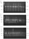
RAPD-PCR revealed the presence of amplicons of a variety of sizes in M. tuberculosis strains. In this study, several fragments were amplified in each sample, and most of these fragments were observed to be common to different strains. However, there were some fragments unique to certain strains. All 44 isolates of M. tuberculosis showed a high degree of polymorphism with RAPD analysis (Fig. 1A, B, C). The number of RAPD patterns generated by each primer is shown in Table 1. All 7 primers revealed discriminating patterns. The primer OPN-01 and the primer BG-66 generated the maximum and minimum number of amplimers, respectively. Among the seven primers, OPN-01 showed maximum discrimination for the ability to type mycobacterium isolates, and produced 10 RAPD patterns (Table 2).
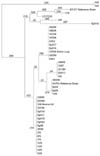
Upon dendrogram analysis, with primer OPN-02, four clusters were formed, the largest cluster consisted of 17 strains, the second largest cluster contained 10 strains and the remaining two smaller clusters contained 5 strains in each; genetic relatedness was closest among strains within clusters (Fig. 2). Five strains were identified in different clusters along with M. tuberculosis strain DT, while the M. tuberculosis strain H37Rv was found in one of the smaller clusters. Two strains, 5/S and 9/S, were in the same cluster but remained separated from the rest of the strains and clusters (Fig. 2). Strain SpS19 demonstrated a unique pattern with all of the primers used (Table 1). Bovine strain 1/86 and swine strain 125/92 were identified within the largest and second largest clusters respectively (Fig. 2).
Discussion
Detailed epidemiological studies of M. tuberculosis have been hampered by difficulties in differential characterization of causative strains. The ability to distinguish strains of M. tuberculosis would be useful for investigating the source of outbreaks of infection, the relatedness of strains recovered from different patient, and the identities of multiple strains recovered from the patients from similar localities. Infections caused by mycobacterium are known to be transmitted from human to human [1], animal to human [6], and animal to animal. [16] In an outbreak investigation of tuberculosis, it is often important to know whether the disease is due to a new strain or relapse of a known strain. This information has a special bearing on our understanding of the emergence of multi-drug resistant disease. In India, the status of M. tuberculosis infection in animals is poorly understood.
In the present study, RAPD showed both similarities and differences among M. tuberculosis strains. All primers amplified scorable fragments in each strain analyzed. The common or monomorphic bands among the different strains likely represent highly conserved regions in the genome. Clusters of M. tuberculosis, consisting of the largest number strains, showed a possible close genetic relationship; these were isolated at different occasions from human sputum and from two animals, one from a bovine lymph node and the other from a swine lung. Therefore, the outcome of our analysis is consolidated, in a comprehensive manner, to draw a phylogenetic relationship, which is consistent with prior reports [13,20,25].
It was interesting to note that a previous study of RFLP using IS 6110 and IS 1081, of these M. tuberculosis strains (13 overlapping M. tuberculosis stains, including H37Rv), showed no polymorphism; [21] this suggested that this RFLP could not differentiate these M. tuberculosis strains. However, in this study using the RAPD analysis, we found differentiation among the M. tuberculosis strains. Standardization of PCR mixtures and conditions are very important for reproducibility of RAPD-PCR results. We found that it was necessary to perform RAPD-PCR in duplicate to obtain valid results. Our findings show that RAPD-PCR yields reliable and reproducible results under precise assay conditions.
Isolation of M. tuberculosis from animals is not common. M. tuberculosis strains from a bovine lymph node and a swine lung were similar to the M. tuberculosis strain from human sputum; this suggests a possible transmission of infection from humans to animals. RAPD analysis may help to establish the molecular relatedness of M. tuberculosis strains, their distribution and zoonotic importance in an agrarian country like India, where there is a close association between livestock and human beings.

 XML Download