

 PDF
PDF ePub
ePub Citation
 Print
Print


Introduction
The production of broiler meat has been on the rise in Kenya and represents an increased opportunity to generate income. Its rise in popularity is based on the availability of a ready market for such meat, low capital requirement, minimal space requirement, proximity to hatcheries, and the availability of a wide selection of animal feeds in urban areas [6]. Production is affected, however, by various diseases, of which coccidiosis is particularly important [3,4]. Coccidiostats are widely used to prevent this condition, to find animals that have the disease and need treatment, and to improve feed conversion efficiency and the rate of animal growth [1,8]. Harmful coccidial residues may appear in foods because of the extensive use of age of anticoccidial drugs in animal husbandry [1,4,7,8].
Despite the common use of antimicrobials, routine monitoring of animals raised for food for anticoccidal residue is lacking. As in most low-income countries, this can be attributed to the high costs of analysis for coccidial residues and the lake of affordable screening methods [11,13].
The tube test, which was developed to detect drug residues levels in milk at the Codex alimentarius maximum residue limit (MRL), is a low-cost microbiological method with potential for use in low-income countries [9,13]. Bacillus stearothermophilus var calidolactis C953 is used as the test organism at a pH of 7.0 to 8.0 to detect a broad spectrum of antimicrobials in milk [13]. The applicability of this method to other food matrices has not been tried yet. This study was determine whether this technique could be used to detect the residue of 3 drugs that are frequently used to control coccidial infevtions: sulfamethazine, furazolidone, and amprolium.
Materials and Methods
Preparation of solutions
A stock solution (1 mg/ml) of sulfamethazine (Sigma, Netherlands) was made by dissolving sulfamethazine in ethanol and adding enough distilled water to create a 100 ml solution. Furazolidone (Cosmos, Kenya) was dissolved in distilled water to produce a stock solution of 1 mg/ml. Amprolium (Cosmos, Kenya) was dissolved in distilled water to produce a stock solution of 1 mg/ml.
The sulfamethazine stock solution was diluted further in either a pH 8.0 phosphate buffer (solution B) or distilled water (solution C) to produce 100, 10, 2.5, and 1.0 µg/ml solutions. The frazolidone and amprolium solutions were diluted further in distilled water to produce solutions of the same concentrations.
Working solutions of the 3 drugs were prepared in using distilled water: sulfamethazine and furazolidone, each at concentrations of 0.1, 0.2, 0.4, 0.6, 0.8, 1.0, 2.0 and 4.0 µg/ml and amprolium at concentrations of 1, 2, 4, 6, 8, 10, 12 and 14 µg/ml. These preparations were tested in the B. sterothermophilus var. calidolactis tube diffusion test using replicates of 10 to determine the limits of detection.
Control samples
Distilled water was used as the negative control and a 50 µg/ml solution of each drug was used as a positive control in our search for the limits of detection of each drug using the tube diffusion test. Liver or kidney tissue that was free of any antimicrobial drugs (ie, "blank tissues") was used as the negative control. Then 5 ml samples of blank kidney and liver tissues was injected with a 50 µg/ml solution of each drug that was prepared by adding 5 ml of the 100 µg/ml drug solution or 5 ml of the negative-control solution.
Tissue spiking
Solutions containing 0.1, 0.2, 0.4, 0.6, 0.8, 1.0, 2.0, and 4.0 µg/ml of sulfamethazine- and furazolidone-contaminated liver and kidney tissues were prepared using appropriate working solutions and blank tissue. Liver tissue was contaminated with sulfamethazine in a pH 8.0 phosphate buffer. Solutions of 1, 2, 4, 6, 8, 10, 12 and 14 µg/ml amprolium-contaminated liver and kidney tissues were prepared for use in the same manner.
Tube test
The tube diffusion was prepared as described by Nouws et al. [9]. In brief, it entailed the addition of 2 ml of bromocresol purple (2.5 µg/ml), 2 ml of a B. stearothermophilius var calidolatcis C953 spore suspension (107 spores/ml) and 0.3 ml of trimethoprim (50 µg/ml ) to a 100 ml of agar count plate (Difco, USA) at 63℃. The pH of the medium was then adjusted to 8.00 ± 0.02 using 1M NaOH solution at 63℃. Subsequently, the medium was distributed among the test tubes in 1 ml portions. The tubes were then placed in an upright position, and the agar was allowed to solidify at room temperature. The prepared tubes were used on the same day or kept at 4℃ to 5℃ for a maximum of 2 days. The detection limit of the tube for each drug was determined by constructing a dose response curve for each. The detection limit was defined as the concentration at which 95% of the test results was positive.
Assay procedure
Each drug was assayed by adding 0.33 ml of the drug solution to the test tubes so that there were 10 tubes for each concentration of the drug. The tubes were allowed to stand for 1 h to allow the drug solution to diffuse into the media. Any drug solution remaining after that time was removed by decanting. The tubes were then covered with an aluminum foil and incubated in a water bath at 63℃ for 4.0 to 4.5 h. The results could be read immediately, because the negative control solutions turned from purple to yellow.
The contaminated tissue samples were centrifuged for 5 min to allow tissue debris to fall out of solution. The supernatant was added to the tubes such that each tube containing a replicate of each drug concentration received 0.33 ml. The tubes were allowed to stand for 1 h to allow the supernatant to diffuse into the media. The supernatant remaining after that time was removed by decanting. The tubes were then covered with aluminum foil and incubated in a water bath at 63℃ for 4 to 5 h. The results could be read immediately the negative controls turned from purple to yellow.
Results
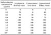
The results for sulfamethazine-contaminated tissue are shown in Table 1. The tube test indicated a detection limit of 0.5 mg/ml whenthe sulfamethazine solution was prepared in distilled water. A detection limit of 0.7 µg/ml was obtained in liver tissue injected with sulfamethazine. In sulfamethazine-contaminated kidney tissue, the limit of detection was 0.54 µg/ml. A 100% positive response was observed with drug concentrations exceeding 0.5 µg/ml.
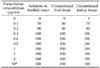
The results for furazolidone-contaninated tissue are shown in Table 2. The tube method was able to detect furazolidone in solutions that had been prepared in distilled water at concentrations smaller than 1 µg/ml. A 100% positive response for furazolidone was obtained at a concentration of 0.3 µg/ml , and it was detected in all liver and kidney tissues samples injected with at least 0.35 µg/ml of this drug. Thus, t-test indicated a limit of detection of 0.35 µg/ml for furazolidone and could detect it a concentration as low as of 0.30 µg/ml in all kidney samples into which it had been injected.
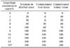
The results for amprolium-contaminated tissue are shown in Table 3. A detection limit of 5.7 µg/ml was observed in amprolium solutions prepared with distilled water. A detection limit of 7.8 µg/ml was observed in contaminated liver tissues and 7.6 µg/ml in contaminated kidney tissues.
Discussion
Drug residues in food animals being raised for human consumption may pose a public health hazard. Consumer protection can be ensured by screening such animals for residues [1,8]. The presence of antimicrobial residue in foods is of particular concern in low-income countries, because legislation regarding maximum tolerance levels for marketed products is often lacking and violation of the time set to terminate drug therapy occurs regularly [11,13].
The tube test is a microbial inhibitor test in which B. stearothermophilus spores are grown in agar with bromocresol purple as the pH indicator. The tubes differ with respect to pH value, supplements and antibiotics [9]. Normal microbial growth causes the pH indicator to change from purple to yellow in solution. Substances that inhibit normal microbial growth cause the color of the pH indicator to remain purple.
B. stearothermophilus has been shown to be sensitive to beta-lactam drugs in milk [10]. The applicability of this method in other foods has not been explored prior to this study. Using the tube diffusion test, we were able to detect sulfamethazine and furazolidone at concentrations smaller than 1 µg/ml. In a previous study using a B. stearothermophilus disk plate, sulfamethazine and furazolidone could only be detected at levels of 1µg/ml and above [8].
The type of organism used to find drug residue influences the detection limit. In the this study, sulfamethazine-contaminated kidney tissue appeared to be better suited for detection of the drug residue compared with liver tissue, because the tube diffusion test indicated lower limits of detection in this tissue. Similarly, furazolidone-contaminated kidney tissue had a lower limit of detection compared with contaminated liver tissues.
The higher limits of detection for the coccidiostasts could be attributed to the insensitivity of B. stearothermophilus to other compounds [13].The growth of B. stearothermophilus is mainly inhibited by beta-lactam drugs and to a lesser extent by other antibiotics [13]. In a other study, a higher sensitivity to salinomycin was reported in chicken tissues using the 4 plate method compared with the disk assay used with B. stearothermophilus [3].
When used to validate the STAR protocol in screening or antibiotics residues in milk, B. stearothermophilus was found to be sensitive to sulfonamides and beta-lactam drugs [5]. When used as the test organism in the inhibitor test, B. stearothermophilus was found to be unsuited for detecting tetracyclines up to the MRL in muscle tissue [12]. A rapid method of detecting sulfonamides in muscle tissue that uses B. stearothermophilus has been described [2]. The investigators were also able to use this method to detect sulfamethazine in tissues and solutions at levels of 75 to 150 ppb.
Our findings are thus in agreement with those of other studies in which B. stearothermophilus was used to find sulfamethazine and furazolidone at concentrations smallwer than 1 µg/ml. In our study, this organism demonstrated a lack of sensitivity to amprolium, however, which it could only detect at concentrations greater than 5 µg/ml, which exceeded the recommended Codex alimentarius MRL of 1 mg/kg in chicken.
The results of this study suggest that the B. stearothermophilus tube test has the potential to useful in detecting anticoccidial residue in poultry. Further studies are recommended to improve its sensitivity to a wider range of drugs at established Codex alimentarius MRLs.
 XML Download