

 PDF
PDF ePub
ePub Citation
 Print
Print


Introduction
A reduction in the number and function of immune cells can lead to a secondary immunological deficency, which manifests as recurrent infections, autoaggresion and cancer diseases. In the last three decades, a wide variety of substances, both natural (calf thymus extract, lysozyme dimer) and synthetic (levamisole, isoprinosine), have been introducted in order to modify the acquired defects of the immune cell activity.
Recently, it was reported that chitin derivatives have immunomodulatory action [5,13]. Chitosan (copolymer of β-91-4)-2-amino-2-deoxy-D-glucopyranose and β-(1-4)-2-acetamide-2-deoxy-D-glucopiranose) is an N-deacetylated product of chitin that is produced by chemical or enzymatic deacetylation resulting mainly in the removal of some or all of the acetyl groups from the acetylamine groups of chitin [1].
In vivo investigations have shown that chitin derivatives are most effective as an immune adjuvant [15,18,20]. van der Lubben et al. [18] reported that chitosan microparticles can enhance both the systemic and local immune responses against diphteria toxoid (DT) after being administered orally and nasally in mice. The trials conducted on SRBC-immunized mice confirmed the potentiating effect of a water insoluble chitosan suspension on the humoral response in CBA mice [8]. This suggests that chitosan injected into mice causes an increase in the number of splenocytes producing haemolytic anti-SRBC antibodies (PFC) and the serum haemagglutinin titre. It was reported that chitin derivatives such as 70% deacetylated chitin (DAC-70) could activate peritoneal macrophages and natural killer (NK) cells, enhance the production of antibodies and delayed-type hypersensitivity in guinea pigs, and enhance the cell-mediated cytotoxicity in mice [12]. However there is little information on the influence of chitin derivatives on the T and B lymphocyte subpopulations. Therefore this study examined the effect of chitosan adipate on the surface marker expression of thymus, spleen and lymph node cells with respect to the treatment schedule.
Materials and Methods
Animals
The male and female Balb/c mice used in this study (weighing 18-20 g, 8-10 weeks of age) were obtained from a breeding laboratory at the Medical University, Wrocław, Poland.
The animals were maintained in accordance with the NIH Guide for the Care and Use of Laboratory Animals (USA).
Drug and treatment
Chitosan adipate at a dose of 20 mg/kg was administered intraperitoneally either once or four times at 24 h intervals. The process for synthesizing the chitosan adipate formed a part of project granted by the Polish Committee for Scientific Research (No 5PO6K04118) [1]. The volume of each chitosan adipate dose was 0.2 ml/mouse. The control mice received 0.2 ml of phosphate buffered saline (PBS) per mouse instead of chitosan adipate. Each experimental group contained eight mice.
Assay of thymocyte, splenocyte and lymphocyte of mesenteric lymph node subpopulations
The mice were anaesthetized with halothane 24 h after the final chitosan adipate injection. The thymus, spleen and mesenteric lymph nodes were removed and placed in disposable Petri dishes containing a sterile, ice-cold PBS. The suspended cells were released from the lymphatic organs by passing them through a nylon mesh and centrifuging them on a layer of Ficoll 400 (Pharmacia, Sweden)/ Uropolinum 75% (diatrizoate sodium and meglumine diatrizoate; Polpharma, Poland) at a 1 : 3 ratio and a density of 1.071. After centrifugation at 4℃, the cells were collected from the interphase and washed twice with PBS supplemented with 1% bovine serum albumin (BSA; Sigma, USA) at 4℃. After the second wash, the cells were suspended in PBS with 1% BSA at 1 × 10⁷ cells/ml. The viability of each cell suspension was 90-94% according to a trypan blue dye exclusion assay. The cells were resuspended in 100 µl PBS solution containing 1% BSA. The thymocytes, splenocytes and lymphocytes of the mesenteric lymph nodes were stained with the FITC-labeled antibody to the mouse CD4+ clone: YTS 177.9 (BioSource, Belgium) and the PE-labelled antibody to the mouse CD8+ clone: KT 15 (BioSource, Belgium) at the dilution recommended by the manufacturer. The splenocytes and lymphocytes of the mesenteric lymph nodes were also stained with the FITC-labelled antibody to the mouse CD3+ clone: KT3 (BioSource, Belgium) and the PE-labelled antibody to the mouse CD19+ clone: 6D5 (BioSource, Belgium) at a dilution recommended by the producer.
The cells were incubated at 4℃ for 30 min. and washed 3 times with an ice-cold PBS buffer. The fluorescence was analysed using a flow cytometer (FACS Calibur; Becton Dickinson, Germany). The distribution of the thymocyte, splenocyte and lymphocyte markers was analysed using the Cell Quest program.
Results
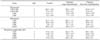
The trials conducted on mice confirmed the modulating effect of chitosan adipate on T and B-cell subpopulations originating from the lymphatic tissue. A relationship was observed between the stimulating effect induced by chitosan adipate and the number of doses applied. The strongest stimulating effect was noted after four injections of chitosan adipate (20 mg/kg) at 24 h intervals. The changes observed in the thymic cell subpopulations highlight the influence of chitosan adipate on thymocyte maturation and differentiation. The administration of chitosan adipate decreased the percentage of immature CD4+CD8+ thymic cells (double-positive cells) with corresponding increases in the percentage of single-positive CD4+ and CD8+ thymocytes. The stimulating effect depends on the number of chitosan adipate doses applied. The strongest effect was noted after four injections of chitosan adipate at a dose of 20 mg/kg (Table 1).
Some changes in the percentage of mesenteric lymph node T cells were also found. The administration of chitosan adipate once at 20 mg/kg led to an increase in the percentage of T CD4+ mesenteric lymph node cells but did not affect the percentage of T CD3+ and CD8+ lymphocytes from the mesenteric lymph nodes. Exposure to four chitosan adipate doses increased the percentage of T CD4+ and CD8+ mesenteric lymph node cells. On the other hand, there were no significant changes in the percentage of T splenocyte subpopulations.
Irrespective of the number of chitosan adipate doses applied, there was an increase in the percentage of B CD19+ mesenteric lymph node cells but there was no effect on the percentage of B CD19+ splenocytes.
Discussion
It is likely that the modulating effect of chitosan adipate on the subpopulations of T lymphocytes in the thymus and peripheral lymphatic organs (lymph nodes) is related to synthesis and release cytokines, particularly interleukin-1 (IL-1) and interleukin-2 (IL-2). Nishimura et al. [11] reported that deacetylated chitin (DAC-70) induces the synthesis and release of cytokines such as IL-1, colony-stimulating factor (CSF) and interferons (IFN) in mice. IL-1 plays a supporting role in the activation of T cells, partly by increasing the expression of the IL-2 receptors on thymocytes [6,19] and enhancing the proliferation of PHA-stimulated thymocytes [3,9]. It is known that the maturation of T lymphocytes not only occurs in the thymus but in other lymphatic organs as well.
The results also show that chitosan adipate administered to mice increases the percentage of B (CD19+) cells in the spleen but does not alter the percentage of B lymphocytes in the lymph nodes. It is likely that the stimulating effect of chitosan adipate on the number of B splenocytes is due to the activation of T lymphocytes and macrophages through the cytokine cascade (IL-1, CSF and IFN-γ) that is enhanced by this agent. These factors activate the functions of polymorphonuclear cells (PMNs) and macrophages [7,10,14,16]. IL-1 can induce the maturation of pre-B-cells [4], This cytokine acts synergistically with various B cell growth factors to augment proliferation, increase the level of immunoglobulin production, and induce or augment T cells for the production of IL-2, IL-4, IL-5 and IL-6, all of which have immunomodulatory effects on B cells [2]. Interferons have a significant effect in B-cell growth and differentiation. IFN-γ acts as a regulatory agent in determining the responses to the immunoglobulin isotype. This agent selectively enhances the production of IgG2a while simultaneously suppressing IgG1, IgG2b and IgG3 synthesis [17]. Iida et al. [5] reported that the intranasal administration of chitin derivatives (DAC-70) enhanced the production of IFN in the lung one day after a Sendai virus infection. In addition, DAC-70 exhibited protective activity against Sendai virus and an E. coli infection in mice. However, they reported that the protective effect of DAC-70 depended on the treatment schedule and the route of drug administration.
In conclusion, chitosan adipate (20 mg/kg) administered either as a single dose or four times induces the maturation and differentiation of thymocytes and regulates the number of specific cluster differentiation antigens on the surface of B splenocytes and lymph node T cells.
 XML Download