

 PDF
PDF ePub
ePub Citation
 Print
Print


Introduction
Anemia and hypoalbuminemia are the most common and consistent accompaniments of chronic fasciolosis. However, the cause of anemia in fasciolosis remains to be determined. It is not known whether the parasite is hematophagus or a tissue dweller or whether the anemia is induced by the metabolites discharged by the parasite in situ or to severe damage to the liver parenchyma and consequent hemorrhage. Levels of the interleukins, IL-6 and IL-8, have been reported to be elevated in the sera of cattle and buffaloes infected with Fasciola gigantica [9], and the roles of the toxic substances emanating from fluke [1,7] have been speculated upon. The infusion of proline, an amino acid released in large quantities by Fasciola hepatica, has been reported to cause a form of anemia resembling that of fasciolosis [5]. However, it has not been determined whether excretory products of F.gigantica have any role in the genesis of anaemia. Therefore, the present investigation was undertaken to study the effect of excretory-secretory antigen on hematological indices in the rat.
Materials and Methods
Experimental animals
Twelve rats of both sexes of body weights 200-300 g, obtained from the Laboratory of Animal Resource Section, Indian Veterinary Research Institute, India, were used in this experiment. Animals were kept in polypropylene cages and acclimatized for a period of 15 days prior to experimentation under standard temperature, humidity and light cycle conditions. Animals were fed on a balanced diet (15 g/head/day), consisting of crushed wheat 62%, maize 30%, wheat bran 7%, and common salt 1%. Fresh potable water was available ad libitum.
Preparation of F. gigantica excretory-secretory antigen
F. gigantica excretory-secretory antigen (Fg-ESA) was prepared from live adult F. gigantica flukes collected in chilled phosphate buffer saline (PBS) from sacrificed buffaloes with fasciolosis. After washing four times with chilled PBS at room temperature, to ensure that flukes were free from host origin material, the washed flukes (40 flukes per 100 ml PBS) were incubated for 2 h at 37℃. Supernatant was centrifuged at 12,000 × g for 30 min at 4℃ to remove particulate material. The supernatant thus collected was designated Fg-ESA. The protein content of Fg-ESA was measured and fixed at 1.8 mg per ml [8].
Animal experimentation
Rats were randomly divided into two groups (A and B) containing 6 rats each. Rats in group-A were injected (i.p. daily for 7 days) with 0.5ml of Fg-ESA (protein concentration 1.8 mg per ml), and rats in group B were injected with 0.5 ml of PBS.
Collection and storage of blood samples
Blood samples (1.0 ml each ) were collected from rats in clean dry vials containing dipotassium EDTA (at 1 mg/ml of blood) from the orbital socket by inserting a heparinized capillary tube into the inner canthus of the eye on days 0, 2, 4, 14 and 21 after the final injection of Fg-ESA.
Hematological procedures
Hemoglobin (Hb) and hematocrit (Ht) levels and total erythrocyte count (TEC) were monitored as previously described [6], and erythrocytic indices, such as, mean corpuscular volume (MCV), mean corpuscular hemoglobin (MCH) and mean corpuscular hemoglobin concentration (MCHC) were calculated using standard formulas [6]. Ht levels were estimated using the microhematocrit method [2].
Results
Effect of Fg-ESA on hemograms
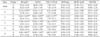
Hematological study results of rats treated with Fg-ESA are presented in Table 1. The Hb values of rats in groups A (14.33 ± 0.14 g/dl) and B (14.25 ± 0.10 g/dl) were initially comparable. However, Hb values started declining significantly (p < 0.01) from day 0 onwards in group A and were lowest on day 14 post-injection (defined as after the completion of treatment), whereas the Hb values of control group animals remained within the normal range with insignificant fluctuations (Table 1). As the experiment progressed mean Ht values of group A animals also started to decline significantly from a mean initial value of 46.48 ± 0.54%), whereas mean Ht values of group B fluctuated insignificantly about it's the mean initial value of 42.00 ± 1.06%. The initial mean total erythrocyte count of rats in group A (7.80 ± 0.11 million per cubic mm) showed a progressive decline from day 0 onward. No significant difference was observed between the mean MCV, MCH and MCHC values of rats in the two groups throughout the experimental period (Table 1).
Discussion
The cause of anemia in fasciolosis is debatable. Various factors, such as, hematophagia [4] and hemorrhages during the migratory phase [3] or metabolites emanating from F. hepatica [5,7] are considered to be of pathogenic significance with respect to the development of anemia in fasciolosis. A rat models was adopted for studying the role of F. hepatica ESA (Fh-ESA) in the genesis of anemia [11]. However, such information is lacking for F. gigantica, which is considerably more pathogenic. The progressive and significant decline in the values of Hb, Ht and TEC without any significant change in MCH, MCHC and MCV in rats injected with Fg-ESA was suggestive of normocytic normochromic anemia, and concurred with earlier observations in fasciolosis due to F. hepatica [11]. In addition, substances such as proline, released by F. hepatica [5] and a substance released by the flukes [11] have also been credited to cause anemia in fasciolosis [5]. Our results suggest Fg-ESA, like Fh-ESA, plays an important role in the genesis of anemia in fasciolosis caused by F. gigantica.
 XML Download