

 PDF
PDF ePub
ePub Citation
 Print
Print


Introduction
Bartonella henselae is a widely distributed, Gram-negative, slow-growing, fastidious, facultative intracellular bacterium causing various diseases including bacillary angiomatosis, bacillary peliosis, and cat scratch disease (CSD) [2]. The transmission of B. henselae to humans is associated with exposure to B. henselae-infected cats and fleas [22]. CSD is usually a self-limiting inflammation of the lymph nodes near the scratch site. In immunocompromized patients, B. henselae causes tumorous proliferation of endothelial cells in internal organs, as well as a recurrent infection that can persist for a prolonged period [22]. Exposure of primary human umbilical vein cells (HUVEC) to B. henselae has been shown to result in bacterial aggregation on the cell surface, and subsequent engulfment and internalization of the bacterial aggregate by the formation of invasomes [13]. One of the most commonly identified pathogenic factors of B. henselae is the Bartonella adhesion A (BadA) protein located in the outer membrane of the bacterium. BadA mediates the mechanism underlying the binding of B. henselae to extracellular matrix proteins and endothelial cells, and it activates hypoxia-inducible factor-1. Moreover, the BadA neck is a major functional domain related to host adhesion, auto-agglutination, and angiogenic reprogramming [20]. On the other hand, B. henselae outer membrane proteins (OMPs), as well as B. henselae itself, can induce adhesion molecule expression in endothelial cells [15]. In the sarcosyl-insoluble fraction of B. henselae lysates, nine proteins were detected, five of which (28, 32, 43, 52, and 58 kDa) were attached to HUVECs [6]. Moreover, Dehio et al. [13] have suggested that the 43-kDa OMP (OMP43) is the major adhesin among B. henselae-derived OMPs that interacts with HUVEC.
The outer membranes of Gram-negative bacteria determine the molecules to be taken in or excreted by the cells. Moreover, in many bacteria, the outer membrane is the predominant layer that interacts with antibodies and other proteins. Porins, which were discovered in 1976, are the major proteins of the outer membrane and are found in every Gram-negative species [26]. Nonspecific diffusion of hydrophilic solutes across the outer membrane usually occurs through porin channels with distinctive diameters. Consequently, porins are the major uptake/excretory route for nutrients, toxins, antibiotics, hydrolytic enzymes, etc. [28]. On the other hand, some researchers were reported that the OMP43 sequence of B. henselae showed 38% identity and 53% similarity to the Omp2b porin of Brucella species [7]. In addition, B. henselae OMP43 showed homology to the proteins of Rhizobium leguminosarum that may possess pore-forming abilities [12].
These data suggest that B. henselae omp43 could be a porin-coding gene and that the OMP43 protein interacts with other cells and molecules. This study was aimed at characterizing the proteome of Δomp43 and comparing it to that of the wild-type (WT) strain by applying proteomic methods, which can help elucidate the pathogenesis of B. henselae. In addition, we performed semi-quantitative reverse transcriptase polymerase chain reaction (RT-PCR) to confirm the proteomic data.
Materials and Methods
Bacterial strains and growth conditions
B. henselae strain Houston-1 (ATCC 49882) was cultured on Columbia blood agar plates containing 5% defibrinated sheep blood (BAP-agar plates) in a humidified atmosphere at 37℃ and 5% CO2. Escherichia coli was grown in Luria-Bertani (LB) broth at 37℃.
Construction of pΔomp43
Primers, plasmids, and bacterial strains used in this study are listed in Table 1 [731]. DNA extraction was performed according to standard protocols. Chromosomal DNA was extracted from B. henselae (Houston-1) by using DNeasy Blood & Tissue Kits (Qiagen, Germany) according to the manufacturer's instructions.
For construction of the omp43 plasmid (pomp43), the omp43 gene was amplified by using BamH-omp43 and omp43-Hind primers. The amplicon was cloned with pGEM-T Easy Vectors (Promega, USA), followed by transformation into E. coli DH5α. Purification of plasmid DNA was performed by using the Wizard Plus SV Minipreps DNA Purification System (Promega) according to the manufacturer's instructions. The pBluescript II KS plasmid and T vector containing omp43 sequences were digested with BamH I and Hind III. The insert (containing the omp43 gene) was ligated into the pBluescript II KS vector and transferred into E. coli DH5α.
A kanamycin resistance gene (Kmr) was amplified from pET-28α by using Sph I-km-F and Sph I-km-R primers, cloned with pGEM-T Easy Vector as previous described. After purifying, the T vector containing Kmr together with pomp43 were digested with Sph I and then the cut Kmr was ligated with pomp43 (middle region of omp43 gene sequences). As a result, the pΔomp43 plasmid was acquired. Additionally, the pΔomp43 sequence was confirmed by dideoxy termination with an automatic sequencer (ABI 3730xl capillary DNA sequencer, Applied Biosystems, USA).
Expression of OMP43
The OMP43 of B. henselae was prepared as described previously [7]. Briefly, omp43 without the signal peptide was amplified from B. henselae (Houston-1) by using Bgl-omp43 and omp43-EcoR primers. The pBAD/His B plasmid and the omp43 PCR product were digested using restriction enzymes (Bgl II and EcoR I). The digested omp43 amplicon was cloned with the digested pBAD/His B vector, followed by transformation into E. coli TOP10. OMP43 was expressed by induction with 0.02% arabinose for 6 h at 37℃ in LB containing 50 mg/mL ampicillin. The denatured samples were separated by 12% sodium dodecyl sulfate-polyacrylamide gel electrophoresis (SDS-PAGE) and transferred to a nitrocellulose membrane. OMP43 protein was detected by western blotting using the anti-Xpress antibody (Invitrogen, USA) and anti-mouse IgG secondary antibody (Cell Signaling, USA) (data not shown).
Purifying and polyclonal antibody production of OMP43
Purified recombinant OMP43 was acquired by Ni2+ affinity chromatography (via request to Bio Basic, USA). Briefly, induced bacteria were harvested by centrifugation and then sonicated on ice in lysis buffer. The OMP43 fusion protein inclusion bodies were diluted into refolding buffer, 1 mM GSSG (oxidized glutathione), and stirred at 4℃ for 24 h. The dissolved OMP43 fusion protein solution underwent dialysis against a buffer solution. The refolded protein was harvested by centrifugation for 20 min at 30,000 × g and then digested with an enterokinase at 4℃ for 24 h. Subsequently, the digested omp43 protein was purified by Ni2+ affinity chromatography.
For production of the polyclonal antibody against OMP43, an OMP43-specific rat antiserum was raised by immunization with purified OMP43 for 9 weeks and purified by affinity chromatography.
Electroporation of B. henselae
Five-day-old B. henselae were harvested from 2 BAP-agar plates with a sterile cotton swab into ice-cold distilled water (DW) containing 10% glycerol. Competent cells were prepared by washing three times with ice-cold DW containing 10% glycerol. The pellet was resuspended in 100 µL of ice cold DW containing 10% glycerol in a cooled electroporation 0.1-cm-gap cuvette (BioRad, USA). Subsequently, 10 µL of the pΔomp43 plasmid solution (2 µg/µL) was added into the cuvette and gently mixed before being allowed to stabilize on ice for 15 min. Electroporation was conducted with a field strength of 1.2 kV/cm and a constant capacitance of 25 µF at 200 Ω. Electroporated cells were immediately transferred into 1 mL super broth (SB) broth at room temperature. Subsequently, cells were incubated for 4 h at 37℃ in 5% CO2 and then seeded on BAP-agar plates containing kanamycin.
Growth curve
For the comparison of growth abilities of WT and Δomp43, counted bacteria (100,000 colony-forming unit [CFU]) were seeded on BAP-agar plates and incubated in a humidified atmosphere at 37℃ and 5% CO2. After harvesting from BAP-agar plates with a sterile cotton swab, the bacteria were resuspended in phosphate buffered saline (PBS). Optical density (OD) was estimated on 600 nm.
Two-dimensional proteomics
Whole cell protein extractions
For whole cell protein extraction, cells were harvested from BAP-agar plates and resuspended in iced PBS. Bacteria were washed three times with iced PBS, centrifuged, and resuspended in lysis buffer. Subsequently, cells were broken by sonication using a Branson sonifier (Thermo Fisher Scientific, USA) until the solution turned an opaque yellow color. By centrifugation at 30,000 × g at 4℃ for 40 min, the debris was pelleted. The supernatant contained solubilized whole cell lysate, which was quantified using the RC/DC Protein Assay kit (BioRad) according to the manufacturer's protocol.
Two-dimensional SDS-PAGE
Whole cell proteins were separated in the first dimension by using immobilized pH 3 to 10 nonlinear gradient strips (Amersham Biosciences, UK). Isoelectric focusing of the protein-containing samples was performed in a protein IEF cell (BioRad). Afterward, seconddimension analysis was performed on 9% to 16% linear gradient polyacrylamide gels, and protein fixation was performed. They were scanned in a Biorad GS710 densitometer (BioRad) and the results converted into electronic files and were analyzed by using the Image Master Platinum 5.0 image analysis program (Amersham Biosciences). Analysis was conducted to identify spots with a minimum 2-fold increased or decreased difference between B. henselae Houston-1 (ATCC49882) and kanamycin-resistant omp43 deficient B. henselae (Δomp43).
Matrix-assisted laser desorption ionization-time of flightmass spectrometry (MALDI-TOF-MS)
Protein processing
Selected spots were excised from stained two-dimensional (2D) gels by using an Ettan spot picker (GE Healthcare, UK) and then transferred into 96-well plates. Tryptic digestion with subsequent spotting on a MALDI-target was carried out automatically with the Ettan Spot Handling Workstation (GE Healthcare). After drying at 37℃ for 15 min, a trypsin solution was added and incubated at 37℃ for 2 h. Subsequently, pieces were covered with 50% CH3CN (0.1% trifluoroacetic acid [TFA]) and incubated for 30 min. The dried samples were dissolved in 50% CH3CN (0.5% TFA) and directly spotted on the MALDI-target. The samples were mixed with saturated a cyano-4-hydroxycinnamic acid solution in 70% CH3CN and allowed to dry on the target before measurement in MALDI-TOF.
Protein measurements
The MALDI-TOF-MS was conducted on 4800 MALDI-TOF/TOF Analyzer (Applied Biosystems) equipped with a 355-nm Nd:YAG laser. The mass spectra were analyzed in the reflectron mode and using the 4700 calibration mixture (Applied Biosystem). The SD of the mass was less than 0.15 Da. After calibration, peak lists were obtained by using the ‘Peak to MASCOT’ script of 4700 Explorer Software (Applied Biosystems). For protein identification, the peptide mass lists were matched against databases proposed by the search engine Mascot (Matrix Science, UK). To select candidate antigens, the highest sequence coverages and Mascot scores were used. According to the Clusters of Orthologus Groups (COGs) classification, the identified proteins were functionally categorized (National Center for Biotechnology Information, USA).
Semi-quantitative RT-PCR
The WT and Δomp43 B. henselae were cultured for 6 days and harvested from BAP. The RNA was extracted from them by using the Ribopure-bacteria kit (Ambion, USA) according to the manufacturer's instructions. The cDNA synthesis was carried out with the Primescript 1st strand cDNA synthesis kit (Takara, Japan) according to the manufacturer's instructions. The primers used for RT-PCR are listed in Table 2. The PCR conditions were 30 sec at 94℃, 30 sec at 60℃, and 30 sec at 72℃ for 20 cycles (16S gene) or 28 cycles (tldD, efp, ntrX, pdhA, and ATPA genes) or 29 cycles (Rho, purB, and yfeA genes). PCR amplicons were analyzed by electrophoresis on 1.5% agarose gels and visualized by ethidium bromide staining. RT-PCR band intensities were measured by performing scanning densitometry with the Kodak 1D image analysis software (Eastman Kodak, USA) and analyzed by normalization to 16S rRNA obtained over the same period. Significance of differences was determined by using a Student's t-test, and p values less than 0.05 were regarded as statistically significant. The data are presented as means ± SDs.
Results
omp43-targeted mutagenesis using homologous recombination in B. henselae
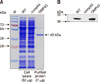
Western blots of OMP43 in E. coli revealed a fusion protein of approximately 45 kDa containing a 3 kDa region resulting from an N-terminal Xpress tag (data not shown). The OMP43 protein was purified using Ni2+ affinity chromatography and the purity was confirmed by using SDS-PAGE (Fig. 1).
Kanamycin-resistant B. henselae grew to form visible single colonies 2 weeks after electroporation. Several PCRs using primers specific for omp43, kanamycin resistance, internal transcribed spacer [34], and 16S rRNA gene sequences were performed to confirm Δomp43 expression (Table 1). Two kanamycin-resistant B. henselae mutants were acquired and the omp43 gene sequences were confirmed by PCR and sequencing (data not shown).
Additionally, to confirm the expression of OMP43, SDS-PAGE gel staining and western blotting were performed. OMP43 expression in the WT cell lysate was detected by using SDS-PAGE gel staining and western blots, but OMP43 expression in the Δomp43 lysate could not be detected (Fig. 1). This result showed that there was a complete loss of OMP43 protein expression in Δomp43.
Δomp43 grows at slower rate compared to the WT bacterium
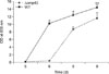
On BAP, Δomp43 grew to form visible single colonies, but at a significantly lower growth rate than that of the WT bacterium. To analyze the difference in the growth rates between Δomp43 and the WT, we cultured the bacteria on BAP and determined the OD600 at 5, 6, 7, 8, and 9 days after seeding. The Δomp43 showed significantly lower OD than the WT at 6 days (Fig. 2). Additionally, over-grown colonies of Δomp43 were slightly smaller than those of the WT. This result indicated that omp43 affected the growth of the B. henselae Houston-1 strain.
Two-dimensional gel electrophoresis profiles of B. henselae
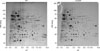
To find new targets associated with omp43, total proteins of Δomp43 and the WT were extracted for 2D gel electrophoresis (2-DE). Representative 2-DE gel images are shown in Fig. 3. In the 2-DE analysis, 422 and 375 protein spots were detected on the 2-DE WT and Δomp43 gels, respectively. Additionally, 282 paired and 233 non-paired protein spots were identified (Fig. 3). Forty-six protein spots showed a 2-fold change in expression levels. Of the 46 protein spots, 26 showed lower protein expression and 20 showed higher protein expression in Δomp43 than in the WT.
Analysis of the expressed proteins in Δomp43
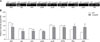
To identify the differentially expressed proteins, 46 of the protein spots selected from the 2-DE gels were excised and subjected to trypsin digestion and subsequent analysis by MALDI-TOF-MS. The 46 protein spots representing 28 different proteins were successfully identified by MALDI-TOF-MS and by MASCOT database searches. Of the identified proteins, 20 showed lower expression and 8 showed higher expression in Δomp43 than in the WT (Tables 3 and 4). Of these proteins, only the tldD-encoded protein displayed both higher and lower expressions, and these two spots were distributed in different parts of immobilized pH gradient (IPG) strips (pH 4.0–5.0 and pH 6.5–7.0, respectively). However, the spot that displayed lower expression and was observed in the part of IPG strip corresponding to pH 6.5–7.0 was more credible than the other spot because of the numerical value of the MASCOT score, the coverage, and the masses matched. This credibility supposition was confirmed by RT-PCR (Fig. 4).
Using categories designated based on the COG database, the differentially expressed proteins could be divided into 4 groups. We found 12 proteins to be associated with metabolism, 7 proteins associated with information storage and processing, 3 proteins associated with cellular processing and signaling, and 6 poorly characterized proteins (Tables 3 and 4). In particular, the number of differentially expressed proteins associated with metabolism indicated that various metabolic processes were affected by the loss of OMP43 expression. Additionally, this phenomenon reflected the changes in the proteins involved in information processing such as replication, translation, and transcription. In addition, the 60 kDa heat-shock protein and protein-L-isoaspartate (D-aspartate) O-methyltransferase (pcm2), which are classified in a cellular processing and signaling group involved in maintaining protein structure and integrity, were also affected. These data indicate global changes in the metabolic pathways in the Δomp43 mutant.
RNA expression analysis of the identified proteins by RT-PCR
To verify the proteomics data, semi-quantitative RT-PCR analysis was performed to correlate gene expression with protein expression. Fourteen of the 27 genes (tldD was duplicated) whose encoded proteins were found in the 2-DE analysis were selected for further investigation (Table 2). Although the mRNA expression of 6 genes was unchanged, that of the other 8 genes showed significant changes (Fig. 4). In RT-PCR analysis, the mRNA expressions of 6 targeted genes (tldD, efp, ntrX, pdhA, purB, and ATPA) were lower, while those of 2 targeted genes (Rho and yfeA) were higher in Δomp43 than in the WT (panel A in Fig. 4). The density of each band was quantified by using scanning densitometry, and the expression was subsequently normalized to 16S mRNA expression (panel B in Fig. 4). The results were consistent with those obtained from the proteins identified in the MALDI-TOF-MS assay.
Discussion
The loss or decrease in OMP expression in several Gram-negative bacteria occasionally results in decreased proliferation and fitness in vitro and in vivo. For example, in Haemophilus species, ompA, which maintains cell structure and functions as a porin regulating the entry of nutrients into the bacterium, has been well established [37]. Additionally, OMP- or porin-deficient mutant strains have shown reduced growth or loss of viability in Mycobacterium species [24], Salmonella enterica [5], Haemophilus ducreyi [10], and E. coli [9].
However, the roles of OMPs in B. henselae have not yet been elucidated. Studies on OMPs in B. henselae have mainly focused on their primary role in host-bacterial interactions, and OMP43 has been suggested as the major adhesion protein in the outer membrane [67]. Burgess et al. [7] produced recombinant E. coli expressing the B. henselae OMP43 as a fusion protein for use in identifying the features of OMP43. Although they successfully determined the amino sequences and characterized the membrane topology of OMP43, as well as the attachment of OMP43 to HUVECs, the function of omp43 has not yet been described [7]. Therefore, in this study, we established a Δomp43 mutant in B. henselae. In order to confirm that proliferation in Δomp43 was significantly lower than that of the WT, protein expression in Δomp43 was investigated by undertaking proteomic analysis and semi-quantitative RT-PCR.
Among the 20 protein spots that showed decreased expression in 2-DE, the mRNA expressions of the ATPA, efp, ntrX, pdhA, purB, and tldD genes decreased in Δomp43. These proteins were mainly categorized based on their role in metabolism, based on their COG assignment (Table 3). This result suggested that the loss of OMP43 expression in Δomp43 disrupted energy metabolism, which might have affected cell growth.
Elongation factor P (efp) is a translation factor that can stimulate ribosomal peptidyl transferase activity and is homologous to the eukaryotic translation factors eIF5A and aIF5A [17]. Although eIF5A may not be absolutely essential for general protein synthesis, several studies have shown that efp is associated with viability, permeability, and fitness of bacteria [11213638]. The results showing downregulation of efp mRNA and protein expressions and the resulting lower growth rate of Δomp43 are consistent with previous data and reports. These results indicate that omp43 might have a role in the integrity of the outer membrane in bacteria through downregulation of efp mRNA levels.
Two-component systems consist of sensor (ntrY) and regulator (ntrX) parts, which are reported to regulate metabolic and respiratory processes [29]. In Azospirillum brasilense, the ntrXY system might be involved in the regulation of nitrate assimilation [19]. The ntrY mutant of Brucella abortus showed lower survival within macrophages, which suggests that the ntrXY system is important for the intracellular viability of bacteria [8]. Atack et al. [3] used phylogenetic analysis to show that the ntrX response regulator is found in five distinct clades of pathogens: Neisseria, Bartonella, Brucella, Ehrlichia, and Anaplasma. It is possible that the ntrXY system is involved in the adaptation and survival of a variety of intracellular pathogens.
The pyruvate dehydrogenase complex (PDHC) converts pyruvate to acetyl-CoA through the serial reactions of three enzymes (E1, E2, and E3). The phdA gene encoding E1 enzymes in various bacteria has been studied and characterized [27]. Moreover, Schreiner et al. [33] showed that inactivation of the aceE (aceE is similar to phdA) gene resulted in an inability to grow in the presence of glucose and the loss of PDHC and E1 activities in Corynebacterium glutamicum. These results, along with the results of our current study, suggest that phdA may influence bacterial growth by altering metabolic pathways.
Adenylosuccinate lyase (purB) catalyzes the conversion of succinylaminoimidazole carboxamide ribotide to aminoimidazole carboxamide ribotide and fumarate or the conversion of adenylosuccinate to adenosine monophosphate and fumarate in the purine-biosynthetic pathway [25]. This indicates that purine nucleotides are essential for cell division. In humans, adenylosuccinate lyase deficiency causes growth retardation and is associated with central nervous system disorders [35]. The structural or chemical features of purB have been studied in bacteria such as Bacillus subtilis, Staphylococcus aureus, and Thermotoga maritime [16]. Although the function of purB in prokaryotes has not been established, our results suggest that purB might be involved in bacterial growth.
The F0F1-ATP synthases, which catalyze the formation of adenosine triphosphate (ATP) from adenosine diphosphate and phosphate in most prokaryotes and eukaryotes, are membrane-bound enzymes that use the energy derived from a transmembrane electrochemical proton gradient. F0F1-ATP synthases consist of two parts, F0 and F1, which contain the subunits α3β3γδε and ab2c10–12 in E. coli. Functionally, the α3β3δab2 subunits act as stators, while the γεc10–12 subunits are rotors [14]. These data suggest that ATPA, which is an F0F1-ATP synthase, might affect ATP synthesis.
The tldD gene is associated with the activity of DNA gyrase in E. coli. Additionally, the protein products of the tldD and tldE genes regulate the stability of the ccdA and ccdA41 antidotes and are essential for MccB17 maturation in E. coli [1]. These results suggest that tldD gene products are involved in protein processing and degradation.
Decreased expression of the above-mentioned genes in Δomp43 might be affected by the loss of OMP43 expression. Therefore, the retarded growth in Δomp43 was possibly induced by metabolic pathways altered as a result of the decrease in the expressions of these genes. In contrast, eight protein spots showed increased expression in the 2-DE analysis, and among those, the mRNA expression levels of Rho and yfeA were increased in Δomp43. Additionally, based on COG assignment, Rho and yfeA are categorized as being important in transcription and metabolism, respectively (Table 4). The increased expression of these genes might contribute to functional compensation for the absence of OMP43 expression in Δomp43.
The colonization and growth of many bacterial pathogens is affected by their ability to invade mammalian hosts to obtain iron. However, the iron in host cells is strongly bound by ferritin, transferrin, and lactoferrin [18]. The ATP-binding cassette (ABC) transport system is a well-characterized uptake system. In Yersinia pestis, the yfe locus is composed of five genes (yfeA–E) and forms a system similar to the ABC transport system associated with the acquisition of inorganic iron and other ions. Moreover, it has been reported that yfe affects the growth of Y. pestis [4]. Although a detailed study on the ABC transport system of B. henselae has not been undertaken, it has been reported that the acquisition of heme compounds such as hemin, erythrocyte membrane fractions, and hemoglobin is essential for the growth and survival of the bacterium [32]. Hence, an upregulated yfeA expression in Δomp43 might be a mechanism to compensate for energy loss due to alterations in the metabolic pathways.
The bacterial transcriptional terminators have two types of pathways: factor-independent (intrinsic) and factor-dependent (Rho-dependent) [30]. The Rho-dependent pathway serves to terminate the synthesis of transcripts. Recently, Leela et al. [23] reported that Rho-dependent transcription termination is necessary to avoid the excessive genome-wide R-loops in E. coli. The observation that Rho expression was increased in Δomp43 suggests that Rho-dependent transcription termination is associated with repair of disrupted transcription occurring because of gene manipulation or other changes. However, further studies are needed to confirm this hypothesis.
In conclusion, the Δomp43 strain, which is the first omp43-deficient bacterial strain generated by using electrophoresis, showed significantly decreased proliferation. The changes in the mRNA expressions of genes in Δomp43 were mainly associated with metabolic processes. Although the possibility that other undetected proteins or genes may display altered expression or function to compensate for the loss of OMP43 cannot be ruled out, our data suggest that the growth retardation in Δomp43 is induced by altered metabolic pathways and will provide useful information for further investigation of the mechanisms underlying this effect.

 XML Download