Abstract
Bovine tuberculosis is a chronic contagious disease responsible for major agricultural economic losses. Abattoir monitoring and trace-back systems are an appropriate method to control bovine tuberculosis, particularly in beef cattle. In the present study, a trace-back system was applied to bovine tuberculosis cases in Korean native Hanwoo beef cattle. Bovine tuberculosis was detected in three index beef cattle during abattoir monitoring in Jeonbuk Province, Korea, and the original herds were traced back from each index cow. All cattle in each original herd were subjected to tuberculin skin test. The positive rates in the tuberculin skin test were 64.6% (62 of 96), 4.8% (2 of 42), and 8.1% (3 of 37) at farms A, B, and C, respectively. On post-mortem examination of 56 tuberculin-positive cattle, 62% had granulomatous lesions, and Mycobacterium bovis was cultured from 40 (71.4%) of the cattle. Molecular typing by spoligotyping and the mycobacterial interspersed repetitive unit-variable-number tandem repeat assay revealed the genotype of the M. bovis strains from the index cattle were same as the M. bovis genotype in each original herd. The results suggest that tracing back from index cattle to the original herd is an effective method to control bovine tuberculosis in beef cattle.
Bovine tuberculosis is a chronic contagious disease caused by Mycobacterium bovis that has been detected in a wide variety of animals [16]. Bovine tuberculosis is responsible for economic losses in agricultural herds. Many countries have adopted bovine tuberculosis control programs and testing, and culling has been used to control and eradicate bovine tuberculosis [15].
Bovine tuberculosis was first reported in 1913 in Korea, and the estimated incidence of bovine tuberculosis in the 1940s was 15% [14]. The National Bovine Tuberculosis Control Program was implemented in 1964 in Korea, and it relies on testing and culling [5]. Bovine tuberculosis is detected by using the intradermal tuberculin skin test with a purified protein derivative (PPD). Through implementation of the National Bovine Tuberculosis Control Program, the incidence of bovine tuberculosis in Korea was only 0.15% in 2005 [1217].
However, the National Bovine Tuberculosis Control Program has been instituted mainly in dairy cattle, whereas bovine tuberculosis control remains unresolved in beef cattle. About three million head of Korean native Hanwoo beef cattle are present in Korea [13]. Nationwide annual bovine tuberculosis testing with the intradermal tuberculin skin test has been performed on all dairy cattle, but only a limited number of beef cattle have been tested because, compared to the number of cattle that need to be tested, there are few authorized and trained personnel available to perform the testing.
A trace-back system could be an alternative to the tuberculin skin test. In such a system, if lesions suggesting bovine tuberculosis are detected on gross inspection at slaughter, all animals in the trace-back herd would be subjected to the tuberculin skin test [78]. A few reports have documented outbreaks of bovine tuberculosis among Korean beef cattle [29]; however, post-mortem examinations and mycobacterial cultures are rarely performed to confirm an M. bovis infection in the original herd traced back from the index cattle.
In the present study, suspected original herds were traced back from the index beef cattle, and the tuberculin skin test was performed on all cattle in that herd. Tuberculin-positive beef cattle were subjected to post-mortem and mycobacterial culture examination to confirm the presence of an M. bovis infection. Spoligotyping and a mycobacterial interspersed repetitive unit-variable-number tandem repeats (MIRU-VNTR) assay were conducted to identify the M. bovis transmission source.
Three index beef cattle were identified via abattoir monitoring in Jeonbuk Province, Korea from July 2011 to October 2012. The index beef cattle had granulomatous lesions in the laryngopharyngeal lymph nodes, and acid-fast bacilli were identified via Ziehl-Neelsen staining. The original herds were traced back by applying the Animal Products Traceability System of the Korean Ministry of Agriculture, Food and Rural Affairs, and a tuberculin skin test was performed on all beef cattle in the original herds. The tuberculin skin test was performed by using the caudal fold tuberculin test according to official Korean national guidelines. In brief, 0.1 mL of bovine PPD was injected intradermally in the skin of the caudal tail fold, and skin thickness of the area where the PPD was injected was measured approximately 72 h later. The reaction was interpreted to be tuberculin-positive if skin thickness increased by > 5 mm, inconclusive if the increase in skin thickness was 1 to 4 mm, and negative if the increase in skin thickness was < 1 mm.
To determine whether the tuberculin-positive beef cattle were infected with M. bovis, they were subjected to post-mortem examination, which was performed by authorized personnel according to the guidelines of the Korean National Bovine Tuberculosis Eradication Program. The laryngopharyngeal lymph node, septal lymph node, diaphragmatic lymph node, lungs, and other tissues were collected for gross observation, histopathological examination, and mycobacterial culture at necropsy. Tissue samples for histopathology were fixed in 10% neutral buffered formalin, and sections were stained with hematoxylin and eosin and Ziehl-Neelsen stain. The tissues were subjected to mycobacterial culture after homogenization, and the mycobacterial cultures were prepared by using the BD BACTEC Mycobacteria Growth Indicator Tube (MGIT) 960 System (Becton-Dickinson, USA). The cultured M. bovis isolates were confirmed by using Ziehl-Neelsen stain and SD Bioline TB AgMPT64 Ag Rapid test (Standard Diagnostics, Korea), as described previously [1]. The M. bovis isolates were subjected to molecular typing.
The index beef cattle were identified at a slaughterhouse in Jeonbuk Province in 2011 to 2012, and a trace-back for bovine tuberculosis was conducted to identify the original herds. All three index beef cattle had granulomatous lesions in laryngopharyngeal lymph nodes (Fig. 1), in which acid-fast bacilli were detected, and M. bovis was isolated from and identified from all three index cattle. Tuberculin skin testing was performed on all cattle of the remaining original herds at the three identified farms. The tuberculin skin test-positive rates were 64.6% (62 of 96), 4.8% (2 of 42), and 8.1% (3 of 37) at farms A, B, and C, respectively. Interestingly, the tuberculin skin test-positive rate was notably high at farm A.
At each farm, all tuberculin-positive cattle were subject to a post-mortem examination, but that examination was only performed on 51 randomly selected cattle from farm A due to a limitation on the number of examinations that could be completed in a day. Of the 56 cattle examined, 35 (62.5%) had granulomatous lesions in various organs (Tables 1 and 2). The major organ affected was the laryngopharyngeal lymph node (45.7%, 16 of 35), followed by the lung (34.3%, 12 of 35). Other affected organs were the septal lymph node (11.6%, 5 of 35), the diaphragmatic wall (11.4%, 4 of 35), the thoracic wall (8.6%, 3 of 35), and the diaphragmatic lymph node (5.7%, 2 of 35).
Mycobacterial cultures were attempted for the affected organs as well as for corresponding lymph nodes with no visible gross lesions. Among the 56 post-mortem-examined tuberculin-positive cattle, 40 M. bovis strains (71.4%) were isolated. Most of the M. bovis strains were isolated from granulomatous lesions (85.0%, 34 of 40); the remaining six strains were isolated from laryngopharyngeal lymph nodes without visible lesions.
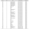
Spoligotyping and MIRU-VNTR assay were performed to investigate the relationships among the isolated M. bovis strains. All M. bovis strains were designated SB0140 for spoligotyping and were divided into two subgroups (Fig. 2). The major spoligotype subgroup was marked by a lack of spacer types 3, 6, 8–12, 16, and 39–43, which comprised 90.7% (39/43) of the isolated M. bovis strains. The other subgroup was marked by the absence of spacers 3, 5–14, 16, 19, and 39–43, to which four M. bovis strains (A36, A37, A38, and A39 strains from farm A) belonged.
Each VNTR allele of the M. bovis strains from the index cattle was detected in the original herd after undertaking MIRU-VNTR analysis. Interestingly, three additional M. bovis genotypes were observed on farm A. The VNTR allele 32531751143411343 and the spoligotype lacking spacers at 3, 6, 8–12, 16, and 39–43 was the major genotype, comprising 87.2% (34/39) of the M. bovis strains. The other genotype, accounting for 10.3% (4/39) of the M. bovis strains, had the same VNTR allele as the major genotype but a different spoligotype subgroup lacking spacers at 3, 5–14, 16, 19, and 39–43. Interestingly, only one M. bovis strain had a VNTR allele different from the major VNTR genotype at farm A.
The M. bovis strains from farms B and C had the same VNTR alleles and spoligotypes as the genotypes of each of the index cows.
The Korean National Bovine Tuberculosis Control Program was implemented in 1964 and has contributed to the reduced incidence of bovine tuberculosis in Korean dairy cattle [14]. However, reducing bovine tuberculosis in Korean beef cattle and other animals remains unresolved. Abattoir monitoring has been adopted in many countries to control bovine tuberculosis in beef cattle [15]. In such programs, cattle are inspected for bovine tuberculosis at the slaughterhouse and those with signs of tuberculosis are traced back to the original herd. There are two reports on bovine tuberculosis at slaughterhouses [911], but a thorough investigation based on the trace-back system, tuberculin skin test, post-mortem examination, and molecular typing has not been reported in Korea.
In the present study, three index beef cattle were found at a slaughterhouse; subsequent to tracing them back to the original herd, tuberculin skin testing was performed on all cattle in the original herds. Tuberculin-positive beef cattle were detected by skin testing at all three farms. The tuberculin-positive rates at farms A, B, and C were 64.6% (62 of 96), 4.8% (2 of 42) and 8.1% (3 of 37), respectively. The farm B and C results were similar to that presented in a previous report [4]. Interestingly, the tuberculin-positive rate at farm A was notably high. Most of the beef cattle at farm A were self-bred, and the cattle had been exposed to self-breeding for a relatively long period. At farm A, calving cows were infected with bovine tuberculosis, which may have been the tuberculosis transmission route for the entire herd.
Of the 56 tuberculin-positive beef cattle that underwent post-mortem examination, 35 (62.5%) had granulomatous lesions, and the most frequently affected organ was the laryngopharyngeal lymph node. At the three farms, the proportions of examined cattle with lesions were higher than those in a previous study, in which 20% to 50% of cattle with tuberculin-positive reactions had pathological lesions [6]. These contrasting results may have been caused by differences in bovine tuberculosis policy implementation. In the report by de la Rua-Domenech et al. [6], post-mortem examination was performed on beef cattle that underwent routine tuberculin testing. In contrast, in this study, the tuberculin study, the tuberculin testing was performed after detecting tuberculosis in index cattle. Although the tuberculin skin testing is routinely performed on dairy cattle in Korea, routine tuberculin testing is rarely conducted on Korean Hanwoo beef cattle, which may contribute to the accumulation or aggravation of bovine tuberculosis incidence in beef cattle.
In this study, 40 M. bovis strains (71.4%) were isolated from among the 56 cattle examined post-mortem. Most were isolated from granulomatous lesions, but eight M. bovis strains (20.0%) were isolated from cattle with no visible lesions, suggesting that M. bovis can be cultured from cattle with or without lesions. Spoligotyping and the MIRU-VNTR assay on the M. bovis strains showed that the genotypes from the index cattle were also detected in the original herds. The M. bovis genotype with the VNTR allele 3253175114341134332 and spoligotype lacking spacers at 3, 6, 8–12, and 39–43 comprised 87.2% (34/39) of the M. bovis strains at farm A, suggesting that this was the major herd-transmitted genotype. On the other hand, the minor M. bovis genotype at farm A had the same VNTR allele but the spoligotype lacked spacers at 3, 5–14, 16, 19, and 39–43, indicating it may have been transmitted from another farm via imported infected beef cattle. Interestingly, there was one M. bovis strain with a unique VNTR allele at farm A, implying the strain might have been imported from another farm.
Nationwide bovine tuberculosis control in beef cattle has not been implemented in Korea because bovine tuberculosis in dairy cattle has taken priority; in addition, tuberculin testing is not mandatory for beef cattle in Korea. However, our results suggest that bovine tuberculosis testing in beef cattle is more urgent than anticipated, particularly at breeding farms where a control policy against bovine tuberculosis in beef cattle should be promoted.
In conclusion, tuberculin-positive beef cattle were detected in the original herds after the herd identity was traced back from the index beef cattle. Bovine tuberculosis in the beef cattle was confirmed by post-mortem and mycobacterial culture examination. Spoligotyping and MIRU-VNTR assay revealed that the genotype of the M. bovis strain from the index cattle were, for the most part, the same as that in the original herd.
Figures and Tables
Fig. 1
Visible lesions in and Ziehl-Neelsen staining of the retropharyngeal lymph node of a beef cow. (A) Visible lesion showing caseous granuloma in a lymph node from a beef cow. (B) Ziehl-Neelsen stained portion of a lymph node showing acid-fast bacilli in a granuloma. 400× (B).

Fig. 2
Genetic relationships among the Mycobacterium bovis strains from beef cattle. The dendrogram is based on spoligotyping and MIRU-VNTR genotypes and created using the unweighted pair group method with arithmetic averages algorithm. MIRU-VNTR, mycobacterial interspersed repetitive unit-variable-number tandem repeat. *M. bovis strains from the index beef cattle.

Acknowledgments
We thank Prof. Sang-Nae Cho for the professional advice and kind comments regarding this study. This work was supported by a grant from the Animal and Plant Quarantine Agency, in part by grants from the Bio-industry Technology Development Program (grant No. 314025-03), the Ministry of Agriculture, Food and Rural Affairs, Korea, and the Basic Science Research Program of the National Research Foundation of Korea (NRF) funded by the Ministry of Education, Science and Technology (No. NRF-2014R1A1A2055172).
References
1. Byeon HS, Ji MJ, Kang SS, Kim SW, Kim SC, Park SY, Kim G, Kim J, Cho JE, Ku BK, Kim JM, Jeon BY. Performance of the SD Bioline TB Ag MPT64 Rapid test for quick confirmation of Mycobacterium bovis isolates from animals. J Vet Sci. 2015; 16:31–35.

2. Byun HS, Lee HJ, Lee SM, Han ST, Quak HK, Choi HY, Cho YS, Ahn BW. Bovine tuberculosis found at slaughtered Korean indigenous cattles. Korean J Vet Serv. 2007; 30:407–414.
3. Cadmus S, Palmer S, Okker M, Dale J, Gover K, Smith N, Jahans K, Hewinson RG, Gordon SV. Molecular analysis of human and bovine tubercle bacilli from a local setting in Nigeria. J Clin Microbiol. 2006; 44:29–34.

4. Cho BJ, Chu KS, Cho YS, Kang MS, Lee JW. Epidemiological studies on bovine tuberculosis in mass outbreak region. Korean J Vet Serv. 2009; 32:119–124.
5. Cho YS, Jung SC, Kim JM, Yoo HS. Enzyme-linked immunosorbent assay of bovine tuberculosis by crude mycobacterial protein 70. J Immunoassay Immunochem. 2007; 28:409–418.

6. de la Rua-Domenech R, Goodchild AT, Vordermeier HM, Hewinson RG, Christiansen KH, Clifton-Hadley RS. Ante mortem diagnosis of tuberculosis in cattle: a review of the tuberculin tests, gamma-interferon assay and other ancillary diagnostic techniques. Res Vet Sci. 2006; 81:190–210.

7. Evans FW. Progress in eradication of bovine tuberculosis and brucellosis in New South Wales and the efficacy of a trace back system. Aust Vet J. 1972; 48:156–161.

8. Hagerman AD, Ward MP, Anderson DP, Looney JC, McCarl BA. Rapid effective trace-back capability value: a case study of foot-and-mouth in the Texas High Plains. Prev Vet Med. 2013; 110:323–328.

9. Jang SJ, Do SH, Ki MR, Hong IH, Park JK, Ji AR, Jeong KS. Bovine tuberculosis of Korean native cattle in an abattoir. J Life Sci. 2009; 19:1847–1850.

10. Je S, Ku BK, Jeon BY, Kim JM, Jung SC, Cho SN. Extent of Mycobacterium bovis transmission among animals of dairy and beef cattle and deer farms in South Korea determined by variable-number tandem repeats typing. Vet Microbiol. 2015; 176:274–281.

11. Kim YH, Ko BRD, Kim HJ, Ji TK, Rho MH, Park SD, Moon YW. Effect of trace back system on the bovine tuberculosis at abattoir. Rep Res Inst Pub Health Environ. 2012; 14:85–86.
12. Ministry for Food, Agriculture, Forestry and Fisheries (MIFAFF). Monthly Reports of Animal Disease. Seoul: MIFAFF;2008.
13. Ministry of Agriculture, Food and Rural Affairs (MAFRA). The Statistical Yearbook of Agriculture, Forestry and Livestock Products. Sejong: MAFRA;2017. p. 376.
14. Moon JB. [Bovine Tuberculosis, in History of Prevent Medicine of Domestic Animals]. Seoul: Korean Veterinary Medical Association;1996. p. 70–74. Korean.
15. Morris RS, Pfeiffer DU. Directions and issues in bovine tuberculosis epidemiology and control in New Zealand. N Z Vet J. 1995; 43:256–265.

16. Thoen CO, LoBue PA. Mycobacterium bovis tuberculosis: forgotten, but not gone. Lancet. 2007; 369:1236–1238.
17. Wee SH, Kim CH, More SJ, Nam HM. Mycobacterium bovis in Korea: an update. Vet J. 2010; 185:347–350.


 PDF
PDF ePub
ePub Citation
 Print
Print


 XML Download