

 PDF
PDF ePub
ePub Citation
 Print
Print


Introduction
Vertebral structures are difficult to evaluate radiographically because of superimposition of complex structures [14]. On the other hand, computed tomography (CT) is an accurate method of measuring vertebral structures as it allows reformatting in a variety of planes [3]; whereas, magnetic resonance imaging (MRI) allows better evaluation of extradural, intradural extramedullary, and intradural intramedullary lesions [1011].
Some investigations have established quantitative and objective anatomic indices of spine by applying these modalities. In dogs with intervertebral disc disease (IVDD), CT-myelography has been performed before and after surgery to allow quantitative analysis of the ratio of the vertebral canal to the spinal cord to demonstrate the effect of decompression [8]. In human medicine, normal cervical spine measurements have been used in the evaluation of patients with cervical spinal disorders [18]. Those studies have provided some important clinical information.
CT allows fast and accurate measurement of vertebral structures. Moreover, it needs a short anesthetic time (even light sedation can be suitable) for image acquisition. Based on these characteristics, CT could be preferred over MRI or examination of sedated patients. Therefore, clinicians should consider using CT for screening tests and for acquiring clinical indices of spinal disorders.
This study was carried out to derive reference MRI and CT values for normal spine indices, and to evaluate the feasibility of using such indices in a CT screening test to discriminate spinal disorder and normal dogs.
Materials and Methods
A 0.4 Tesla MRI scanner (APERTO 0.4; Hitachi, Japan) was used to image all dogs. MRI was undertaken to acquire T1-weighted (T1W) and T2-weighted (T2W) image spin echo sequences. The MRI repetition time (TR) and time to echo (TE) were adjusted for each particular dog. T1W (TR = 680, TE = 20) and T2W (TR = 2,500, TE = 100) transverse plane images with 2 to 3 mm thicknesses were obtained. CT images were acquired by using a dual-channel CT scanner (Somatom Emotion; Siemens, Germany). The scanning parameters were 110 kVp, 70 mAs, 2.0 mm slice thickness, and 0.95 pitch. The reconstruction parameters were 2.0 mm slice thickness, 2.0 mm interval, and H70s kernel. The window was established with 1,500 Hounsfield units (HU) of width and level centered on 450 HU. All indices were measured at an offline workstation (OsiriX 3.8; Pixmeo, Switzerland).
Scanning regions were: cervical region (C2–T1 at the vertebral body [VB] and intervertebral disc space [IVDS] level), thoracic region (T1–T13 at the VB, and T1–T2 and T13 to L1 IVDS levels), and lumbar region (L1–L7 at the VB and IVDS levels). The VB level measurements were obtained from the middle of VB, while the IVDS level measurements were obtained from the middle of IVDS.
Twelve healthy Beagle dogs were recruited to the normal group; eight males and four females with a mean age of 5 years (range, 3–8 years) and a mean weight of 12.6 ± 3.5 kg (SD). All procedures were approved by the Institutional Animal Care and Use Committee of Gyeongsang National University, and the dogs were taken care of according to the Guidelines for Animal Experiments (GNU-150804-D0035) of Gyeongsang National University. All dogs were judged to be normal based on physical examination, neurologic examination, and laboratory test results (complete blood cell count and serum chemistry). All dogs were preanesthetized with glycopyrrolate 0.01 mg/kg (0.2 mg/mL; Myungmoon, Korea) given subcutaneously general anesthesia (Isoflurane 100% liquid; Kyongbo, Korea) was induced by administering propofol 6 mg/kg (10 mg/mL; Myungmoon). All dogs were placed in a dorsal recumbent position. After positioning, curvature of the vertebral column was minimized by using sponge pads. The number of measurements at the cervical, thoracic, and lumbar region were 84, 48, and 72 at the VB level and 72, 48, and 60 at the IVDS level, respectively.
The medical record database at the Veterinary Medical Teaching Hospital of the Gyeongsang National University was searched for dogs with spinal disease that were examined between 2013 and 2015. Patients with both MRI and CT of the same spinal lesions were included. Based on the inclusion criteria, 50 dogs (28 males and 22 females) with 56 spinal diseases were included in the study. Breeds included Maltese (n = 10), mixed breed (n = 6), Shih-tzu (n = 8), Pekingese (n = 6), Poodle (n = 4), Cocker Spaniel (n = 4), Beagle (n = 3), Dachshund (n = 2), Spitz (n = 2), Miniature Pinscher (n = 1), French Bulldog (n = 1), Greyhound (n = 1), Schnauzer (n = 1), and Yorkshire Terrier (n = 1). The mean age of dogs was 7.6 ± 3.4 years (range, 0.5–13 years). Final diagnoses based on MRI, CT, surgical procedure and histopathologic examination were IVDD (n = 34), vertebral fracture (n = 5), vertebral tumor (n = 5), hematoma (n = 3), myelomalacia (n = 3), syringomyelia (n = 3), spinal cord contusion (n = 2) and arachnoid cyst (n = 1). The numbers of cervical, thoracic, and lumbar lesions were 15, 9, and 10 at the VB level, and 50, 14, and 25 at the IVDS level, respectively.
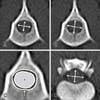
In this study, the obtained indices were: the ratios of width to height of the spinal cord (CR) and of the vertebral foramen (FR), and the ratio of cross-sectional area of the spinal cord to that of the vertebral foramen (CFAR) (Fig. 1). In the case of CFAR, the spinal cord cross-sectional area on MRI was measured on T2W images and the vertebral foramen cross-sectional area was measured on T1W images; this difference is because we considered T2W to be better than T1W for depiction of soft tissue, and T1W was better than T2W for depiction of bone.
Statistical analysis was performed by using the IBM SPSS statistical program (ver. 19.0; IBM, USA). Reliability analysis was performed to determine the intermodality agreement between CT and MRI. Receiver operating characteristic curve analysis was used to compare the sensitivity and specificity of indices for discriminating normal dogs from patients with spinal disorders on CT. An area under curve (AUC) of more than 0.5 and a p value of less than 0.05 were considered to indicate statistical significance.
Results
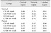
All intermodality agreements between the normal and patient groups were acceptable to excellent (Cronbach's α 0.74–0.96 for the normal group and 0.70–0.99 for the patient group) (Table 1).
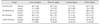
The mean CR of the patient group was higher than that of the normal group (Table 2). In addition, there were significant differences between normal and patient group in CR at both the VB and IVDS levels.
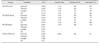
The AUC values for the CR were fair to good at the VB level (0.73–0.82) and excellent at the IVDS level (0.93–0.95) (Table 3). The AUCs for the FR and CFAR indices were good only in the cervical region (0.81 and 0.82, respectively).
Discussion
A main use of MRI and CT is in the diagnosis of spinal diseases. Although MRI has a great advantage in distinguishing anatomic structure, it requires a long anesthetic period and has expensive setup costs. In contrast, CT can acquire a diagnostic index without an invasive procedure, and it requires a relatively short imaging time. Although MRI and CT have long been used to provide clinical information, few investigations into developing quantitative and objective anatomic indices of vertebrae have been undertaken in veterinary medicine [578141718]. Diagnosis of spinal disease is based on the normal spinal anatomy. If normal reference values for spinal morphometric indices are established, they can be useful during screening and/or for diagnosis. On that basis, this study was undertaken to derive indices for discriminating normal dogs from patients with spinal disorder.
Many comparisons of T1W and T2W images have been reported in human and small animal medicine [1246912131516]. Both T1W and T2W images can provide a high degree of structure depiction for use in morphometric analyses [413]. In addition, some reports have indicated that T2W scanning produces better depiction of soft tissue than that from T1W scanning [29]. Therefore, we measured spinal cord area in T2W images and vertebral foramen area in T1W images when determining CFAR.
We searched the records of patients who were diagnosed based on MRI in order to overcome the disadvantage that CT is not completely reliable in diagnosing some spinal disorders. This study evaluated patients who had undergone both MRI and CT in order to obtain index measures of the same lesion on both MRI and CT. Reliability analysis showed acceptable to excellent intermodality agreement in the normal and patient groups, indicating that differences between modalities may not significantly affect the index result.
The mean CR value of patients with spinal disorder was higher than that of normal dogs at both the VB and IVDS levels. In addition, CR had sufficient sensitivity and specificity to indicate a reference cut-off value. The possible cause of the significant difference between the normal dog and patients with spinal disorder groups may be a swelling or mass effect of intradural extramedullary, intramedullary, or extradural lesions, which can lead to compression of the spinal cord. However, the indices of this study were not evaluated under the separate classifications of intramedullary, intradural extramedullary, and extradural lesions. Based on the results, the CR index could be applied in a screening test for patients with a possible spinal disorder. In addition, CR at the IVDS level had a higher AUC than that at the VB level, which suggest that CR is a better method to identify a lesion at the IVDS level than at the VB level. The FR and CFAR indices of the patient group were higher than those of the normal dogs at the cervical region and had sufficient AUC results, indicating that FR and CFAR indices can be useful in the cervical region.
We are aware of the limitations imposed on this study by the relatively low number of cases and the use of normal dogs of a single breed; thus, the results may not be representative of all dogs. In addition, increasing the number of measurements and ratios may be needed to increase objectivity. Among the disorders in this study, IVDD comprised a large portion (34 IVDD/56 spinal disorders), and the CR index could be affected by lesions inducing extradural compression, such as intervertebral disc material. Thus, the higher CR of the patient group than that of the normal dogs could have been influenced by the high IVDD proportion. Further study may be necessary to develop the indices for use in a screening test.
Among the patients in this study, lesions in the cervical region contained many diseases that caused vertebral foramen remodeling, whereas the thoracolumbar region lesions did not. To validate the feasibility of using FR and CFAR indices, studies including additional diseases that may cause remodeling of the vertebral foramen of the thoracolumbar region will be necessary. Moreover, our results may not be completely representative because small to middle-sized breed made up the majority of the spinal disorder patient group.
This study obtained objective and quantitative information on spinal morphometry, and the results were used to suggest index reference ranges for normal dogs. Patients with spinal disorders had a higher CR index than that of normal dogs. The highest discriminating levels of CR at the VB and IVDS levels were 1.25 and 1.44, respectively. In addition, the FR and CFAR indices could be applied to the cervical region with discrimination levels of 1.24 and 0.86, respectively. These indices may be useful during CT screening of canine patients with possible spinal disorders.
 XML Download