

 PDF
PDF ePub
ePub Citation
 Print
Print


Introduction
Brucella abortus is a bacterium that cause brucellosis and is the causative agent of a worldwide zoonotic disease that infects both humans and animals. Brucellosis generally causes persistent abortion and infertility in infected animals and can cause serious economic damage. In the case of human infection, symptoms include undulant fever and arthritis [311]. B. abortus has a small, non-motile, non-spore forming bacterium with a rod shape. Due to the survival characteristics of bacteria at the intracellular level, they are very difficult to isolate. Unlike other pathogenic bacteria, B. abortus does not produce exotoxins, toxic lipopolysaccharide, fimbriae, or plasmids [10]. B. abortus is a Gram-negative facultative intracellular pathogen that can enter to the host macrophage and survive. Due to its intracellular survival characteristics, its isolation is complicated. B. abortus does not utilize the classical pathogenicity factors mentioned above; rather, it invades and proliferates in host phagocytic cells, thereby avoiding the host's cell-mediated immune reaction. This infection mechanism allows it to survive for a long time in the host and can result in chronic infection [21].
Intracellular pathogens are commonly exposed to a variety of environmental factors, including stresses such as pH, oxidation, and nutrition. The survival of B. abortus in macrophages is closely related to the production of various proteins. For example, in the case of heat-shock protein, produced when cells are exposed to high temperatures or other stresses, intracellular bacteria can adapt to a stressful environment [30]. In addition, Hfq, an RNA-binding protein, also has a role in adapting to various stressful environments. It has been reported that, when hfq mutation occurs, several genes associated with the cellular processes are not properly regulated [7]. Several attempts have been made to solve the mechanism of Brucella infection [821]. Although several proteins have been proposed as potentially pathogenic, the basic mechanism of B. abortus infection still needs to be described [21].
Post-genomic technology provides a new and exciting opportunity for further research into and understanding of B. abortus [13]. Various studies on the immunogenic components of B. abortus can be conducted based on proteomic studies for the detection of various antigens [14]. The post-genome approach of proteomics using two-dimensional gel electrophoresis (2-DE) can be used to discover, isolate, and identify new antigens that are different from previously reported ones [142229].
Five mutants were selected for study after analyzing the molecular characteristics of B. abortus wild-type and several mutants [20]. The purpose of this study was to investigate protein expression levels of the selected B. abortus mutants. In addition, we examined the relationships between protein expression level changes and the growth features of B. abortus mutants.
Materials and Methods
Sample selection and preparation for 2-DE
B. abortus mutants were prepared by transposon mutagenesis using wild-type and kanamycin resistance (rKan) genes and the EZ-Tn5 transposon system (Epicentre Biotechnologies, USA). The mutants were used in experiments to assess changes in characteristics such as growth feature and pathogenic factors following rKan insertion. Biochemical tests were performed on B. abortus wild-type and 24 B. abortus mutants. Five mutants were selected based on growth features [20]. The selected B. abortus mutants C3, C8, and C13 had a slower growth pattern than B. abortus wild-type, whereas selected B. abortus mutants C24 and C30 had a faster growth pattern than B. abortus wild-type.
B. abortus wild-type cultured in 10 mL Brucella broth (BD, USA) for 24 h was used as a seed in 250 mL Brucella broth (BD) for 24 h. B. abortus mutants C3, C8, C13, C24, and C30 from 10 mL cultures grown in Brucella broth (BD) with kanamycin (100 µL/500 mL; Sigma, USA) for 24 h were used as seeds in 250 mL Brucella broth (BD) with kanamycin (100 µL/500 mL; Sigma) for 24 h. These were harvested via centrifugation at 8,000 × g for 30 min at 4℃. The pellets were washed twice with PBS. All procedures were approved by the Seoul National University Institutional Biosafety Committee (SNUIBC-R160314-1).
2-DE analysis
The 2-DE was carried out essentially as described. B. abortus wild-type and 5 mutants in sample buffer (7 M urea, 2 M thiourea, 4.5% CHAPS, 100 mM DTE, 40 mM Tris, pH 8.8) were applied to immobilized pH 3 to 10 nonlinear gradient strips (Amersham Biosciences, Sweden) for isoelectric focus (IEF). Similar amounts of each protein from B. abortus wild-type and mutants underwent 2-DE analysis after quantification. IEF was performed at 80,000 Vh. The second dimension was analyzed on 9% to 16% linear gradient polyacrylamide gels (18 cm × 20 cm × 1.5 cm) at constant 40 mA per gel for approximately 5 h. After protein fixation in 40% methanol and 5% phosphoric acid for 1 h, the gels were stained with Coomassie brilliant blue G-250 for 12 h. The gels were then destained with distilled water, scanned in a Bio-Rad GS710 densitometer (Bio-Rad, USA) and converted to electronic files, which were then analyzed by using the Image Master Platinum 5.0 image analysis program (Amersham Biosciences) [1617].
Liquid chromatography tandem mass spectrometry (LC-MS/MS) for peptide analysis
According to the results of 2-DE analysis, we selected 30 spots from each image of the B. abortus mutants that had higher than 2-fold changes (increase or decrease) from those of B. abortus wild-type. LC-MS/MS for peptide analysis was performed on the selected spots.
Nano LC-MS/MS analysis was performed with an Easy n-LC chromatograph (Thermo Fisher, USA) and a LTQ Orbitrap XL mass spectrometer (Thermo Fisher) equipped with a nanoelectrospray source. Protein samples were separated on a C18 nano bore column (150 mm × 0.1 mm, 3 µm pore size; Agilent, USA). Mobile phase A for LC separation was 0.1% formic acid, 3% acetonitrile in deionized water, while mobile phase B was 0.1% formic acid in acetonitrile. The chromatography gradient was designed for a linear increase from 5% B to 30% B in 23 min, 30% B to 60% B in 3 min, 95% B in 3 min, and 3% B in 6 min. The flow rate was maintained at 1,500 nL/min. Mass spectra were acquired by using data-dependent acquisition with a full mass scan (350–1,200 m/z) followed by 10 MS/MS scans. For MS 1 full scans, the orbitrap resolution was 15,000 and the AGC was 2 × 10⁵. For MS/MS in the LTQ, the AGC was 1 × 10⁴ [15].
Database searching
Analysis and interpretation of the LC-MS/MS data were conducted according to the Mascot algorithm (Matrix Science, USA), which was used to identify peptide sequences present in a protein sequence database. Database search criteria were: taxonomy, B. abortus bv. 1 str. 9-941 (downloaded 12 April 2016; National Center for Biotechnology Information, USA); fixed modification; carbamidomethylated at cysteine residues; variable modification; oxidized at methionine residues; maximum allowed missed cleavage, 2; MS tolerance, 10 ppm; MS/MS tolerance, 0.8 Da. The peptides were filtered with a significance threshold of p < 0.05.
Results
For this study, five mutants, among 24 mutants showing differences in growth features from those of B. abortus wild-type [20], were selected. The mutants were commonly resistant to kanamycin and were expected to show differences in protein expression following insertion of the rKan gene.
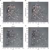
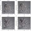
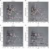
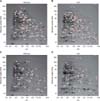
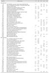
The 2-DE analysis, using the same concentrations of prepared proteins from B. abortus wild and mutant strains, revealed 814 spots in B. abortus wild-type and 541 spots in mutant C3. Among them, 444 paired spots were identified after comparison of the mutants with B. abortus wild-type. Ninety-four spots showed increases and 30 showed decreases from B. abortus wild-type (Fig. 1). In case of mutant C8, the total number of spots was 577, and 418 spots were paired with B. abortus wild-type. Among them, 78 spots showed relatively increased expression and 65 spots showed decreased expression (Fig. 2). In mutant C13, the total number of spots were 491, of which 256 spots formed pairs with B. abortus wild-type. Among them, 87 spots were relatively increased and 43 spots were decreased (Fig. 3). The total spot number of mutant C24 was 516, of which 246 spots formed pairs with B. abortus wild-type. Of these, 87 spots increased in relative expression and 45 spots had decreased expression (Fig. 4). A total of 560 spots were identified in mutant C30, of which 282 spots formed pairs with B. abortus wild-type. In 96 spots of those spots, expression was relatively increased, while in 50 spots, expression was decreased (Fig. 5, Table 1). All spots were detected on 2-DE gels and had pI (isoelectric point) and molecular weight ranges of 4.0 to 10.0 and 10 to 250 kDa, respectively. Based on the 2-DE image analysis results, 30 spots of each mutant strain with greater than 2-fold changes (increases or decreases) from those of the spots of B. abortus wild-type were selected. The selected spots underwent LC-MS/MS for peptide analysis (Table 2). The spot number, gene name, gene ID, protein identification, protein ID, accession number, sequence length, locus tag, experimental molecular weight, theoretical molecular weight, pI, sequence coverage (%), subcellular location, and Clusters of Orthologous Groups (COG) functional category of these proteins are presented in Table 3.
The results confirmed that expressions of pathogenic factors such as ClpB and its interacting DnaK, RpsE, and cold-shock proteins were increased; particularly in mutants C24 and C30. In some mutants, an increase in proteins related to energy metabolism such as Zwf, TrpA, and Pgk were identified, and an increase in proteins associated with ATP-binding protein or ABC transporter were also detected.
The SecA, GAPDH, and GNAT family have been shown to reduce expression in energy metabolism-related factors. In particular, expressions of mutants C3 and C8, which have a slower growth rate than that of B. abortus wild-type, were observed to decrease in common. Additionally, in mutants C3 and C8, expression of AcnA, which is involved in bacterial growth, was present but was decreased. Expression of Hfq, which is related to stress resistance and pathogenicity, metabolism, and also important in intracellular survival in B. abortus, was also decreased in mutants C3 and C8.
Discussion
Brucellosis is a re-emerging zoonosis that can infect not only animals but also humans throughout the world. At present, the pathogenesis of the disease is reported to have greatly increased [1924]. Despite many studies, however, eradication of brucellosis remains a difficult task, and many potential effective diagnostics and vaccines are under study to prevent the spread of this disease [618].
In this study, B. abortus mutants were generated by using the EZ-Tn5 transposon system. The resulting mutants were commonly resistant to kanamycin, and the expression of their proteins was different due to the insertion of the related rKan gene. The expression patterns of the proteins of the B. abortus mutants were generated, compared to B. abortus wild-type, and found to vary. We identified various proteins that were changed following transposon insertion and confirmed functions of each protein.
ClpB, shown to increase in expression commonly in B. abortus mutants C13, C24, and C30, is associated with energy-related metabolism including ATP-binding and peptidase activity. In addition, ClpB is a heat-shock protein that allows pathogens to adapt to the intracellular environment; moreover, ClpB works in conjunction with DnaK, DnaJ, and GrpE to suppress protein aggregation, thereby affecting pathogenicity [31]. DnaK (Hsp70), which increased in expression in all of the studied B. abortus mutants, is a chaperone protein. Protein folding is also performed by DnaK, and ATP is essential for this role. It is also reported that structural change in DnaK occurs through ATPase activity [4]. Moreover, DnaK is involved in replication of phage lambda DNA, is related to hyperosmotic shock, and has a role in ATP-dependent resolubilization of aggregations of protein in conjunction with ClpB. Binding of ClpB occurs according to a previous DnaK association with the protein aggregation. The bi-chaperone network of DnaK and ClpB consists of three stages. When DnaJ is attached, the co-chaperone forms an aggregation with DnaK, and DnaK interacts with ClpB on the surface on the aggregation. The TRP domain protein also has an important role in stimulating the molecular chaperone complex, particularly in the formation of bridges between Hsp70 (DnaK) and Hsp90 [1252331].
RpsC was identified as ribosomal protein S3, which is involved in the expression of the nuclear factor kappa B (NF-κB)-induced reporter gene when T cell stimulation occurs. When the T cell is stimulated, RpsC is transferred into the cell to transmit the NF-κB signal within the cytoplasm. In particular, RpsC has an important role in signal transduction of NF-κB though p65, where NF-κB has an important role in the expressions of genes that bind to regulatory sites or are regulated by p65 [28]. This suggests that not only growth but also T cell stimulation may occur more actively in mutants C24 and C30, which are faster growing than B. abortus wild-type.
The SecA, GAPDH, and GNAT family, which are commonly expressed in the mutants C3 and C8 that have slower growth than that in B. abortus wild-type. It is thought that the growth rate is lower than that in B. abortus wild-type due to expression decreases in metabolic function-related factors active in obtaining the energy required for the normal growth of the strains [927]. In addition, the decrease in the expression of AcnA, which is directly related to the growth feature of the strain, in related to regulation networks, such as CRP, ArcA, Fur, and SoxR5, and seems to have affected the growth features of the mutants [12]. Hfq, showing decreased expression in mutants C3 and C8, is an RNA-binding protein that is common to bacteria and is important in gene expression control. Hfq acts on pairs of small RNA and mRNA and is involved in translation, transcription, and post-transcription networks. It acts on small RNA to inhibit the translation of the 30S and 50S ribosomal subunits and inhibits degradation of small RNA. In addition, Hfq induces cleavage by ribonuclease E (RNase E) of target mRNA. It is also associated with resistance to environmental stressors such as oxidation, acid, and heat [72526]. It is thought that the observed decrease of Hfq expression may be a cause of the slower growth rate in mutants C3 and C8. Furthermore, transcription in these mutants may cause abnormal expression of several genes.
It is well-known that all the biological processes within living organisms are controlled by the various proteins present within the organism. Protein is not only a basic biological component, but also constituents of enzymes, antibodies, and hormones. In addition, proteins are deeply involved in all cell processes, such as DNA replication, RNA transcription, and protein translation, as well as in determining the function of the organism. Protein analysis of the major pathogenic factors of bacteria is of great importance in research into disease eradication or prevention.

 XML Download