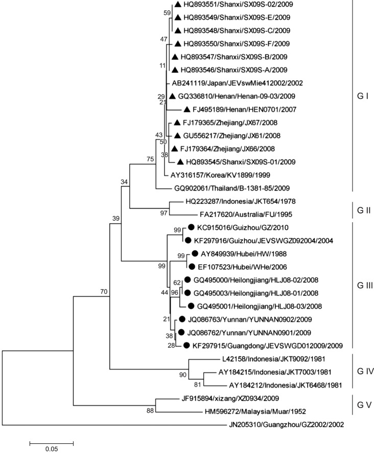
 | Fig. 2Results of phylogenetic analysis of Japanese encephalitis virus strains isolated from Chinese swine herds based on assessment of the capsid/premembrane protein (C/PrM) nucleotide sequences. The multiple sequence alignments were obtained by using MEGA software (ver. 5.0; Molecular Evolutionary Genetics Analysis). The tree was constructed by applying the neighbor-joining method. Scale bar indicates number of nucleotide substitutions per site. Bootstrap confidence limits are shown at each node. The Dengue 1 virus (strain GZ2002) was used as an outlier group. Those strains (marked by ▲ or ●) were isolated and identified from pigs between 2006 and 2012 in China. |
Abstract
Japanese encephalitis virus (JEV) is a mosquito-borne, zoonotic flavivirus causing viral encephalitis in humans and reproductive disorder in swine. JEV is prevalent throughout China in human; however, spatiotemporal analysis of JEV in Chinese swine herds has not been reported previously. Herein, we present serological and molecular epidemiological results and estimates of prevalence of JEV infections among swine herds in various regions of China. The results suggest that JEV infections are widespread and genotype I and III strains co-exist in the same regions. Therefore, there is an urgent need to monitor JEV infection status among swine herds in China.
Go to : 
Japanese encephalitis virus (JEV) is a zoonotic flavivirus that causes viral encephalitis in humans and reproductive disorder in swine in eastern and southern Asia [2]. In China, JEV strain P3 was isolated in 1949, and JE has been epidemic for succeeding 60 years; moreover, it has been reported that JEV is prevalent throughout China, including its presence in Tibet [4]. In 2015, 624 human JE cases with 19 deaths were reported in China.
JEV can infect a variety of animals through its natural replication cycle. Pigs act as an amplifying host and have a key role in JEV transmission in China; therefore, human infections are closely associated with the prevalence of JEV in pigs. To evaluate the potential risk of JEV infections among swine on human health, we examined all of the available data on JEV infections in China and investigated the dataset's geospatial epidemiologic features.
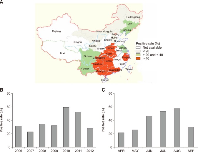
The JEV epidemiological data from 2006 to 2012 for swine herds in China were collected from the China National Knowledge Infrastructure database. A total of 22,343 serum samples were collected from pigs in 18 provinces and autonomous regions of China, including Guangxi, Guangdong, Zhejiang, Hunan, Jiangsu, Guizhou, Yunnan, Sichuan, Chongqing, Tibet, Hubei, Henan, Gansu, Hebei, Inner Mongolia, Liaoning, Jilin, and Heilongjiang (panel A in Fig. 1). Among those samples, 8,807 (39.4%) were JEV-positive, and prevalence of JEV-positive samples varied by year (range, 23.0%–59.2%); the highest (59.2%, 3,286/5,548) occurring in 2010 and the lowest (23.0%, 417/1,810) in 2007 (panel B in Fig. 1). The distribution of JEV-positive cases also varied among the assessed geographic regions (range, 9.6%–60.6%) with the highest (60.6%, 1,575/2,597) in Guangxi province and the lowest (9.6%, 35/365) in Hebei province (panel A in Fig. 1). Geographically, JEV was more prevalent in southern China; the JEV-positive rate gradually decreased from south to north, similar to the drop in average daily temperature along the same path.
Numerous analyses have indicated that the incidence of JEV infection is seasonal and closely related to geographical distribution and climate [2]. Generally, peaks in human JEV epidemics in China occur for 7–9 months each year, which varies geographically from 5–6 months in southernmost Hainan province, to 6–7 months in southern China, to 8–9 months in northern China (panel C in Fig. 1), further indicating that the prevalence of JEV infections coincides with changes in average temperatures and the seasonal distribution of mosquitoes. Analyses of JEV serological data from Chinese swine herds showed that the prevalence and distribution of JEV in swine also displays seasonal and geographical distribution changes; JEV infections appear 1 to 2 months earlier in southern China than in northern China, and new JEV infections occur 1 to 2 months earlier in swine than in humans. Therefore, by monitoring JEV epidemic trends in swine, relevant and timely information can be acquired and used to better predict JEV epidemics among humans, thereby promoting effective prevention and control of human JEV infections.
Previous nucleotide sequencing of the capsid/premembrane protein (C/PrM) and envelope (E) genes of JEV revealed five genotypes [3], and genetic analysis of JEV strains isolated from humans and mosquitoes showed that JEV genotypes I and III co-exist in China [3]. To uncover the status of JEV in swine in China, mosquito samples were collected and viruses isolated and sequenced. The results demonstrated that JEV genotypes I and III were prevalent in Chinese swine herds, and the JEV isolated from mosquitoes shared high homology with those isolated from humans and pigs [345].
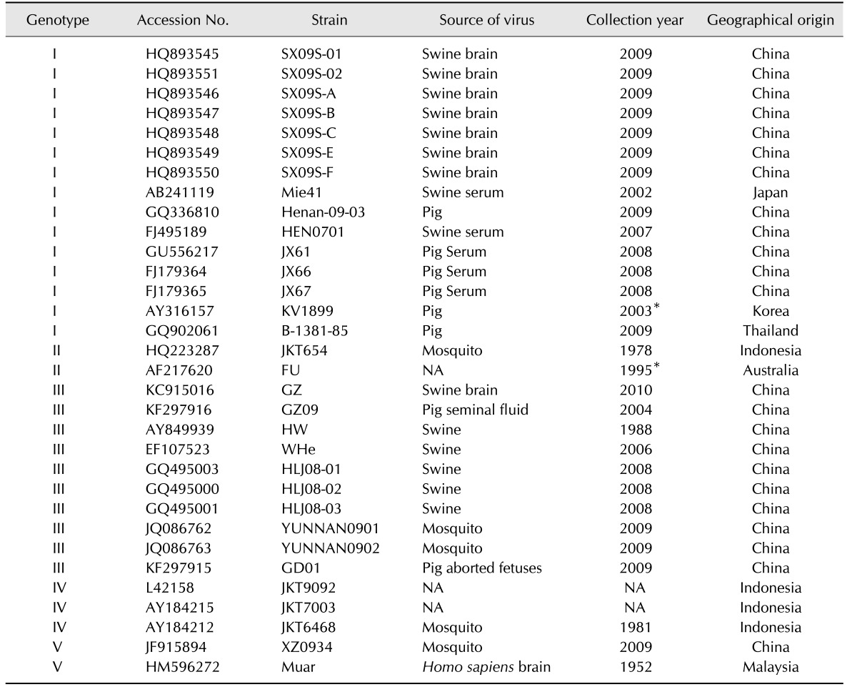
To understand the genetic relationships among JEV strains isolated from pigs in China, 18 available JEV sequences were extracted from the NCBI database (National Center for Biotechnology Information, USA), and phylogenetic analysis based on the C/PrM and E nucleotide sequences was performed (Table 1). Of the 18 JEV strains isolated between 2006 and 2012 in China, most were isolated from aborted fetuses; however, a few were isolated from piglets with confirmed viral encephalitis. Our results indicated that 12 of the isolated strains belonged to genotype I and the remaining 6 strains were identified as genotype III, indicating that these two genotypes co-exist within the swine herds in China (Fig. 2). In China, the first JEV strain (AY849939), which belongs to genotype III, was isolated from swine in 1988 [23]. In 2007, a genotype I strain (FJ495189) was isolated from a pig herd in China [2]. Thereafter, several genotype I strains were isolated from pigs, indicating that JEV genotype I is predominant in China [1678].
In this study, we present the results of our serological and molecular epidemiological analyses of data obtained from previously reported JEV infections in swine herds in China. The results indicate that JEV infections are widespread throughout the sampled regions of China and confirm that genotype I and III strains co-exist at pig farms in the same region. The results indicate that increased surveillance of JEV among Chinese swine herds is warranted.
Acknowledgments
This study was supported by the Special Fund for Agroscientific Research in the Public Interest in China (No. 201203082).
Go to : 
References
1. Cao QS, Li XM, Zhu QY, Wang DD, Chen HC, Qian P. Isolation and molecular characterization of genotype 1 Japanese encephalitis virus, SX09S-01, from pigs in China. Virol J. 2011; 8:472. PMID: 21999532.

2. Erlanger TE, Weiss S, Keiser J, Utzinger J, Wiedenmayer K. Past, present, and future of Japanese encephalitis. Emerg Infect Dis. 2009; 15:1–7. PMID: 19116041.

3. Huanyu W, Shihong F, Ying H, Jiguang M, Xiaoling P, Guodong L. [Co-prevalence of two genotypes of Japanese encephalitis virus in Shanghai, China]. Chin J Vector Biol Control. 2012; 23:398–401. Chinese.
4. Li YX, Li MH, Fu SH, Chen WX, Liu QY, Zhang HL, Da W, Hu SL, Mu SD, Bai J, Yin ZD, Jiang HY, Guo YH, Ji DZ, Xu HM, Li G, Mu GG, Luo HM, Wang JL, Wang J, Ye XM, Jin ZM, Zhang W, Ning GJ, Wang HY, Li GC, Yong J, Liang XF, Liang GD. Japanese encephalitis, Tibet, China. Emerg Infect Dis. 2011; 17:934–936. PMID: 21529419.

5. Liu H, Lu HJ, Liu ZJ, Jing J, Ren JQ, Liu YY, Lu F, Jin NY. Japanese encephalitis virus in mosquitoes and swine in Yunnan province, China 2009-2010. Vector Borne Zoonotic Dis. 2013; 13:41–49. PMID: 23199264.

6. Liu WJ, Zhu M, Pei JJ, Dong XY, Liu W, Zhao MQ, Wang JY, Gou HC, Luo YW, Chen JD. Molecular phylogenetic and positive selection analysis of Japanese encephalitis virus strains isolated from pigs in China. Virus Res. 2013; 178:547–552. PMID: 24045128.

7. Wang LH, Fu SH, Wang HY, Liang XF, Cheng JX, Jing HM, Cai GL, Li XW, Ze WY, Lv XJ, Wang HQ, Zhang DL, Feng Y, Yin ZD, Sun XH, Shui TJ, Li MH, Li YX, Liang GD. Japanese encephalitis outbreak, Yuncheng, China, 2006. Emerg Infect Dis. 2007; 13:1123–1125. PMID: 18214202.

8. Zheng H, Shan T, Deng Y, Sun C, Yuan S, Yin Y, Tong G. Molecular characterization of Japanese encephalitis virus strains prevalent in Chinese swine herds. J Vet Sci. 2013; 14:27–36. PMID: 23388434.

Go to : 


 PDF
PDF ePub
ePub Citation
 Print
Print


 XML Download