

 PDF
PDF Citation
 Print
Print


It is widely accepted that calcium deposition in cellular degenerated areas can be associated with infection and ischemic changes [710]. Calcification in the uterine cavity is an uncommon finding in veterinary medicine. In human, results from a few cases have indicated that uterine calcification could be caused by the presence of osseous fragments associated with a previous history of abortion [9]. Alternative etiologies have included osseous metaplasia from multipotent cells, secondary osteogenesis after retention of fetal bone, implantation of embryonic parts, dystrophic calcification of retained necrotic tissue, continuous estrogenic stimulation of the endometrium, and chronic endometrial inflammation. [23]. However, there are no reports of uterine calcification in pregnant pig without a history of abortion.
The aim of this study was to present the first case of uterine calcification in a pig with normal piglet delivery through routine Cesarean section. A naturally synchronized female pig was prepared for cloned embryo transfer (ET). Somatic cell nuclear transfer was performed as described in our previous study with slight modification [6]. Cumulus-oocyte complexes were collected and cultured in medium containing TCM-199 with 0.57 mM cysteine, 0.91 mM sodium pyruvate, 5 µL/mL insulin transferrin selenium solution 100X (Invitrogen, USA), 10 ng/mL epidermal growth factor, 10% porcine follicular fluid (vol/vol), 10 IU/mL (IU, international unit) equine chorionic gonadotropin, and 10 IU/mL human chorionic gonadotropin. After 44 h of maturation, cumulus cells of matured oocytes with a first polar body were denuded in 0.1% hyaluronidase. After enucleation in TALP containing 5 µg/mL cytochalasin B and 5 µg/mL Hoechst 33342, a single fibroblast was injected and electrically fused. A total of 260 reconstructed embryos were electrically activated and cultured in vitro. Reconstructed embryos with normal morphology were surgically transferred into both oviducts of a surrogate pig. The female pig used in this study was raised at our private facility. During the ET, progesterone concentration of the naturally synchronized surrogate pig was 7.14 ng/mL. Pregnancy was identified via ultrasonography at 28 days after ET. At 112 days after ET, 5 piglets with no gross structural abnormalities were delivered by Cesarean section. During the Cesarean section surgery, the sow's measured progesterone concentration was 3.78 ng/mL. The surgical procedure was performed under general anesthesia and efforts were made to minimize any potential suffering of the surrogate. The 5 cloned piglets were produced normally, but abnormal tissue attached to the maternal right uterine horn cavity was observed. All protocols involving animal use were approved by the Institutional Animal Care and Use Committee of Seoul National University (SNU-151019-4) in accordance with the Guide for the Care and Use of Laboratory Animals of Seoul National University, Korea.
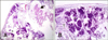
The abnormal uterine tissue was observed to expand outward from the serosal surface of the right uterine horn and attach to the right uterine horn cavity near the bifurcation. A sample of the tissue was examined after staining with hematoxylin and eosin. Histological examination revealed typical porcine epitheliochorial placental tissue with an area of calcified tissue (panel A in Fig. 1). Around the area of dystrophic calcification, inflammatory cells, mainly histiocytes, had infiltrated (panel B in Fig. 1).
In guinea pigs, it has been reported that calcification can be induced by hormone administration, including administration of prolactin, human chorionic gonadotropin, estradiol, estrone, and growth hormone [11]. In the present study, pregnancy was maintained with a normal progesterone concentration, and the surgery was performed two days before the due date. Although we do not know why calcification was identified in the uterine cavity, we assume that the phenomenon may be related to metabolic and hormonal changes secondary to the pregnancy.
Although causes of ectopic ossification and mineralization are well described, little has been reported on the pathogenesis of uterine calcification in pregnant animals. In this report, normal cloned piglets were produced and there was no accumulation of mucus within the uterine horns, which is in contrast to observations reported for a case of uterine lithiasis in a dog [5].
In humans, calcification of placenta is considered a part of maturation and aging [12]. Pathogenesis of endometrial calcifications is involved in uterine trauma during instrumentation and/or uterine infection. However, in the present case, we surgically transferred cloned embryos to both oviducts of a sow without a history of uterine trauma or instrumentation. There are few similar reports of uterine calcification; one case of a uterine stone in an 8 years old female child without a history of uterine trauma, and two other cases of uterine stones, one reported in a 28 years old woman with a history of pregnancy loss and the second in a 73-year-old women [14]. The female pig in the present study had no history of pregnancy loss in its previous two natural deliveries. Although it has been reported that endometrial calcification in the uterine cavity can induce secondary infertility [8], it did not affect the maintenance and mortality of the cloned litter piglets in the present case.
In summary, herein, we have described a case of uterine calcification, a rarely reported phenomenon in a pregnant farm pig without pregnancy loss.
 XML Download