Abstract
Age-related involution in dogs involves loss of muscle mass and changes in connective tissue and articular cartilage. The aim of this study was to examine whether an age-related influence on joint mobility can be detected in the absence of disease. Five young (mean age 2.0 years) and five old (mean age 10.4 years) healthy and sound Beagle dogs underwent computer-assisted gait analysis during locomotion on a treadmill. Shoulder, elbow, carpal, hip, stifle, and tarsal joint angles including joint angle progression curves, minimum and maximum joint angles, and range of motion (ROM) in degrees were analyzed. The old group had a smaller maximum joint angle (p = 0.037) and ROM (p = 0.037) of the carpal joint; there were similar tendencies in the shoulder, elbow, and carpal joints. Descriptive analysis of the progression curves revealed less flexion and extension of the forelimb joints. The results indicate restricted joint mobility of the forelimb in old dogs, primarily of the carpal joint. Results in the joints of the hindlimb were inconsistent, and the contrasting alterations may be due to a compensatory mechanism. As most alterations were found in the distal joints, these should receive particular attention when examining elderly dogs.
It is only since the end of the 20th century that research on pet geriatrics has been reported. In recent years, interest in this topic has grown as pets today reach a greater age than previously, and the population of elderly pets is increasing [2733]. During the progressive and irreversible process of aging, an animal's adaptability to internal and external stresses decreases. This reduces physiological functions, increases multimorbidity, and finally leads to death [17]. The onset and progression of the aging process in dogs, as well as their longevity, depend on various factors, with body size being the most important one; in large dogs, signs of aging occur earlier and their lifespan is shorter compared to small dogs [34]. According to Bellows et al. [3] small- and medium-breed dogs may be classified as senior at 7–10 years of age and as geriatric at ≥ 11 years of age, whereas large- and giant-breed dogs may be classified as senior at 6–8 years of age and as geriatric at ≥ 9 years of age.
The aim of this pilot study was to examine whether, and if so to what extent, an influence of the aging process on the locomotor system in the absence of disease can be detected. Signs of aging in dogs are, among other things, a loss of muscle mass and strength as well as changes in connective tissue and articular cartilage [20293739]. Such changes can lead to primary degenerative joint disease [35]. Moreover, such processes can affect locomotion and restrict joint mobility in elderly dogs. More precise knowledge of the influence of aging on the locomotor system could support the need for regular orthopedic health examinations as well as the provision of physiotherapy and exercise therapy in elderly dogs. Knowledge of which joints are most frequently or most intensely affected by restricted joint mobility could enable targeted therapy. In addition, determining base values for old dogs compared to those of young dogs of the same breed could be helpful in further studies in this field.
The human eye cannot perceive the complex, rapidly executed elements of locomotion in detail, and it has been shown that observers cannot reliably score induced lameness in dogs, not even observers with orthopedic experience [3844]. This demonstrates that visual examination of locomotion is neither objective nor sufficiently accurate. Computer-assisted gait analysis systems can record hundreds of observations per second and enable quantification [22]. As a result, minute differences can be captured and various components of locomotion, such as the range of motion (ROM) of joints, can be examined. Most previous kinematic studies have analyzed the gait of healthy adult dogs at different ages [1328] or of dogs with various orthopedic diseases [48], focusing in particular on investigation of different therapeutic treatments [1016]. In one case, alterations of limb angles in growing puppies were analyzed [25], but not the effects associated with aging. For the present pilot study, we hypothesized that joint mobility is restricted and that consequently joint ROM is decreased in elderly dogs. Therefore, joint angles of five young and five old Beagle dogs during trotting were examined by computer-assisted gait analysis.
Ten Beagle dogs participated in this study. Five Beagle dogs from the same litter, owned by the Small Animal Clinic, University of Veterinary Medicine Hannover, Foundation, Germany, formed the young group. The young dogs were 2.0 ± 0.0 years of age (mean ± SD), male, neutered, and had a body weight of 18.3 ± 2.4 kg. Five more Beagle dogs, two from another institute within the above-mentioned university and three from private owners, formed the old group. The old dogs were 10.4 ± 0.9 years of age; three of the dogs were male and two female and all were neutered, except for one male dog. The old dogs had a body weight of 15.5 ± 2.4 kg. The difference in mean body weights between these two groups was not statistically significant. All experiments were performed in accordance with the relevant statutory provisions. The study was reported to the Lower Saxony State Office for Consumer Protection and Food Safety in Oldenburg, Germany (reference No. 33.9-42502-05-13A369).
All participating dogs were to be free of orthopedic disease; therefore, detailed anamnesis, as well as general and orthopedic examinations, were performed. There were no signs of general or orthopedic disease in the participating dogs, and none of the dogs were receiving any medication. In a companion study by Willen et al. (unpublished paper), vertical ground reaction force was recorded as an objective measure to determine lameness. Kinetic and kinematic data were recorded in parallel, except for one young dog for which the kinetic recordings had to be retaken; in that case, the later results were used for kinetic analysis. Willen et al. (unpublished paper) found no statistically significant differences between the groups in vertical impulse, peak vertical force, or mean vertical force. In both groups, body weight distribution, calculated according to the method reported by Steiss et al. [41], was approximately 60% for the forelimbs and 40% for the hindlimbs, which is considered physiologically normal in the Beagle dog [1]. The symmetry indices for the forelimbs and hindlimbs were calculated according to the method of Herzog et al. [26]; a symmetry index of < 6% is considered physiologically normal [11]. The symmetry indices of both groups revealed an almost symmetrical gait pattern without signs of lameness.
Kinematic recordings were performed in the gait analysis laboratory of the Small Animal Clinic in a manner similar to those in previous kinematic studies in this laboratory [1016212425]. An instrumented four-belt treadmill (TM-07-B; Bertec, USA) was used. Six high-speed infrared cameras (recording frequency 100 Hz, MX3+; Vicon Motion Systems, UK) placed around the treadmill detected three-dimensional movements of reflective markers during locomotion. A digital high-speed video camera (pilot piA640-210gc; Basler, Germany) recorded locomotion of the dogs from a lateral position. All devices were managed and controlled with Vicon Nexus software (ver. 1.8.5; Vicon Motion Systems) and Bertec Treadmill Control Panel software (ver. 1.7.12; Bertec).
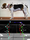
Prior to recording, a training phase was carried out until the dogs were perceived to be running at a relaxed and loose trot. The young dogs, which were accustomed to the treadmill due to participation in a previous study, had a predetermined training phase of 5.0 ± 0.0 min; the old dogs required 18.2 ± 14.5 min of training. With double-sided adhesive tape and hair clips, 35 retroreflective passive markers (16 mm diameter) were attached, by the same person, to all four limbs and the back of each dog. Joint-determining markers were attached above clearly defined and easily palpable anatomical landmarks; additional markers were attached to locations in the intermediate segments (panel A in Fig. 1) [2124]. For each dog, treadmill speed was adjusted until the dog was able to trot smoothly and regularly. The young group trotted at 1.8 ± 0.0 m/sec and the old group at 1.7 ± 0.1 m/sec. For each dog, around ten recordings of approximately 30 sec each were obtained; therefore, for each dog the complete duration of the recordings including preparations between the takes was about 10 min.
For each dog, a sequence of ten consecutive and representative strides in which the dog ran straight and uniformly was chosen. The marker positions, recorded by the infrared cameras, were processed by Vicon Nexus software (Vicon Motion Systems) with a deposited kinematic model of the dog's four limbs and back. Each marker position was linked to an anatomical location of the kinematic model. Thus, a rod model was created (panel B in Fig. 1) in which the respective joint angles were determined from the location coordinates of three joint-determining markers. The times when the dog's feet touched the ground and lifted off were determined manually. The measured vertical ground reaction force, which indicates the beginning and end of the stance phase and, as a result, also the duration of the swing phase, and the parallel recorded video were used.
Angles of the shoulder, elbow, carpal, hip, stifle, and tarsal joints were projected two-dimensionally in the sagittal plane and exported to Excel 2010 software (Microsoft, USA). To improve comparability among dogs, the data output was time normalized to a 100% stride duration and 50% stance and swing phase durations [2124]. The analysis of each joint angle included the joint angle progression curve in degrees, the minimum joint angle in degrees (MIN, i.e., maximum flexion), the maximum joint angle in degrees (MAX, i.e., maximum extension), and the range of motion of the joint in degrees (ROM, i.e., difference between MIN and MAX) during each stride [101621]. For each joint angle, the data for 10 strides were averaged. To compensate for possible differences between marker positions, standardization, in which the mean of the progression curve was subtracted from every value, was performed for each joint angle [7831]. As all dogs were measured while trotting, which is considered a symmetrical gait [23], and as no statistically significant differences were detected between the joint angles of the left and right side, the joint angles for the two sides were averaged (Table 1).
Due to the small group sizes (n = 5), a normal distribution of data was not assumed. Therefore, the non-parametric Wilcoxon rank-sum test for two independent samples was used to assess differences in body weight and standardized joint angles between the young and the old group (differences with p < 0.05 were considered significant, differences with p < 0.10 were considered to indicate a tendency). To compare joint angles between left and right sides, the non-parametric Wilcoxon signed-rank test for two paired samples was used (differences with p < 0.05 were considered significant). The sides were compared within a group and across both groups. Statistical analyses were performed by using SAS Enterprise Guide software (ver. 7.1; SAS Institute, USA). To validate the results, the test's statistical power was analyzed retrospectively. For this purpose, a standard deviation of 10% was assumed, which is an average value observed in this study and reported in previous kinematic analyses [228]. In addition, a biologically relevant difference between the groups of 15% was assumed. In sound dogs with borderline hip dysplasia, Bockstahler et al. [8] reported alterations of the ROM of the hindlimb joints of about 10%, whereas, in slightly lame dogs with bilateral degenerative joint disease of the hips alterations of the ROM of the hindlimb joints of about 30% were reported [6]. Therefore, 15% was deemed an appropriate value that could be indicative of a clinically apparent orthopedic disease. The α-risk was 0.05 and the hypothesis testing was one-tailed. To perform this test, PASS software (ver. 14; NCSS, USA) was used.
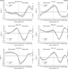
The results of the descriptive analysis of joint angle progression curves (D; Fig. 2) and of the statistical analysis of MIN, MAX, and ROM (S; Fig. 3) are presented in each of the following joints:
During the stance phase, the joint flexed slowly. In the young group, there was a short extension at the end of the stance phase. At the beginning of the swing phase, the point of greatest flexion, the joint of the old group showed less flexion than that in the young group. This point occurred earlier during the stride in the old group than in the young group. The joint then extended rapidly, moving the limb forward. At the point of greatest extension, the joint of the old group showed less extension than that in the young group. This extension was associated with a high standard deviation in the old group, indicating high variability among the old dogs.
After a small flexion, the joint extended during the stance phase. At the point of greatest extension, the joint of the old group showed slightly less extension than that in the young group. The joint then flexed intensely while the limb moved forward. At the point of greatest flexion, approximately in the middle of the swing phase, the joint of the old group showed slightly less flexion than that in the young group. Next, the joint extended a second time, during which the old group again achieved slightly less extension than that in the young group. Differences between the progression curves in the two groups were slight in the elbow joint.
During the stance phase, the joint angle was nearly constant; however, at the beginning of the stance phase, there was less extension in the old group than in the young group. The start of the stance phase was also the time of greatest extension. At the end of the stance phase, the joint flexed intensely. At the beginning of the swing phase, the point of greatest flexion, the joint of the old group showed less flexion than that in the young group. The carpal joint extended when the limb was moved forward.
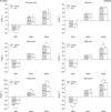
The old group showed a significantly smaller MAX (p = 0.037) at the beginning of the stance phase and a smaller ROM (p = 0.037) than those in the young group. There was also a tendency toward a larger MIN (p = 0.060) at the beginning of the swing phase in the old group compared to that in the young group.
During the stance phase, the joint extended slowly. At the beginning of the stance phase the joint was less flexed in the old group than in the young group. The point of greatest extension was at the shift from the stance phase to the swing phase; at that point, the joint was less extended in the old group than in the young group. Subsequently, the joint flexed, moving the limb forward. At the point of greatest flexion, the joint of the old group showed greater flexion than that in the young group.
At the beginning of the stance phase, the joint flexed. Subsequently, it extended, and during extension, the joint of the old group achieved slightly less extension than that in the young group. In addition, the extension point occurred slightly later during the stride in the old group than in the young group. Afterwards, the joint flexed again as the limb moved forward. At the point of greatest flexion, almost in the middle of the swing phase, both groups attained approximately the same amount of flexion. The joint then extended again, and at the point of greatest extension, the joint of the old group showed slightly more extension than that in the young group. Differences between progression curves were slight in the stifle joint.
At the beginning of the stance phase, the joint flexed; at that point, both groups reached approximately the same level of flexion. The joint then extended, and at the point of greatest extension (i.e., at the shift from the stance phase to the swing phase), the joint of the old group showed more extension than that in the young group. Compared to the young group, this point occurred slightly later during the stride in the old group. Afterward, the joint flexed, thus moving the limb forward. At the point of greatest flexion, the joint of the old group showed more flexion than that in the young group. The joint then extended a second time, thereby the joint of the old group achieved a slightly greater extension than that in the young group.
Compared to the young group, the old group showed a tendency toward a smaller MIN (p = 0.095) in the middle of the swing phase and a greater ROM (p = 0.095). Although the MAX occurred later in the stride in the old group than in the young group, the difference in extension between the two groups was neither statistically significant nor indicative of a tendency.
The statistical analyses revealed a smaller maximum extension and ROM of the carpal joint in the old group than in the young group. Additionally, there were tendencies toward smaller maximum flexion of the shoulder and carpal joints and a smaller ROM of the elbow joint in the old group compared to the young group. This indicates the old dogs had restricted joint mobility in the forelimb, primarily in the carpal joint. The progression curves also revealed reduced flexion and extension of the joints of the forelimb in the old group. In addition, there was reduced extension of the hip joint in the old group, and stifle joint extension was reduced during the stance phase in the old group. There are several reasons why joint mobility can become restricted with age. Aging involves a reduction of nearly all tissue types in the body. This includes sarcopenia, a progressive loss of muscle mass and strength [2937]. Furthermore, there are changes in connective tissue and, therefore, in joint capsules, ligaments, tendons, and fasciae [39]. Articular cartilage becomes thin, and a reduction of chondrocytes leads to decreased production of extracellular matrix [320]. This can finally lead to age-related primary degenerative joint disease, which is chronic, progressive, and irreversible; and is associated with degeneration and loss of articular cartilage and subchondral bone, formation of periarticular osteophytes, joint capsule thickening, and synovitis [35]. At the onset of primary degenerative joint disease, there may be no or slight clinical signs.
Notably, in the present study, age-related restriction of joint mobility was more striking in the forelimb than in the hindlimb. In dogs, the greatest anatomical difference between forelimbs and hindlimbs is the different orientation of the tri-segmented z-shaped limbs in which the elbow joints face posteriorly and the stifle joints face anteriorly; furthermore, the scapulae have no bony connection with the trunk [1319]. During level locomotion with consistent speed, the hindlimbs are primarily propulsive while the forelimbs primarily brake; additionally, the forelimbs act more like a strut, while the hindlimbs act more like a lever [1215]. Both forelimbs together bear about 60% of the body weight while both hindlimbs bear about 40% [12]. It can be hypothesized that, in this study, age-related restriction of joint mobility was more striking in the forelimb due to the forelimbs bearing the greater percentage of the body weight, which may, over time, lead to increased attrition of the joints and decreased ROM.
In contrast are the tendencies toward a greater maximum flexion of the tarsal joint and a greater ROM of the stifle and tarsal joint in the old group compared to the young group. The progression curves revealed greater flexion and extension of the tarsal joint and greater flexion of the hip joint in the old group than in the young group, while, in the stifle joint, flexion and extension were barely greater. This may be due to a compensatory mechanism for the restriction of joint mobility in the forelimb, which was seen in the present study. Some researchers have reported that restriction of the forelimbs can lead to increased mobility of the hindlimbs [1830]. Another explanation may be a compensatory mechanism for a restricted function of the hip joint. Several studies have found that hip joint restrictions can be compensated via increased mobility of the stifle and tarsal joints, even in subclinical diseases without lameness [489]. In the present study, the hip joint angle progression curves were different between the groups. However, differences in MIN, MAX, and ROM were not statistically significant.
This study used external markers because they are non-invasive and, therefore, practicable even in a clinical setting. However, they are associated with problems concerning reproducibility of marker position and related to soft tissue artifacts [4043]. Nonetheless, in humans, it was reported that intrasubject repeatability is excellent if the data are projected in the sagittal plane, standardization is performed, and the subjects walk at their preferred speed [31]. The first two specifications were implemented in this study, and the treadmill speed was adjusted for each dog. Locomotion varies with body size as well as body types [2]. Also, different skin and coat conditions can result in variability. However, only one breed participated in this study, and the slight difference in body weight between the groups was not significant.
Kinematic parameters are reported to be slightly influenced by gait speed [1442]. In the present study, all dogs were of the same breed and their individually adjusted trotting speeds were very similar. In the old group, trot speed was slightly lower than that in the young group, mainly due to the trotting speed of the two smaller female dogs. However, in both groups, the results were similar for all dogs and, therefore, standard deviation values were of an average size for kinematic analyses, except for the values for the shoulder joint in the old group. With increasing speed of locomotion, the ROM of joints mainly increases [1332], whereas the stance phase duration and, therefore, dynamic stability decrease [1442]. The trot gait was used because it seemed more suitable than the walk gait for determining ROM differences. In addition, it appeared to be difficult to record stable gait sequences at a galloping gait. As the trot is considered a symmetrical gait [23], and as there were no significant differences between the left- and right-side results, the results for the two sides were averaged. This similarity of results on the two sides provides further evidence that the locomotion of the dogs was sound.
This study confirms the importance of objective and quantifiable kinematic gait analysis to detect slight alterations in joint mobility. Although there were no differences between the groups in the anamnesis, the general and orthopedic examinations, and the analysis of vertical ground reaction force, there were significant differences in joint kinematics. This shows that clinical evaluations may be insufficient, and that measurement of ground reaction forces provides information limited to the complete limb during the stance phase.
A limitation of this pilot study is its small group size (n = 5). A larger group size would reduce the influence of the results from a single dog as well as inter- and intra-individual differences. However, a small group size may be more tolerable due to the use of computer-assisted gait analysis [3136]. Regardless, the small group size in this study may have prevented statistical validation of the tendencies detected (a type II statistical error). To evaluate the validity of the results, statistical power was analyzed retrospectively. A statistical power of approximately 70% was calculated, whereas, in general, an 80% power level is desired. Moreover, the standard deviations of the shoulder joint in the old group were broad, which may have prevented statistical validation of this joint's results. Consequently, the results of this study should be interpreted with appropriate caution. Further studies with larger sample sizes would be useful. Additionally, it would be advantageous to perform a radiological examination of all joints to gain more detailed information about the status of the joints.
Another consideration is that Beagle dogs are a medium-sized, chondrodystrophic breed. Age-related changes in the articular cartilage may be greater in a large- or giant-sized breed, and they may be smaller in breeds that are not chondrodystrophic. Furthermore, the kinematic recordings were made following a short period in which the markers were attached, and the total duration of the recordings for each dog was about ten minutes. Changes in locomotion may be greater after a period of physical stress (e.g., after rapid or prolonged running on a treadmill) as has been reported after moderate exercise in dogs with degenerative joint disease [5].
The old group showed restricted joint mobility of the forelimb joints, but only in the carpal joint were the differences between the groups significant. Results of the hindlimb joints were inconsistent and differences were not significant. Contrasting differences between the groups, most apparent in the tarsal joint, may be due to a compensatory mechanism. Regular orthopedic health examinations as well as physiotherapy and exercise therapy are advisable for elderly dogs and should include all joints. Altered joint mobility leads to adaptations in the muscles of the limbs and the trunk; therefore, the entire musculoskeletal system should be taken into consideration. As the most striking alterations were found in the carpal and tarsal joint, the distal limb segments should receive particular attention when examining elderly dogs. The methods and results of this study may form a basis for further studies in this field; for example, in the assessment of therapeutic treatments of the locomotor system in elderly dogs. It would be useful to verify the results of this study by undertaking a study with a larger sample size.
Figures and Tables
 | Fig. 1Positions of the retroreflective passive markers on a representative Beagle dog (A) and the processed kinematic rod model (B). (A) Positions of joint-determining markers (asterisks) and additional markers. A, cervicothoracic transition; B, margo dorsalis scapulae; C, thoracolumbar transition; D, crista iliaca ossis illi; E, os sacrum; F, trochanter major ossis femoris; G, tuber ischiadicum ossis illi; H, femur; I, epicondylus lateralis ossis femoris; J, tibia; K, malleolus lateralis fibulae; L, distal at os metatarsale quintum; M, distal at os metacarpale quintum; N, processus styloideus ulnae; O, radius; P, epicondylus lateralis humeri; Q, humerus; R, tuberculum majus humeri; S, scapula. (B) The joint-determining markers are framed in white and the calculated joint angles are marked and labeled. S, shoulder joint; H, hip joint; E, elbow joint; ST, stifle joint; C, carpal joint; T, tarsal joint. |
 | Fig. 2Joint angle progression curves. Comparative depiction of the standardized joint angle means ± SD in degrees in the progression of one stride for young (n = 5) and old (n = 5) Beagle dogs. The vertical dashed line marks the shift from the stance phase to the swing phase, located at exactly 50% of the stride through time normalization. Due to standardization, the horizontal zero line depicts the mean of the progression curve. Increasing values depict extension, decreasing values indicate flexion. The movement patterns are very similar in dogs with the same morphology and are specific for each joint and gait. Joint angles are projected in the sagittal plane and, therefore, are viewed from a lateral position. |
 | Fig. 3Comparative depiction of the standardized minimum joint angle in degrees (MIN; maximum flexion), maximum joint angle in degrees (MAX; maximum extension) and range of motion in degrees (ROM) of the various joints for young (n = 5) and old (n = 5) Beagle dogs. The means ± SD are depicted. *Statistically significant difference between the groups (p < 0.05). †Tendency toward statistically significant difference between the groups (p < 0.10). |
Table 1
Minimum joint angle in degrees (MIN; maximum flexion), maximum joint angle in degrees (MAX; maximum extension) and range of motion in degrees (ROM) of the left and right joints and the average of the left- and right-side data before standardization for young (n = 5) and old (n = 5) Beagle dogs

Acknowledgments
The authors wish to thank Alexandra Anders for her assistance with the kinematic recordings as well as Laura Darracott and Frances Sherwood-Brock for proofreading the translation.
References
1. Abdelhadi J, Wefstaedt P, Galindo-Zamora V, Anders A, Nolte I, Schilling N. Load redistribution in walking and trotting Beagles with induced forelimb lameness. Am J Vet Res. 2013; 74:34–39.

2. Agostinho FS, Rahal SC, Miqueleto NSML, Verdugo MR, Inamassu LR, El-Warrak AO. Kinematic analysis of Labrador Retrievers and Rottweilers trotting on a treadmill. Vet Comp Orthop Traumatol. 2011; 24:185–191.

3. Bellows J, Colitz CMH, Daristotle L, Ingram DK, Lepine A, Marks SL, Sanderson SL, Tomlinson J, Zhang J. Common physical and functional changes associated with aging in dogs. J Am Vet Med Assoc. 2015; 246:67–75.

4. Bennett RL, DeCamp CE, Flo GL, Hauptman JG, Stajich M. Kinematic gait analysis in dogs with hip dysplasia. Am J Vet Res. 1996; 57:966–971.
5. Beraud R, Moreau M, Lussier B. Effect of exercise on kinetic gait analysis of dogs afflicted by osteoarthritis. Vet Comp Orthop Traumatol. 2010; 23:87–92.

6. Bockstahler B, Kräutler C, Holler P, Kotschwar A, Vobornik A, Peham C. Pelvic limb kinematics and surface electromyography of the vastus lateralis, biceps femoris, and gluteus medius muscle in dogs with hip osteoarthritis. Vet Surg. 2012; 41:54–62.

7. Bockstahler B, Müller M, Henninger W, Mayrhofer E, Peham C, Podbregar I. Kinetic and kinematic motion analysis of the forelimbs in sound Malinois dogs – sampling of basic values. Wien Tierarztl Monatsschr. 2008; 95:127–138.
8. Bockstahler BA, Henninger W, Müller M, Mayrhofer E, Peham C, Podbregar I. Influence of borderline hip dysplasia on joint kinematics of clinically sound Belgian Shepherd dogs. Am J Vet Res. 2007; 68:271–276.

9. Bockstahler BA, Prickler B, Lewy E, Holler PJ, Vobornik A, Peham C. Hind limb kinematics during therapeutic exercises in dogs with osteoarthritis of the hip joints. Am J Vet Res. 2012; 73:1371–1376.

10. Böddeker J, Drüen S, Meyer-Lindenberg A, Fehr M, Nolte I, Wefstaedt P. Computer-assisted gait analysis of the dog: comparison of two surgical techniques for the ruptured cranial cruciate ligament. Vet Comp Orthop Traumatol. 2012; 25:11–21.

11. Budsberg SC, Jevens DJ, Brown J, Foutz TL, DeCamp CE, Reece L. Evaluation of limb symmetry indices, using ground reaction forces in healthy dogs. Am J Vet Res. 1993; 54:1569–1574.
12. Budsberg SC, Verstraete MC, Soutas-Little RW. Force plate analysis of the walking gait in healthy dogs. Am J Vet Res. 1987; 48:915–918.
13. Catavitello G, Ivanenko YP, Lacquaniti F. Planar covariation of hindlimb and forelimb elevation angles during terrestrial and aquatic locomotion of dogs. PLoS One. 2015; 10:e0133936.

14. Colborne GR, Walker AM, Tattersall AJ, Fuller CJ. Effect of trotting velocity on work patterns of the hind limbs of Greyhounds. Am J Vet Res. 2006; 67:1293–1298.

15. Deban SM, Schilling N, Carrier DR. Activity of extrinsic limb muscles in dogs at walk, trot and gallop. J Exp Biol. 2012; 215:287–300.

16. Drüen S, Böddeker J, Meyer-Lindenberg A, Fehr M, Nolte I, Wefstaedt P. Computer-based gait analysis of dogs: evaluation of kinetic and kinematic parameters after cemented and cementless total hip replacement. Vet Comp Orthop Traumatol. 2012; 25:375–384.

17. Egenvall A, Bonnett BN, Olson P, Hedhammar A. Gender, age, breed and distribution of morbidity and mortality in insured dogs in Sweden during 1995 and 1996. Vet Rec. 2000; 146:519–525.

18. Eward C, Gillette RL, Eward W. Effects of unilaterally restricted carpal range of motion on kinematic gait analysis of the dog. Vet Comp Orthop Traumatol. 2003; 16:158–163.

19. Fischer MS, Blickhan R. The tri-segmented limbs of therian mammals: kinematics, dynamics, and self-stabilization—a review. J Exp Zool A Comp Exp Biol. 2006; 305:935–952.

20. Francuski JV, Radovanović A, Andrić N, Krstić V, Bogdanović D, Hadžić V, Todorović V, Lazarević Macanović M, Sourice Petit S, Beck-Cormier S, Guicheux J, Gauthier O, Kovačević Filipović M. Age-related changes in the articular cartilage of the stifle joint in non-working and working German Shepherd dogs. J Comp Pathol. 2014; 151:363–374.

21. Galindo-Zamora V, Dziallas P, Wolf DC, Kramer S, Abdelhadi J, Lucas K, Nolte I, Wefstaedt P. Evaluation of thoracic limb loads, elbow movement, and morphology in dogs before and after arthroscopic management of unilateral medial coronoid process disease. Vet Surg. 2014; 43:819–828.

22. Gillette RL, Craig Angle T. Recent developments in canine locomotor analysis: a review. Vet J. 2008; 178:165–176.

23. Gillette RL, Zebas CJ. A two-dimensional analysis of limb symmetry in the trot of Labrador retrievers. J Am Anim Hosp Assoc. 1999; 35:515–520.

24. Goldner B, Fuchs A, Nolte I, Schilling N. Kinematic adaptations to tripedal locomotion in dogs. Vet J. 2015; 204:192–200.

25. Helmsmüller D, Anders A, Nolte I, Schilling N. Ontogenetic change of the weight support pattern in growing dogs. J Exp Zool A Ecol Genet Physiol. 2014; 321:254–264.

26. Herzog W, Nigg BM, Read LJ, Olsson E. Asymmetries in ground reaction force patterns in normal human gait. Med Sci Sports Exerc. 1989; 21:110–114.

27. Hoskins JD. Preface. Geriatrics and Gerontology of the Dog and Cat. 2nd ed. St. Louis: Saunders;2004.
28. Hottinger HA, DeCamp CE, Olivier NB, Hauptman JG, Soutas-Little RW. Noninvasive kinematic analysis of the walk in healthy large-breed dogs. Am J Vet Res. 1996; 57:381–388.
29. Hutchinson D, Sutherland-Smith J, Watson AL, Freeman LM. Assessment of methods of evaluating sarcopenia in old dogs. Am J Vet Res. 2012; 73:1794–1800.

30. Jarvis SL, Worley DR, Hogy SM, Hill AE, Haussler KK, Reiser RF 2nd. Kinematic and kinetic analysis of dogs during trotting after amputation of a thoracic limb. Am J Vet Res. 2013; 74:1155–1163.

31. Kadaba MP, Ramakrishnan HK, Wootten ME, Gainey J, Gorton G, Cochran GVB. Repeatability of kinematic, kinetic, and electromyographic data in normal adult gait. J Orthop Res. 1989; 7:849–860.

32. Kim SE, Jones SC, Lewis DD, Banks SA, Conrad BP, Tremolada G, Abbasi AZ, Coggeshall JD, Pozzi A. In-vivo three-dimensional knee kinematics during daily activities in dogs. J Orthop Res. 2015; 33:1603–1610.

33. Kraft W. [Introduction]. [Geriatrics in Dogs and Cats]. 2nd ed. German: Parey Verlag, Stuttgart;2003. p. 23–31.
34. Michell AR. Longevity of British breeds of dog and its relationships with sex, size, cardiovascular variables and disease. Vet Rec. 1999; 145:625–629.

35. Morgan JP, Pool RR, Miyabayashi T. Primary degenerative joint disease of the shoulder in a colony of beagles. J Am Vet Med Assoc. 1987; 190:531–540.
36. Off W, Matis U. [Gait analysis in dogs. Part 2: Installation of a gait analysis laboratory and locomotor studies]. Tierarztl Prax. 1997; 25:303–311. German.
37. Pagano TB, Wojcik S, Costagliola A, De Biase D, Iovino S, Iovane V, Russo V, Papparella S, Paciello O. Age related skeletal muscle atrophy and upregulation of autophagy in dogs. Vet J. 2015; 206:54–60.

38. Quinn MM, Keuler NS, Lu Y, Faria MLE, Muir P, Markel MD. Evaluation of agreement between numerical rating scales, visual analogue scoring scales, and force plate gait analysis in dogs. Vet Surg. 2007; 36:360–367.

39. Schofield JD, Weightman B. New knowledge of connective tissue ageing. J Clin Pathol Suppl (R Coll Pathol). 1978; 12:174–190.

40. Schwencke M, Smolders LA, Bergknut N, Gustås P, Meij BP, Hazewinkel HA. Soft tissue artifact in canine kinematic gait analysis. Vet Surg. 2012; 41:829–837.

41. Steiss JE, Yuill GT, White NA, Bowen JM. Modifications of a force plate system for equine gait analysis. Am J Vet Res. 1982; 43:538–540.
42. Tian W, Cong Q, Menon C. Investigation on walking and pacing stability of german Shepherd dog for different locomotion speeds. J Bionic Eng. 2011; 8:18–24.


 PDF
PDF Citation
 Print
Print


 XML Download