

 PDF
PDF ePub
ePub Citation
 Print
Print


Zoonotic transmissions can cause severe disease in different mammals, including cats, dogs, horses, pigs, and humans [2714] due to a lack of pre-existing immunity in these species to a new influenza virus strain. In particular, highly pathogenic avian influenza (HPAI) viruses have the potential to infect a diverse range of animal species, which can contribute to a high level of viral genetic diversification. According to a report from the Korea Ministry of Agriculture, Food and Rural Affairs, between January and April 2014, 255 positive cases of HPAI H5N8 infection were reported and approximately 14 million farm poultry were slaughtered in Korea. During the outbreak, a layer farm in Chungnam province was contaminated by HPAI H5N8 [9], and investigations indicated that one of three dogs reared on the farm was seropositive for HPAI H5N8 [10]. In order to investigate the role of dogs in the transmission and adaptation of HPAI H5N8 to mammals, we undertook the experimental infection of dogs with HPAI H5N8.
The HPAI H5N8 virus (A/baikal teal/Korea/K14-E016/2014) was propagated once in specific pathogen-free (SPF) embryonated chicken eggs. The sequences of the HPAI H5N8 virus (GenBank Nos. KP851843 and KP851844) showed high (99.9%) identity with that of a virus isolated from Chungnam province (GenBank Nos. KJ509028.1 and KJ509036.1) [4]. Infectivity titers of virus were determined by calculating the 50% egg infectious dose (EID50). Twelve three-month-old SPF beagles (Orient Bio, Korea) were determined to be avian influenza seronegative by using a competitive enzyme-linked immunosorbent assay (ELISA) (Bionote, Korea) directed to the anti-nucleoprotein antibodies. A challenge study with live virus was conducted in a biosafety level 3 facility located in the Konkuk University Laboratory Animal Research Center under the supervision of the Institutional Animal Care and Use Committee of Konkuk University (accreditation No. KU14051).
Prior to virus inoculation, four dogs were anesthetized by using a solution of Zoletil 50 (Virbac Lab, France) and 2% Rompun (Bayer, Korea) mixed in a 1:1 ratio. To evaluate the intranasal virus infection, the four dogs (nasal inoculation group, Nos. 1–4) were intranasally inoculated with 107 EID50 of HPAI H5N8 virus. To determine if intranasally inoculated dogs could transmit the virus to naïve animals, four uninoculated dogs (contact-exposed group, Nos. 5–8) were housed in the same containment cage. In addition, two uninoculated dogs (mock control, Nos. 9, 10) were held in a separate cage. Clinical signs were observed and body temperature was measured by using a digital thermometer (Becton Dickinson, UK) for 14 days after infection. For statistical analysis of body temperature changes, ANOVA with the Tukey-Kramer post hoc test was used; p values < 0.05 were considered statistically significant.
To detect systemic infection and viral shedding from the respiratory tract, nasal swab samples and blood were collected on 2, 3, 5, 7, 10, and 14 days post-inoculation. RNA was extracted from nasal swabs by using the RNeasy Mini Kit (Qiagen, USA) and from blood serum by using the RNeasy MinElute Cleanup kit (Qiagen) according to the manufacturer's instructions. Viral RNA was quantified using the cycle threshold (Ct) method and a matrix gene-based real-time reverse transcription polymerase chain reaction (rRT-PCR) technique [13]. Serial 10-fold dilutions of known H5N8 virus titers from egg allantoic fluid, measured in EID50, were performed to extrapolate the Ct values to infectious units. Viral RNA was extracted from these dilutions and quantified by rRT-PCR as described above. For generating a standard curve, Ct values of each viral dilution were plotted against viral titers. The resulting standard curve was highly correlated (r2 > 0.99) and was used to convert Ct to EID50. In addition, identification of the shed virus from Ct value-positive samples was confirmed by re-isolation of the virus using five SPF embryonated eggs per sample. To identify serological evidence of virus infection, serum hemagglutination inhibition (HI) antibody titers were assayed according to the World Organisation for Animal Health (OIE) manual [15], with some modifications. Briefly, prior to the HI test, all serum samples were treated overnight with receptor-destroying enzyme (Denka Seiken, Japan) at 37℃ to eliminate nonspecific HI factors. In order to improve test sensitivity, homologous antigen was mixed with Tween 80 to give a 0.125% (v/v) concentration of Tween 80. After mixing gently at room temperature for 5 min, diethyl ether was added to give a final concentration of 33.3% by volume. After separating the aqueous antigen layer as previously described [5], further procedures were executed as per the OIE manual [15]. A competitive ELISA (Bionote) directed to the anti-nucleoprotein antibodies was performed at the same time.
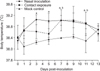
There were no prominent clinical features except in one inoculated dog (No. 3) that showed depressed activity with nasal discharge at 2 and 3 days post-inoculation. Two of four nasal swab samples from dogs in the nasal inoculation group were positive for the presence of viral RNA at 2 days post-inoculation (Table 1). In the contact-exposed group, one nasal swab sample was positive for viral RNA at 5 and 7 days post-inoculation. The three positive samples were confirmed by SPF egg re-isolation, and viral hemagglutinin gene sequences were observed to be the same as the inoculated sequence. However, there was no evidence of viral RNA in blood serum samples. Severe elevation of body temperature (> 40℃) did not develop in inoculated, contact-exposed, or mock control dogs (Fig. 1). However, mild body temperature elevations (> 39℃) developed in dogs in the nasal inoculation and contact-exposed groups. When body temperature following inoculation was compared with body temperature of mock control dogs, those in the inoculation group showed significantly elevated body temperatures on 3, 5, 7, and 11 days post-inoculation. Significant differences in body temperature between dogs in the contact-exposed and mock control groups were observed on 7 and 11 days post-inoculation. Average body temperatures above 39℃ developed on 3, 5, and 7 days post-inoculation in dogs in the nasal inoculation group and on days 7 and 9 in the contact-exposed dogs.
HI antibody titers against homologous antigen were detected in two of the four dogs with nasal inoculation (Nos. 2 and 3) and one of the four dog with contact exposure (No. 5). Two serum samples from dogs in the nasal inoculation group, one from a dog in the contact-exposed group showed seropositive results in the competitive ELISA. Serum from the mock control group exhibited no responses in the HI and ELISA tests (Table 2).
In marked contrast to other carnivores experimentally infected with HPAI H5N1 viruses [712], dogs experimentally infected with H5N8 did not present obvious clinical signs, other than a mild elevation in body temperature. Serological evidence presented in this study suggests that, when compared with other canine influenza virus infections, intranasal infection with the H5N8 virus does not induce a significant antibody response. This could be explained by inefficient viral replication due to a host species barrier [6]. However, early viral shedding found in two dogs in the intranasally inoculated group and two dogs in the contact-exposed group, and detection of antibodies in serum raises the possibility that, although it is much less efficient than the H5 subtype canine influenza, the H5N8 virus can be transmitted weakly between dogs without clinical sign [11]. Evidence of low antibody detection in serum was insufficient to prove that exposure to the H5N8 virus infects the host systemically.
Because of the intermingling between domestic poultry and other animals in Korea, as seen in live-bird markets or in small-scale backyard livestock operations [8], dogs are likely to have contact with poultry infected with avian influenza virus. Although the H5N8 virus does not seem to cross the host species barrier completely, several passages might result unexpected adaptation to ferret with only a few amino acid mutations [3]. Furthermore, adaptation of the virus could increase the possibility of recurrent infection from dogs to poultry [1].
In this experiment, although strong evidence for viral transmission and shedding between dogs was not demonstrated, the detection of virus in nasal swab and seroconversion results alerted us to doubt whether the H5N8 virus could cause a silent infection in dogs. Our observations suggest that, although the H5N8 virus does not seem to adapt fully to canine species, dogs should continue to be monitored as a species in which avian influenza virus may acquire adaptive changes, thereby enabling efficient replication and transmission in mammals.

 XML Download