Abstract
Swine vesicular disease (SVD) is a highly contagious viral disease that causes vesicular disease in pigs. The importance of the disease is due to its indistinguishable clinical signs from those of foot-and-mouth disease, which prevents international trade of swine and related products. SVD-specific antibody detection via an enzyme-linked immunosorbent assay (ELISA) is the most versatile and commonly used method for SVD surveillance and export certification. Inactivated SVD virus is the commonly used antigen in SVD-related ELISA. A recombinant SVD virus-like particle (VLP) was generated by using a Bac-to-Bac baculovirus expression system. Results of SVD-VLP analyses from electron microscopy, western blotting, immunofluorescent assay, and mass spectrometry showed that the recombinant SVD-VLP morphologically resemble authentic SVD viruses. The SVD-VLP was evaluated as a replacement for inactivated whole SVD virus in competitive and isotype-specific ELISAs for the detection of antibodies against SVD virus. The recombinant SVD-VLP assay produced results similar to those from inactivated whole virus antigen ELISA. The VLP-based ELISA results were comparable to those from the virus neutralization test for antibody detection in pigs experimentally inoculated with SVD virus. Use of the recombinant SVD-VLP is a safe and valuable alternative to using SVD virus antigen in diagnostic assays.
Swine vesicular disease (SVD) is a contagious viral disease of pigs caused by an enterovirus of the Picornaviridae family. SVD is characterized by a mild fever and vesicles on the coronary band, the bulbs of the heel, skin of the limbs, and less frequently on the snout, lips, tongue, and teats. The morbidity rate may be as high as 100%, but mortality is very low. Although SVD was not listed by the World Organization for Animal Health (OIE) after 2014, its clinical signs are indistinguishable from foot-and-mouth disease, which is one of the world's most widespread epizootic and highly contagious animal diseases. Therefore, rapid and accurate diagnosis of SVD is considered essential in countries that are free of vesicular diseases.
A competitive enzyme-linked immunosorbent assay (cELISA) is one of the serological tests used frequently for SVD surveillance and certification for export of swine and related products. In the cELISA, the binary ethyleneimine (BEI)-inactivated SVD virus (SVDV) was used as an antigen [2]. Whole virus antigen production is cumbersome and timeconsuming, and using noninfectious recombinant virus particles as a diagnostic antigen would avoid those drawbacks and help reduce the handling of live viruses.
SVDV is a non-enveloped virus with a single positive-stranded RNA genome containing a long open reading frame that encodes a polyprotein of 2,185 amino acids [7815]. The polyprotein has been divided into three regions: P1, P2, and P3. The P1 region encodes the viral capsid proteins of VP1, VP2, VP3, and VP4, whereas the P2 and P3 regions encode proteins involved in protein processing (2A, 3C, and 3CD) and genome replication. In cells infected with enteroviruses, the 3D sequence within 3CD allows efficient processing of P1 into VP0, VP3, and VP1 by the 3C part of the enzyme [920]. These polypeptides will self-assemble into empty capsid particles if viral RNA is not encapsulated [114]. The native virion consists of VP1, VP3, VP2, and VP4, the last two being cleaved from VP0. In contrast, VP0 is not cleaved in empty capsids, which allows in vitro construction of recombinant SVDV empty capsid particles.
Recombinant SVD virus-like particles (VLP) have been described previously [11]. However, the particles have not been fully characterized and their applications have not been fully explored. The use of SVD-VLP as an antigen for SVD isotype-specific antibody detection has never been reported. A major problem with current serological tests for SVD is the generation of false-positive results, so called singleton reactors (SR), which are commonly reported worldwide [310]. Singleton reactor sera are anti-SVDV antibody positive sera from pigs without a history of contact with SVDV [3]. Although the percentage of SR is low, it is a problem for international trade. It has previously been shown that the positive reaction is due to immunoglobulin (Ig) M antibodies [12]. Therefore, the detection of SVD-specific IgG and IgM may reduce the occurrence rate of SR [3].
In this study, a recombinant SVD-VLP was constructed by using a Bac-to-Bac baculovirus expression system. The properties of the baculovirus-expressed SVD-VLP were characterized. Applications of recombinant SVD-VLP as an antigen for the detection of antibodies against SVDV were evaluated in cELISA and isotype-specific ELISA.
All animal-related work was carried out in compliance with Canadian Council on Animal Care guidelines and was approved by the Animal Care Committee at the Canadian Science Centre for Animal and Human Health (approval No. A5972-01).
SVDV strain UK 27/72 was grown in swine testicular (ST) cells in alpha minimal essential medium (Wisent, Canada) supplemented with 2 mM L-glutamine, 50 µg/mL gentamycin, and 10% fetal bovine serum (FBS; Hyclone, USA). BEI inactivation of SVDV was performed as previously described [19]. The Sf9 (Spodoptera frugiperda) and High Five (Trichoplusia ni BTI-Tn-5B1-4) insect cells were cultured in a protein-free ESF 921 insect cell culture medium (Expression Systems, USA) and maintained as suspensions at 27℃.
Genomic RNA was extracted from amplified SVDV UK 27/72 with QIAzol and RNeasy mini kit (Qiagen, Canada) according to manufacturer's instructions. Primers used to amplify the P1 and 3CD regions of SVDV UK 27/72 were designed based on the genomic sequences from the GenBank database (accession No. X54521). The primers were designated as follows: P1F, 5′-ATTGGATCCATGGGAGCTCAAGTGTCA-3′, P1R, 5′-CCCAAGCTTCTAAGTGGTTTTCATGGTTGTTA-3′, and 3CDF, 5′-ATTCTCGAGATGGGTCCAGCGTTTGAGTTC-3′, and 3CDR, 5′-CGCGGTACCTTAAAAGGAGTCCAACCAC-3′. The incorporated Bam HI, Hind III, Xho I, and Kpn I restriction enzyme sites are underlined. The cDNAs of the P1 and 3CD regions were synthesized by using SuperScript III Reverse Transcriptase (Invitrogen, USA). The cDNAs were then used as templates for amplification of the full lengths of the P1 and 3CD regions by using an Expand High FidelityPLUS PCR system (Roche, USA). The amplified PCR products were purified by using a QIAquick gel extraction kit (Qiagen) and separately cloned into pCR 4-TOPO vector by using a TOPO TA cloning kit (Invitrogen).
To generate a recombinant baculovirus expressing both P1 and 3CD polyproteins, the P1 and 3CD regions were subcloned into the pFastBac Dual vector (Invitrogen) using the Bam HI and Hind III sites for P1 and the Xho I and Kpn I sites for 3CD in order to generate pFBD-P1-3CD. This cloning strategy positioned the P1 and 3CD regions for transcription from the baculovirus polyhedrin and p10 promoters, respectively. The dual clone was then used to generate a recombinant baculovirus as per the Bac-to-Bac system (Invitrogen). Briefly, purified pFBD-P1-3CD plasmid DNA was transformed into DH10BacTM Escherichia coli for generation of a recombinant bacmid by using site-specific transposition. The recombinant bacmid DNA containing SVDV P1 and 3CD regions was then used to transfect Sf9 insect cells to produce recombinant baculoviruses.
To optimize conditions for production of VLP in Sf9 or High Five cells, various multiplicities of infection (MOI) and harvest times were assessed. Expression analysis was performed in a 24-well tissue culture plate format. Sf9 or High Five cells were infected with various MOI for 1 h. After adsorption, the inoculum was removed, and the monolayers were washed and overlaid with insect cell culture medium ESF 921 (Expression Systems). At various times post-infection, cells were harvested and disrupted by freeze-thawing three times. Cell debris was removed by low-speed centrifugation, and supernatants were saved for later analysis. The yields of VLP were determined by double antibody sandwich ELISA (DAS-ELISA) and dot blot analyses. For DAS-ELISA, polystyrene microtiter plates were coated with anti-SVD rabbit sera at a dilution of 1:10,000 in carbonate buffer overnight at 4℃. All subsequent incubations were performed at 37℃ for 1 h, using 100 µL of reagent added to each well. Clarified supernatants from infected insect cells were two-fold diluted with diluent buffer consisting of 2% normal rabbit serum, 2% normal bovine serum (Sigma-Aldrich, USA), 0.004% phenol red, and 0.05% Tween-20 in 0.01 M phosphate-buffered saline (PBS) and added to each well. Inactivated SVDV particles and mock-infected insect cell lysates were used as positive- and negative-control antigens, respectively. Plates were then incubated with anti-SVDV guinea pig sera (1:10,000 diluted), followed by donkey anti-guinea pig IgG conjugated with horseradish peroxidase (HRP, 1:5,000 diluted; Jackson ImmunoResearch Laboratories, USA). Following the washing step, ortho-phenylenediamine dihydrochloride substrate (OPD; Sigma-Aldrich) was added to the plate, and sulphuric acid was used to stop the substrate/chromogen reaction. The optical density at 490 nm of each well was determined in an automated ELISA plate reader (Photometer Multiskan Reader; Labsystems, USA).
After optimization of expression of VLP in monolayer cultures of insect cells, large-scale expression was performed in shaker flasks. Cells were infected at an MOI of 5 plaque forming unit (PFU)/cell and harvested at 5 days post-infection. Cells were frozen and thawed three times and cell debris was removed by centrifugation at 5,000 × g for 30 min in a Beckman JLA-10500 rotor (Beckman Coulter, USA). Supernatants were collected and VLPs were precipitated by adding PEG 8000 (Sigma-Aldrich) to a final concentration of 7.5% stirring overnight at 4℃. VLPs were pelleted by centrifugation at 8,000 × g for 30 min in a Beckman JA-14 rotor (Beckman Coulter) and resuspended in Tris-NaCl buffer (50 mM Tris, pH 7.5, 150 mM NaCl). The VLP suspension was sonicated with addition of 0.1% of Triton X-100 and centrifuged at 2,543 × g for 15 min in a Beckman JA25.5 rotor (Beckman Coulter). The supernatant was collected for use in ELISA. For electron microscopy, the VLP were further purified by overlaying 30% sucrose cushion (6 mL) in an Ultra-Clear centrifuge tube (40 mL; Beckman Culter). The sucrose cushion was centrifuged in a Beckman Optima XL-100 K ultracentrifuge (Beckman Coulter) using a SW28 rotor at 89,527 × g for 2.5 h. The pellet was resuspended in Tris-NaCl buffer (50 mM Tris, pH 7.5, 150 mM NaCl).
The purified VLP underwent morphological examination via electron microscopy. The samples were mounted on 400-mesh formvar-coated nickel grids by means of the drop method [5], negatively stained with 2% phosphotungstic acid, pH 7.0, and examined by using a Philips CM120 transmission electron microscope (Philips, Holland).
The Sf9 cells were grown on glass slides and infected with the recombinant baculovirus at 0.1 MOI. At 24 h post-infection, infected cells were fixed with 10% buffered formalin phosphate (Fisher Scientific, USA) for 30 min at 37℃ and permeabilized with 20% acetone in PBS for 10 min at room temperature. The slides were blocked with 1× blocking buffer (Sigma-Aldrich) for 1 h at 37℃ and incubated with an in-house SVDV-specific Mab F44SVD [18] (1:100 in blocking buffer) for 1 h at room temperature. Slides were washed three times with PBS containing 0.05% Tween 20 and then incubated with Alexa Fluor 594 goat anti-mouse IgG (Invitrogen; 1:400 diluted in PBS) for 1 h at room temperature. After washing and air drying, slides were mounted with SlowFade antifade reagent (Invitrogen) and covered with coverslips. Microscopy was performed by using an Olympus FV1000 confocal microscope (Olympus, USA). Image acquisition and analysis were performed by using Fluo-View software (Olympus).
SVD-VLP, mock-infected Sf9 cell lysates, and SVDV grown in ST cells prepared in the same manner as SVD-VLP were separated with 10% NuPAGE Novex Bis-Tris gels (Invitrogen) followed by protein transfer to nitrocellulose membranes using a iBlot gel transfer device (Invitrogen). Membranes were incubated in PBS-T with 5% skim milk at room temperature for 1 h then blotted with anti-mouse SVD serum (1:500) in blocking buffer overnight at 4℃, followed by incubation with HRP-conjugated anti-mouse antibody (1:2,000; Jackson ImmunoResearch Laboratories) for 1 h at room temperature. Blots were developed by using 3, 3′-diaminobenzidine (Sigma-Aldrich).
SVD-VLP and whole virus samples were electrophoresed and stained with imperial protein stain (Fisher Scientific). Candidate protein bands were excised from the gel. The gel slices were washed four times at room temperature, first with 100 mM ammonium bicarbonate and then with three consecutive washes of a 100 mM ammonium bicarbonate/50% acetonitrile solution. Gel slices were dehydrated with 100% acetonitrile. Proteins were reduced by incubation with 10 mM of dithiothreitol dissolved in 100 mM ammonium bicarbonate (45 min at 57℃). Protein alkylation was performed by incubation with 55 mM iodoacetamide dissolved in 100 mM ammonium bicarbonate (45 min at room temperature in the dark). Gel pieces were then subjected to the acetonitrile/ammonium bicarbonate wash cycle once and allowed to shrink with acetonitrile. Sequencing grade trypsin (Promega, USA; 5 ng/L) dissolved in 100 mM ammonium bicarbonate was added into the gel to digest the peptide overnight at 37℃. Tryptic peptides were extracted from the gel slices by washing with 0.1% trifluoroacetic acid and then in 0.1% trifluoroacetic acid/50% acetonitrile solutions. The tryptic peptide extracts were lyophilized and stored at −80℃ before analysis by performing liquid chromatography tandem mass spectrometry (LC-MS/MS) on an ABI Qstar Pulsar instrument with an LC Packings nano LC system (Applied Biosystems, USA). The Mascot (version 2.2; Matrix Science, UK) search engine was used to search the NCBI database with the obtained MS/MS data for protein identification.
The monoclonal antibody (Mab) 5B7 [2] is the OIE reference antibody used in the SVD cELISA. Both 5B7 catching Mab and 5B7 Mab conjugated with peroxidase were purchased from Istituto Zooprofilattico Sperimentale della Lombardia e dell'Emilia Romagna, Italy.
The serum samples from experimentally inoculated pigs were generated at the National Centre for Foreign Animal Disease (Canada). Pigs 1 to 5 were inoculated with SVDV strain POR/1/2003 and pigs 6 to 10 were inoculated with SVDV strain UKG/27/1972. All pigs were inoculated through intradermal injection with a high dose (> 107 TCID50/pig) into the bulb of the heel and nostrils. Sera were collected at 0, 1, 2, 3, 4, 5, 7, 9, 11, 14, 21, and 28 days post-inoculation (dpi). The negative pig sera (n = 739) were obtained from SVD-free farms in Canada. A panel of SVD reference sera obtained from the Pirbright Institute, UK contained a normal pig serum (S01), one strongly positive serum and four weak positive pig sera (S03-S06) collected from pigs infected with SVDV.
The virus neutralization test (VNT) was performed by using SVD UK 27/72 and ST cells [17]. Serum samples were heat inactivated (56℃ for 30 min) before testing. Sera with titers ≥ 1.8 log10 (1:64) were considered positive.
The goat anti-pig IgM antibody (Bethyl, USA) diluted 1:2,000 in 0.06 M carbonate buffer was coated onto microtiter plates overnight at 4℃. Plates were washed, blocked with 1×Sigma blocking buffer (Sigma-Aldrich), and incubated for 1 h at 37℃. Plates were washed, and sera were diluted 1:200 in blocking buffer, added to four consecutive wells, and incubated for 1 h at 37℃. After washing, SVD-VLP and Sf9 negative-control antigen (1:1,500), or SVDV-antigen (SVDV-Ag) and ST cell culture supernatant, a negative control (1:5) were added to duplicate wells for each serum and incubated for 1 h at 37℃. After washing, HRP-Mab 5B7 diluted in blocking buffer (1:500) was added to wells containing VLP or virus antigen and incubated for 1 h at 37℃. Then OPD substrate solution was added and the mixture incubated for 15 min at room temperature. The reaction was stopped by addition of 2.0 M sulphuric acid, and the optical intensity (OD) was determined photometrically. The isotype ELISA results were expressed as P/N ratios, defined as the average difference in measured OD values between duplicate wells of positive- (P) and negative- (N) control antigens for each serum sample. The cutoff P/N ratio was determined based on the average P/N ratio of the negative sera plus three standard deviations. A P/N ratio ≥ 2.4 was considered positive.
Antibody 5B7 was diluted 1:300 in 0.06 M carbonate buffer, pH 9.6, and coated onto microtiter plates overnight at 4℃. Plates were washed, blocked with 1×Sigma blocking buffer, and incubated for 1 h at 37℃. After washing, SVD-VLP and Sf9 negative-control antigen, diluted 1:1,500 each in blocking buffer, or whole virus antigen and ST negative-control antigen, diluted 1:5 each in blocking buffer, were added to duplicate wells of a microtiter plate, and incubated for 1 h at 37℃. After washing, serum samples diluted 1:200 in blocking buffer were added and incubated for 1 h at 37℃, then goat anti-swine IgG-HRP (1:2,000; Jackson ImmunoResearch Laboratories) was added and incubated for 1 h at 37℃. After a final wash, the OPD substrate solution was added, and OD was determined as previously described. A P/N ratio ≥ 3.4 was considered positive.
A cytoplasmic extract was prepared from insect cells infected with a recombinant baculovirus designed to express SVD-VLP. The expression of SVD capsid proteins was confirmed by performing an ELISA using an anti-SVDV rabbit serum as the capture antibody and anti-SVDV guinea pig sera as the detecting antibody (data not shown). Assembly of capsid proteins to form VLP was demonstrated by a dot blot assay using the Mab F44SVD. Expression of SVD-VLP was 2- to 3-fold higher in Sf9 cells than in High Five cells. Peak expression in Sf9 cells was observed at 5 dpi with MOI of 5 or 10 (data not shown).
The SVD-VLP was examined using a SVD-specific Mab F44SVD in an immunofluorescent assay. This Mab binding site was conformational and located in the VP1 region by using the Mab mutant selection method (data not shown). The infected cells stained red, indicating VP1 expression (panel A in Fig. 1). In order to morphologically examine VLP, the precipitated and purified particles were negatively stained, and visualized via electron microscopy. The VLP were detected and morphologically resembled native virions with a diameter of approximately 25 nm (panel B in Fig. 1).
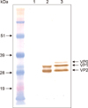
SDS-PAGE and western blotting analyses were used to provide evidence that SVD-VLP contain SVDV capsid polypeptides. Western blotting analysis of the SVDV protein showed that two significant bands represent VP1 (~32 kDa), and a band with a molecular weight between 26 to 28 kDa corresponded to both VP2 and VP3 (lane 2 in Fig. 2). The individual protein band for VP2 and VP3 were unable to be resolved because the molecular weight difference between them is small. Western blotting results for SVD-VLP revealed three bands with molecular weights ~36 kDa, ~32 kDa, and ~26 kDa, representing VP0, VP1, and VP3, respectively (lane 3 in Fig. 2). VP0 is the uncleaved precursor of VP2 and VP4.
A proteomic approach was performed to identify the sequences of the protein bands. The stained protein bands were excised, digested, and analyzed by LC-MS/MS. The identified peptide SMPALNSPSAEECGYSDR (Fig. 3) contained the VP2 cleavage site, which was evidence that SVD-VLP contains the uncleaved polypeptide VP0. A total of four peptides from the VP1 band and four peptides from the VP3 band were identified (Fig. 3).
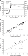
The suitability and performance of the SVD-VLP as an antigen in the SVD cELISA was assessed by testing known SVDV-negative sera (n = 468) and positive sera of foot-and-mouth disease virus (FMDV, n = 40) and vesicular stomatitis virus (VSV, n = 2). The determined diagnostic specificities were 97.2% for the SVD-VLP and 97.4% for the SVDV antigen, based on a predetermined cutoff of 50% inhibition in the cELISA. None of the FMDV- or VSV-positive sera demonstrated positive results when using either the SVD-VLP or the SVDV-Ag in the cELISA. The SVD-VLP were evaluated by using sera collected from experimentally inoculated pigs and compared with those of SVDV-Ag in the cELISA. An antibody response first appeared at 5 dpi, and all pigs demonstrated seroconversion at 9 dpi with both antigens (panel A in Fig. 4). The correlation coefficient between the SVDV-Ag and SVD-VLP cELISA results was 0.96 with p < 0.0001 for 120 serum samples collected from 10 pigs (panel B in Fig. 4). The cELISA was compared with the gold standard serological test (i.e., the VNT) by using 24 serial blood samples (5, 7, 9, and 28 dpi) from six of 10 pigs. All pigs demonstrated positive responses at 7 dpi in the VNT (panel A in Fig. 4). The results indicate that the VNT detected seroconversion two days earlier than the cELISA. The correlation coefficient between the SVD-VLP cELISA and VNT was 0.64 with p < 0.0001 (panel C in Fig. 4).
Determination of IgG and IgM may be of great value in reducing the occurrence of SR. Thus, SVD-VLP-based isotype-specific ELISAs were developed for the detection of SVDV-specific IgM and IgG. The diagnostic specificity of the SVD-VLP IgM-specific ELISA was 98.16% based on a P/N ratio cutoff value of less than 2.4 following testing of 327 SVDV-negative sera. Sera from SVDV experimentally infected pigs were examined by using the IgM-specific ELISA. The IgM response first appeared at 5 dpi, and all pigs showed positive response at 9 dpi. The IgM response peaked at 14 dpi and then declined near the end of the experiment when using either SVD-VLP or SVDV-Ag (panel A in Fig. 5). The correlation coefficient between the SVDV-Ag and the SVD-VLP IgM-specific ELISAs was 0.87 with p < 0.0001 (panel B in Fig. 5).
The diagnostic specificity of the SVD-VLP-based IgG ELISA was determined by using SVDV-negative sera (n = 739). The diagnostic specificity was 99.19% based on a P/N ratio cutoff value of less than 3.4. The sera from experimentally infected pigs were tested using both antigens in the IgG-specific ELISA. The antibody responses appeared positive at 5 dpi and all pigs demonstrated positive results at 9 dpi (panel A in Fig. 6). The IgG antibody responses steadily increased to the end of the experiment at 28 dpi. The correlation coefficient between the SVDV-Ag and the SVD-VLP IgG-specific ELISAs was 0.72 with p < 0.0001 (panel B in Fig. 6). A panel of SVD reference pig sera was evaluated by using VLP-based IgM and IgG ELISAs. One negative serum and 5 positive sera were correctly identified by the SVD-VLP-based IgM and IgG ELISAs (Table 1).
To evaluate IgG and IgM isotype-specific ELISAs for the reduction of the occurrence of SR, 59 reactor sera used in the SVDV-Ag cELISA were examined by using the IgG and IgM isotype-specific ELISAs in parallel with SVD-VLP and SVDV as the antigens. Four of the 59 (6.8%) sera were positive for IgG when using SVD-VLP, while 22 of 59 (37.3%) were positive when using SVDV-Ag. Fifteen of 59 (25.4%) sera were positive in the IgM-specific ELISA when using SVD-VLP and 24 of 59 (40.7%) sera were positive when using SVDV-Ag.
A recombinant baculovirus containing the SVDV capsid protein P1 region and the viral proteinase 3CD region was generated and shown to induce the synthesis of SVD-VLP. The SVD-VLP morphologically resembled authentic SVD virions under electron microscopic examination. This is a new addition to a previous report [11], in which Ko et al. [11] were unable to detect the SVD structural capsid proteins (VP0, VP1, and VP3) by performing western immunoblotting. In the current study, the capsid proteins of the SVD-VLP were successfully detected in western blotting by using a polyclonal anti-SVD serum that recognized linear epitopes. Western blotting analysis of SVD-VLP showed three bands corresponding to the molecular weights of VP0, VP1, and VP3, respectively, while the native virion consists of VP1, VP2, VP3, and VP4. Mass spectrometric analyses of corresponding bands excised from the SDS-PAGE gel confirmed that the SVD-VLP consisted of VP1, VP3, and uncleaved VP0. One of identified peptides, DTPFIKQDNFFQ, confirmed previous findings that VP1 is cleaved at the FQ/GP junction [7].
The expression of SVDV capsid proteins was detected by using a SVDV-specific Mab in dot blot and immunofluorescent assays. This Mab's binding site was conformational and located at the VP1 capsid protein of SVDV. The results indicated that the conformational epitope on the VP1 protein of the native virion was retained on the recombinant SVD-VLP. Meanwhile, the binding of Mab 5B7 [13] to the SVD-VLP proved that the antigenic epitope on the VP2 protein of the native SVDV was reserved on the recombinant SVD-VLP. Based on all of the characterization results for SVD-VLP, the SVD-VLP has potential to be used as an antigen to replace the native virus in serodiagnosis.
Currently, the VNT is considered the gold standard and has remained the most reliable test in serodiagnosis. However, both the VNT and ELISA are OIE-accepted standard tests for SVD antibody detection [17]. The 5B7 MAb cELISA is a reliable technique for detecting SVD antibodies and is recommended by the OIE [26]. Thus, the VLP-based cELISA results were compared with those from the 5B7 MAb cELISA in this study. The diagnostic specificities are 97.2% for the SVD-VLP cELISA and 97.4% for the SVDV-Ag cELISA. When testing sera collected from experimentally inoculated pigs with the VLP-cELISA, antibody response kinetics were similar to those observed with VNT and SVDV-Ag cELISAs. However, the VNT results detected seroconversion two days earlier than the cELISAs using both antigens (SVDV-Ag and SVD-VLP). It is possible that the VNT detects neutralizing antibody activity and ELISA may detect different antibody subsets [16].
Similar to the SVDV-Ag results, a low percentage of the negative sera showed false-positive results using SVD-VLP in the cELISA. These false-positive sera cannot be ruled out completely by the golden standard VNT with up to approximately 50% of these sera estimated as positive by the VNT [17]. The origins of SR remain unclear. Serological cross-reactivity with SVDV may due to antibodies raised from other virus infections in sera. Identification of antibody isotypes presented in SVDV-positive sera can help identify SRs because sera from uninfected pigs usually contain IgM, but not IgG [23]. Thus, isotype-specific ELISAs were developed in order to reduce the false-positive rates and serve as complementary tests for cELISA. The capture IgM-specific ELISA described in this study is similar in configuration to that reported by Dekker et al. [4], except that the polyclonal anti-pig IgM was used instead of monoclonal anti-pig IgM as the capture antibody. The IgG-specific ELISA procedure was similar to that reported by Brocchi et al. [2], where polyclonal anti-pig IgG was used as the detecting antibody. Using the VLP as the antigen in IgM and IgG ELISAs demonstrated accurate results for a panel of SVD reference sera. The isotype-specific SVD-VLP ELISAs detected SVDV-specific antibodies in all positive sera from experimentally infected pigs, and the results were significantly correlated with those obtained from the isotype-specific ELISA and the cELISA using SVDV-Ag. The earliest IgM response was detected at 5 dpi, which is 2 days later than previous reported by Brocchi et al. [2] and Dekker et al. [4]. This later development of an IgM response may be correlated with a lack of clinical signs observed from experimentally infected pigs. In the current study, IgM responses peaked between 11 to 14 dpi and then started to decline, while the IgG responses were detected as early as 5 dpi and steadily increased toward the end of experiment.
The 59 SR sera previously identified using the SVDV-Ag cELISA were tested using the IgG and IgM ELISAs with SVD-VLP and SVDV-Ag as the antigens. Of the 59 SR sera, 6.8% and 37.3% demonstrated IgG positive results using the SVD-VLP and SVDV-Ag, respectively. Whereas 25.4% and 40.7% of the SR sera demonstrated IgM positive results using SVD-VLP and SVDV-Ag, respectively. The results are in agreement with those previously reported in studies that showed that identification of antibody isotypes, especially IgG, would reduce the SR rate [3]. The results from this study indicate that replacing the whole SVDV antigen with SVD-VLP in isotype-specific ELISAs could further reduce the SR percentage, which would benefit international trade in swine and related products. Currently, the recombinant SVD-VLP-based isotype-specific ELISAs and cELISA are under full evaluation with more serum samples.
The recombinant SVD-VLP is morphologically and antigenically similar to the native SVDV, as shown by the results of immunofluorescent, western blot, and MS/MS analyses. The recombinant SVD-VLP and the SVD-Ag showed comparable results in cELISA and isotype-specific ELISAs. Thus, the SVD-VLP can be used as an alternative antigen for SVD serodiagnosis.
Figures and Tables
 | Fig. 1Swine vesicular disease (SVD) virus-like particles (VLP) detection using fluorescence (A) and electron microscopy (B). (A) Sf9 cells were grown on glass slides, infected with recombinant baculovirus at multiplicities of infection 0.1, and harvested at 24 h post-infection. SVD-VLP were stained with the Mab F44-SVD and then with Alexa Fluor 594 (red; Invitrogen). (B) Sf9 cells were infected with recombinant baculovirus and then lysed. SVD-VLP were purified, negatively stained, and examined via transmission electron microscopy. Scale bar = 100 nm. |
 | Fig. 2Western blot analysis of swine vesicular disease virus (SVDV) and swine vesicular disease (SVD) virus-like particles (VLP) capsid proteins. The mock-infected Sf9 cell lysates (lane 1), purified SVDV (lane 2), and SVD-VLP (lane 3) were separated in SDS-PAGE and transferred to nitrocellulose membranes. The SVD capsid proteins were probed with an anti-mouse SVD serum and visualized with horseradish peroxidase-conjugated goat anti-mouse antibody and then 3, 3′-diaminobenzidine. |
 | Fig. 3Liquid chromatography tandem mass spectrometry identification of swine vesicular disease virus-like particles capsid proteins. The protein bands corresponding to positions indicated in Fig. 2 were separately excised from SDS-PAGE, destained, processed with trypsin, and identified by mass spectrometry peptide fingerprinting as the VP0, VP1, and VP3 proteins. Fig. 3 shows the amino acid sequence of the SVD virus capsid polyprotein and indicates the 9 unique tryptic peptides identified within the protein sequence. Cleavage sites are shown by arrows. |
 | Fig. 4Comparison of swine vesicular disease virus (SVDV) antigen (Ag) competitive enzyme-linked immunosorbent assay (cELISA), swine vesicular disease (SVD) virus-like particles (VLP) cELISA, and virus neutralization test (VNT) results for SVD-specific antibody detection. The sera were collected at different times in the infection course of ten pigs experimentally inoculated with SVDV. (A) Results are expressed as means of percent inhibitions ± SD from ten pigs for cELISA. Samples at 5, 7, 9, and 28 dpi from six pigs were analyzed using the VNT. (B) Scatter plot representing the percentage inhibition in SVDV-Ag cELISA vs. SVD-VLP cELISA for 120 serial blood samples from ten pigs. The line is fitted by linear regression (r2 = 0.96, p < 0.0001). (C) Scatter plot representing the percentage inhibition in SVD-VLP cELISA vs. VNT titers for 24 serial blood samples from six pigs. The line is fitted by linear regression (r2 = 0.64, p < 0.0001). |
 | Fig. 5Swine vesicular disease (SVD) immunoglobulin (Ig) M-specific antibody detection using swine vesicular disease virus (SVDV) antigen (Ag) and SVD virus-like particles (VLP) in IgM-specific enzyme-linked immunosorbent assay (ELISA). The sera were sequentially collected from ten pigs experimentally inoculated with SVDV. (A) Results are expressed as means of P/N absorbance ratio ± SD from 10 pigs. (B) Scatter plot representing the P/N ratio in SVDV-Ag-IgM ELISA vs. SVD-VLP-IgM ELISA for 120 serial blood samples from ten pigs. The line is fitted by linear regression (r2 = 0.87, p < 0.0001). |
 | Fig. 6Swine vesicular disease (SVD) immunoglobulin (Ig) G-specific antibody detection using swine vesicular disease virus (SVDV) antigen (Ag) and SVD virus-like particles (VLP) in IgG-specific enzyme-linked immunosorbent assay (ELISA). The sera were sequentially collected from 10 pigs experimentally inoculated with SVDV. (A) Results are expressed as means of P/N absorbance ratio ± SD from ten pigs. (B) Scatter plot representing the P/N ratio in SVDV-Ag-IgG ELISA vs. SVD-VLP-IgG ELISA for 120 serial blood samples from ten pigs. The line is fitted by linear regression (r2 = 0.72, p < 0.0001). |
Acknowledgments
This work was supported by Canadian Food Inspection Agency (WIN-1-1103). The authors thank Lynn Burton for the expert electron microscopy assistance, Dr. Garrett Westmacott and David Lee, Proteomics Core Facility, Public Health Agency of Canada for the mass spectrometry analyses, and Dr. Soren Alexandersen and Dr. John Copps for critical review of the manuscript.
References
1. Basavappa R, Syed R, Flore O, Icenogle JP, Filman DJ, Hogle JM. Role and mechanism of the maturation cleavage of VP0 in poliovirus assembly: structure of the empty capsid assembly intermediate at 2.9 Å resolution. Protein Sci. 1994; 3:1651–1669.

2. Brocchi E, Berlinzani A, Gamba D, De Simone F. Development of two novel monoclonal antibody-based ELISAs for the detection of antibodies and the identification of swine isotypes against swine vesicular disease virus. J Virol Methods. 1995; 52:155–167.

3. De Clercq K. Reduction of singleton reactors against swine vesicular disease virus by a combination of virus neutralisation test, monoclonal antibody-based competitive ELISA and isotype specific ELISA. J Virol Methods. 1998; 70:7–18.

4. Dekker A, van Hemert-Kluitenberg F, Baars C, Terpstra C. Isotype specific ELISAs to detect antibodies against swine vesicular disease virus and their use in epidemiology. Epidemiol Infect. 2002; 128:277–284.

5. Hammond GW, Hazelton PR, Chuang I, Klisko B. Improved detection of viruses by electron microscopy after direct ultracentrifuge preparation of specimens. J Clin Microbiol. 1981; 14:210–221.

6. Heckert RA, Brocchi E, Berlinzani A, Mackay DK. An international comparative analysis of a competitive ELISA for the detection of antibodies to swine vesicular disease virus. J Vet Diagn Invest. 1998; 10:295–297.

7. Inoue T, Suzuki T, Sekiguchi K. The complete nucleotide sequence of swine vesicular disease virus. J Gen Virol. 1989; 70:919–934.

8. Inoue T, Yamaguchi S, Kanno T, Sugita S, Saeki T. The complete nucleotide sequence of a pathogenic swine vesicular disease virus isolated in Japan (J1'73) and phylogenetic analysis. Nucleic Acids Res. 1993; 21:3896.

9. Jore J, De Geus B, Jackson RJ, Pouwels PH, Enger-Valk BE. Poliovirus protein 3CD is the active protease for processing of the precursor protein P1 in vitro. J Gen Virol. 1988; 69:1627–1636.

10. Karakas M, Palfi V. [Diagnostic difficulties during the serological examination of swine vesicular disease]. Magy Allatorvosok Lapja. 1996; 51:595–597. Hungarian.
11. Ko YJ, Choi KS, Nah JJ, Paton DJ, Oem JK, Wilsden G, Kang SY, Jo NI, Lee JH, Kim JH, Lee HW, Park JM. Noninfectious virus-like particle antigen for detection of swine vesicular disease virus antibodies in pigs by enzyme-linked immunosorbent assay. Clin Diagn Lab Immunol. 2005; 12:922–929.

13. Nijhar SK, Mackay DK, Brocchi E, Ferris NP, Kitching RP, Knowles NJ. Identification of neutralizing epitopes on a European strain of swine vesicular disease virus. J Gen Virol. 1999; 80:277–282.

14. Racaniello VR. Picornaviridae: the viruses and their replication. In : Knipe DM, Howley PM, editors. Fields Virology. 5th ed. Philadelphia: Lippincott Williams & Wilkins;2007. p. 795–838.
15. Seechurn P, Knowles NJ, McCauley JW. The complete nucleotide sequence of a pathogenic swine vesicular disease virus. Virus Res. 1990; 16:255–274.

16. Vratskikh O, Stiasny K, Zlatkovic J, Tsouchnikas G, Jarmer J, Karrer U, Roggendorf M, Roggendorf H, Allwinn R, Heinz FX. Dissection of antibody specificities induced by yellow fever vaccination. PLoS Pathog. 2013; 9:e1003458.

17. World Organisation for Animal Health (OIE). Swine vesicular disease. OIE Biological Standards Commission. Manual of Diagnostic Tests and Vaccines for Terrestrial Animals. Volume 2:6th ed. Paris: Office International des Epizooties;2008. p. 1139–1145.
18. Yang M, Clavijo A. Monoclonal antibody against swine vesicular disease virus. Hybridoma. 2008; 27:325.


 PDF
PDF ePub
ePub Citation
 Print
Print


 XML Download