

 Citation
 Print
Print


Introduction
While numerous studies have demonstrated a misbalance of pulmonary hemostasis and fibrinolysis in various forms of pulmonary disease in humans, little related data has been reported for the horse. Respiratory diseases, in particular, recurrent airway obstruction (RAO) and inflammatory airway disease (IAD), affect a large number of racing and performance horses and may cause exercise insufficiency; therefore, such diseases can have a high impact on the career of a sport horse. In chronic pulmonary disease, remodeling results in a decreased airway lumen, increased smooth muscle mass, peribronchial fibrosis, epithelial cell hyperplasia, and impaired airway function [2427]. Fibrosis formation is the ultimate result of a misbalance between pulmonary hemostasis and fibrinolysis, which is the result of procoagulatory factors favoring hemostasis, downregulation of fibrinolysis, or a combination of both. Regulation of remodeling may be critical for developing new therapeutics and managing respiratory diseases [26].
Previous authors [1718] have reported that the cell-free supernatant of respiratory secretions from horses affected by RAO may induce coagulation of plasma. This procoagulatory activity was associated with the severity of the respiratory disease and the neutrophil percentages in tracheobronchial aspirate (TBA) and bronchoalveolar lavage fluid (BALF). As shown for various forms of pulmonary disease in humans [192140], in the horse, the procoagulatory state is a result of increased tissue factor-factor VII complex activity [1718] and, therefore, the extrinsic coagulation pathway.
Fibrin and its derivation products may have a central role in equine pulmonary disease. Increases in soluble fibrinogen derivates are present in the majority of cell-free supernatants of respiratory secretions from horses suffering from chronic pneumopathies, which again is associated with neutrophil percentages and severity of disease [45]. Based on immunohistochemical analysis, fibrin and fibrinogen were observed in TBAs and in histologic preparations of thickened alveolar septae and bronchial mucus accumulations. These results were observed in cases of RAO as well as in cases with chronic broncho-interstitial and granulomatous interstitial pneumopathies [45].
Although not directly coagulation and fibrinolysis associated, serum amyloid A (SAA) was also evaluated as part of the present study. As fibrinogen is not only a product of coagulation but is also an acute-phase protein, we decided to evaluate SAA as it is another parameter known for its sensitivity to inflammatory processes [232534]. SAA has been reported to be involved in pulmonary inflammation in humans, and increased SAA concentrations are present in asthma and chronic obstructive pulmonary disease (COPD) [123032]. In addition, reported increased SAA concentrations in plasma of horses affected by RAO. To our knowledge, SAA has not been evaluated in equine BALF.
In addition, there are no reports of studies focusing on equine pulmonary fibrinolysis, although some information is available for other organ systems. In colic, sepsis, and septic and non-septic arthritis, there is evidence of alterations in coagulation. Moreover, changes in fibrinolysis have been reported in humans [382936]. The affected parameters may be measured systemically in plasma, but local changes in fluids from affected body cavities can be even more pronounced, such as in the peritoneal fluid of horses with colic or synovia in septic and non-septic arthritis [91135]. Therefore, in the present study, we examined BALF to compare markers of hemostasis and fibrinolysis in different equine chronic pneumopathies.
In dogs, pulmonary fibrinolysis associated with micro thromboembolism caused by heartworm infection has been studied, and the results showed that D-dimer deposits in lungs and kidneys and increased plasma values of D-dimers were associated with pulmonary thromboembolism and microfilaremic status [5].
A study in cats investigated the effects on lungs of inhibiting the fibrinolytic system by using tranexamic acid. Inhibition of the fibrinolytic system appears to have initiated emphysematous alterations, alveolar wall destruction, and collagen accumulation possibly by causing microthromboses leading to mechanical blockage and ischemic changes, or by causing secondary fibrinolysis as a result of fibrin degradation products affecting local plasminogen activators and proteases. An injury-repair process also appears to have occurred [20].
Pulmonary hemostasis and fibrinolysis has been studied extensively in mice, and mouse models have been used for research focusing on human asthma and COPD. In mouse, proteins of the hemostasis and fibrinolysis systems have been shown to be involved in the pathomechanisms related to airway hypersensitivity. After inhalation of an aerosol containing fibrinogen and followed by thrombin, mice were found to be hypersensitive in a methacholine provocation test compared to the sensitivity in mice after inhalation of saline [44]. The presence of thrombin, fibrinogen, and fibrin in the airways of asthmatic subjects indicates inhibition of fibrinolysis as well as increased hemostatic activity [3344]. Yuda et al. [46] found increased thrombin and soluble tissue factor levels in BALF of exacerbated mice after ovalbumin challenge. Treatment with anticoagulant protein C inhibited the type 2 T helper cells mediated immune response and airway obstruction.
Coagulation factor Xa has a central role in pulmonary remodeling. Increased concentrations of factor Xa mRNA have been observed in pulmonary tissue and BALF of mice after allergen challenge, and the application of a factor Xa inhibitor has resulted in decreased respiratory mucosa thickness and collagen deposition in pulmonary tissue. Factor Xa-dependent mucin production has also been reported [39].
The aim of this study was to compare clinical and cytological data with fibrinogen, SAA, plasminogen activator inhibitor-1 (PAI-1), and D-dimer concentrations between healthy horses and different groups of horses with chronic pneumopathy including horses with RAO, IAD, and chronic interstitial pneumopathy (CIP). The study was undertaken to evaluate the possibility of a misbalance between the two branches of hemostasis: coagulation and fibrinolysis.
Materials and Methods
Preparticipation examination
Horses were examined for 3 successive days from the day of admission until date of discharge. A total of 61 horses (height 154 ± 12 cm, age 12 ± 5 years, bodyweight 469 ± 96 kg) were examined, of which 15 had no clinical signs or history of respiratory disease (controls) and 46 had presented to the Equine Clinic of the Free University of Berlin with a history of chronic lower airway disease. Sampling of horses affected by respiratory disease was not classified as animal experimentation by the State Office of Health and Social Affairs Berlin, but sampling of control horses was approved by that group (reference No. L0294/13). In addition, owners gave permission to involve their horses in the study.
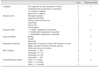
A preparticipation examination was performed and included clinical examination of the respiratory tract, blood gas analysis, endoscopy, and cytology of BALF. A clinical scoring system recommended by an international workshop was used (Table 1) [153137]. The scoring system was modified by including the determination of neutrophil percentages in BALF instead of TBAs.
The experimental groups were established as follows: (1) Control group, no history of respiratory disease, clinical score < 2, no tracheal secretions, low cellular density and neutrophils ≤ 10% in BALF, alveolar-arterial gradient (AaDO2) ≤ 7 mmHg, and exclusion of acute signs of infection (leukocytosis, fever, depression); (2) RAO group, history of recurrent cough or dyspnea, clinical score > 6, high amount or viscosity of tracheal secretion, high cellular density and neutrophils ≥ 25% in BALF, AaDO2 > 7 mmHg, and exclusion of acute signs of infection (leukocytosis, fever, depression), as previously described [37]. (3) IAD group, history of cough or exercise insufficiency, clinical score 2–6, low to moderate amount or viscosity of tracheal secretions, increased cellular density and neutrophils ≥ 10% or mast cells ≥ 2% or eosinophils ≥ 0.1% in BALF, AaDO2 > 7 mmHg, and exclusion of acute signs of infection (leukocytosis, fever, depression), as previously described [10]; and (4) CIP group, history of exercise insufficiency, clinical score 2–6, low to moderate amount or viscosity of tracheal secretions, increased cellular density and ratio of macrophages to neutrophils ≥ 2.6:1 in BALF, increased interstitial opacity of thoracic radiographs, and exclusion of acute signs of infection (leukocytosis, fever, depression), as previously described [12].
BALF collection and processing
Following endoscopy, 20 mL of 2% lidocaine (bela-pharm, Germany) were infused around the carina of the trachea. Bronchoalveolar lavage (BAL) was performed by using a 300 cm silicone BAL catheter (Smiths Medical, USA), which was passed nasally into the distal respiratory tract and wedged into the bronchus by expansion of an air balloon. Five hundred milliliters of pre-warmed phosphate buffered saline (Lonza, Belgium) were infused into the bronchi through a BAL catheter as recommended by the International Workshop on Equine Chronic Airway Disease [37] and immediately aspirated.
After aspiration, the recovered sample volume was recorded and divided into two portions, one for cytological examination and the second for biochemical analysis. For cytological examination, the samples were centrifuged at 250 × g for 10 min, then a direct smear was obtained, air-dried, stained by using the May-Grünwald Giemsa method (Sigma-Aldrich Germany), and 500 cells were examined via oil immersion microscopy at 1,000 × magnification. For biochemical analysis, BALF was centrifuged in a table top refrigerated centrifuge (Hermle Z326K; HERMLE Labortechnik, Germany) at 250 × g for 10 min at 4℃. The cell-free supernatant was collected and stored at −80℃ until assayed. Samples were transported to external laboratories on dry ice.
Group classification
Based on the results of the preparticipation examinations, the 61 horses presented for participation in this study were grouped as follows: Control group, 15 horses (24.5%); RAO group, 18 (29.5%) horses; IAD group, 14 (23.0%) horses; and CIP group, 11 (18.0%) horses. Three horses (5.0%) suffered from acute respiratory infection and were excluded from the study. Among the remaining 58 horses, there were insignificant differences in the severity of dyspnea, amount and viscosity of tracheal secretions, and AaDO2 estimates among the disease groups. The overall results of the clinical examinations are presented in Table 2. Percentages of macrophages, lymphocytes, mast cells, and neutrophils were similar among the groups, except for significant differences between the control and RAO groups in macrocyte, lymphocyte, and neutrophil percentages and between control and IAD groups in neutrophil percentages (Table 3).
Evaluation of fibrinogen
Fibrinogen concentrations in the 58 horses were measured in samples of BALF supernatant by using automatized immune turbidimetry (C701 module, Cobas 8000 series; Roche Diagnostics, Germany). The specific limit of fibrinogen detection was 0.001 g/L.
Evaluation of SAA
SAA was evaluated in BALF supernatant samples from 58 horses by using a multispecies sandwich enzyme-linked immunosorbent assay (ELISA) kit (Phase SAA Assay; Tridelta Development, Ireland). All BALF supernatant samples were tested undiluted and the limit of SAA detection was 0.01 µg/mL.
Evaluation of D-dimers
D-dimer concentrations in BALF supernatant were measured in samples from 58 horses by using an automatized analyzer (Sysmex CA-1500 system; Sysmex, Germany) and performing immune turbidimetry (INNOVANCE D-dimer test; Siemens Healthcare Diagnostics, Germany). The limit of D-dimer detection was 0.19 mg/L fibrinogen equivalent units. As most samples were below the limit of detection, 3 samples from control horses and 18 samples from RAO horses were concentrated by ultrafiltration at 2,000 × g for 60 min by using a Rotina 420R centrifuge (Andreas Hettich, Germany).
Evaluation of PAI-1
For PAI-1 measurements, three ELISA kits from different manufacturers were used a competitive ELISA kit (Horse Plasminogen Activator Inhibitor 1 ELISA Kit, Cusabio Biotech, USA), a sandwich ELISA kit with equine antibodies (Nori Equine Serpin E1/PAI-1 ELISA, Genorise Scientific, USA), and a human ELISA kit (Quantikine ELISA, Human Serpin E1/PAI-1; R&D Systems, USA). All kits were tested for their suitability for measuring PAI-1 in equine BALF.
Statistical analysis
Data were statistically analyzed and graphically presented by using SPSS Statistics (ver. 22.0 SPSS Campus Edition; IBM, USA). The data were tested for normal distributions by using the Kolmogorov-Smirnov and Shapiro Wilks tests and are expressed as mean ± SD values for normally distributed data and as median and 25%/75% quartiles for non-normally distributed data. The level of significance was set at p < 0.05. Kruskal Wallis H testing followed by post hoc testing using the Dunnett's test was used to compare the control group results with those of the different disease groups.
Results
Assays for the quantification of fibrinogen, SAA, D-dimer, and PAI-1 concentrations were performed in BALF supernatant obtained from 58 horses. The results are summarized as follows.
Fibrinogen
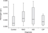
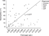
Concentrations of samples below the limit of detection at 0.001 g/L were defined as 0.0005 g/L for statistical analysis purposes. Although no significant differences were found between disease groups (Fig. 1), descriptive data analysis showed a trend toward a lower value in the control group (median 0.0024 g/L) than those in the combined chronic pneumopathy groups (RAO, IAD, and CIP; median 0.0052 g/L) and, in particular, lower than that in the RAO group (median 0.0062 g/L). Forty percent of the control samples were below the limit of detection, while among the combined chronic pneumopathies only 16% were below the limit of detection; percentages below the limit of detection were 5.6%, 21.4%, and 27.7% in the RAO, IAD, and CIP groups, respectively. The individual fibrinogen concentrations in the 58 horses were positively correlated with their percentage of neutrophils in BALF (rs = 0.377, p = 0.004) (Fig. 2).
SAA
Sample SAA concentrations that were below the limit of detection (0.01 µg/mL) were defined as 0.005 µg/mL for statistical analysis purposes. Overall, the SAA concentrations were notably low and in the majority of samples (65.5%) were below the limit of detection. No significant differences were found between the control group and the combined chronic pneumopathies group or each of the individual disease groups.
D-dimers
Immune turbidimetric analysis of D-dimers revealed concentrations below the test's specific limit of detection in 94.8% of samples. The only samples in which concentrations could be quantified were from horses in the RAO group. Therefore, 18 samples of this group and 3 control samples were subjected to ultrafiltration, which yielded quantifiable concentrations in all but one of the RAO samples; however, the D-dimer concentrations in the control samples remained below the limit of detection. The median concentration in the ultrafiltrated RAO samples was 0.1 (0.07/0.185) mg/L with a range of 0.035 to 0.33 mg/L.
PAI-1
PAI-1 concentrations were measured by using three types of ELISA kits supplied by three different manufacturers. Unfortunately, none of the assays produced reliable standard curves or quantifiable results in plasma or BALF samples. Therefore, no results were available for statistical analysis.
Discussion
The aim of this study was to characterize the fibrinolytic activity in equine BALF in horses with different chronic pneumopathies. Specific activators of fibrinolysis are tissue plasminogen activator and urokinase-type plasminogen activator (u-PA), while the system is inhibited by plasminogen inhibitors (PAI-1 and -2), α2-antiplasmin, and the degradation products of fibrinolysis (D-dimers). Several factors need to be evaluated to determine whether the system is ultimately activated or inhibited. For example, increased D-dimer concentrations might lead to the suspicion of increased fibrinolytic activity, but increased D-dimer levels may also occur after severe coagulation, massive fibrin production, and increased fibrinolysis, indicating that the level of D-dimers is insufficient to counteract coagulation and may lead to misbalance and, ultimately, hypercoagulation. Therefore, it is essential to evaluate other specific regulators of the fibrinolytic system as well the D-dimer level. PAI-1 was chosen as a parameter of interest in this study, as pulmonary fibrinolysis in other species is strongly dependent on u-PA and PAI-1 [72238]. PAI-1 concentrations were measured by using three types of ELISA, but unfortunately, none of the available assays produced reliable standard curves or quantifiable results in plasma or BALF samples. Therefore, we focused on examining fibrinogen and D-dimer levels and the ratio between those two. In a case of overwhelming coagulative activity, a high ratio caused by a marked increase in D-dimers might be suspected, while a low ratio would be expected in a case of overwhelming fibrinolysis. Unfortunately, even after ultrafiltration, D-dimer levels could not be quantified in control horses, which made comparisons of such ratios among the study group impossible; however, after ultrafiltration, D-dimer levels could be quantified in horses in the RAO group.
In the present study, the results of fibrinogen concentrations are in agreement of those in previous reports, which have shown that fibrinogen, an acute-phase protein and a key factor in coagulation, is present in TBAs and immune histopathologic samples of horses suffering from chronic respiratory disease [4345]. Fibrinogen increases may be explained by increased permeability of the capillary endothelium and the alveolar epithelium as well as plasma exudation and leakage of plasma proteins from the vasculature into the alveolar lumen, features that have been reported in humans and suspected in equine disease [4164345]. The positive correlation of fibrinogen concentrations with the percentage of neutrophils in BALF supports the role of fibrinogen in the pathogenesis of RAO, a disease in which neutrophils have a central role as a causative agent, or as a consequence of different inflammatory mechanisms [14]. This suggestion is supported by the results of Winder et al. [45], which showed a correlation of fibrinogen concentration and neutrophil percentages in TBAs.
As D-dimer concentrations could only be quantified after ultrafiltration and only in samples of horses exhibiting RAO, this product of increased coagulation and fibrinolysis may be produced in a considerable amount but only in more severe inflammatory processes. In this study, D-dimers were not produced above the limit of detection in minor stages of inflammation, such as in horses with IAD or RAO in remission; moreover, they may not be produced to a detectable extent in chronic interstitial pneumopathies in which the exudative phase of inflammation is over and where fibrosis formation has already occurred. Studies in human asthma, a disease resembling many features of equine RAO, as well as in acute respiratory distress syndrome (ARDS) and pneumonia have reported increased D-dimer concentrations in respiratory secretions like sputum and BALF [4]. In that study, a clear correlation was detected between the grade of inflammation and the D-dimer concentration; concentration levels that were in range similar to that in our study. Coagulation and fibrinolysis may be triggered in the horse by inflammatory stimuli that are minor compared to those in humans, as equine RAO has a more chronic nature than ARDS or acute pneumonia; regardless, the measured concentrations were still similar. Inflammatory processes in other organ systems of the horse, such as intestinal disease and arthropathies, are associated with notable increases in D-dimers [6133536]. Studies in human and equine arthropathies have shown that D-dimer concentrations in synovial fluids compared to those in plasma were higher in horses than in humans [36]. Moreover, the basal D-dimer value in plasma also seems to be higher in horses than in humans [41], therefore, there may be species-specific differences in D-dimer production, with horses showing more pronounced increases during inflammatory processes. Based on the low levels of D-dimers observed in our study, more sensitive assays are needed in order to evaluate the role of D-dimers in horses with IAD, RAO in remission, or CIP. Ultrafiltration was not performed in our IAD and CIP groups as all samples were below the limit of detection; nevertheless, if undertaken, ultrafiltration may have yielded quantifiable concentrations. Ultimately, it cannot be ruled out that D-dimers have a more central role in respiratory disease than that revealed by our data.
As expected, the results of our study do not support an important role of SAA in equine chronic pneumopathies. In the majority of samples in our study, SAA was below the limit of detection, although significant increases have been observed in plasma of horses with severe RAO [25]. It seems logical that plasma exudation during inflammation would also lead to leakage of SAA into the alveolar space, but there are several reasons that SAA may have remained below the limit of detection in our study. First, there is the dilution effect associated with BALF. Second, SAA levels can rise quickly, and SAA has a short half-life. In the study reported by Lavoie-Lamoureux [25], high SAA concentrations were measured in plasma at 7 and 30 days after antigen challenge, and the SAA levels were easy to quantify. Most horses presented for our study had a history of chronic respiratory disease and most had not shown acute deterioration of clinical signs shortly before examination; therefore, it can be suspected, that their SAA concentrations had fallen prior to sampling.
Partly insurmountable weak points of this study were the group definitions and assay feasibilities. Although IAD and RAO in remission were initially planned as two distinct groups, it was not possible to differentiate clearly between the two. The available anamnestic information related to respiratory distress was often unreliable and the majority of owners did not agree to a natural challenge test. Reports describing equine CIP are rare, and there is no international consensus statement on CIP features; thus, definition of the CIP group was based on a relatively old clinical case series of only 12 horses [12]. Thoracic radiography showing interstitial patterns have not produced very specific results for CIP and some features may also be found in RAO and IAD [2842]. Finally, the majority of owners did not agree to lung biopsies. We attempted to reduce these problems by calculating correlations between clinical and cytological parameters for all 58 horses.
Regarding the assays, typically, one would choose a method with the highest sensitivity, thus we principally preferred ELISA-based systems, but due to the fact that many of these have not been validated for horses, we used other validated or at least commonly used assays in our study. A validated ELISA assay was available for SAA and has been used in horses [2534], however, many of our SAA samples produced results below the limit of detection. For D-dimers, none of the available ELISAs provided quantifiable results, thus we undertook immunoturbidimetry, and, with ultrafiltration, we were able to produce quantifiable concentrations. Fibrinogen was also evaluated by using immunoturbidimetry, and that method is commonly used in horses and is offered by a commercial laboratory. The biggest assay-related quantification problem was PAI-1; none of the three assays tested produced reliable results even though two species-specific tests were attempted.
In conclusion, this is the first study in which fibrinogen, D-dimer, and SAA concentrations were evaluated in BALF of healthy horses and compared to horses affected by different chronic pneumopathies. Although the aims of the study could only be realized in part, the results of the study lead us to suspect a misbalance between coagulation and fibrinolysis in respiratory disease in horses, with a tendency within that misbalance toward fibrin formation. Further studies and improvements in assays are essential to further characterize respiratory disease pathophysiologic background mechanisms and their influencing factors in horses.


 XML Download