Abstract
Salmonella (S.) enterica and Shiga toxin-producing Escherichia coli (STEC) are foodborne pathogens. Here, we report the prevalence of S. enterica and STEC in feces of 316 zoo animals belonging to 61 species from Chile. S. enterica and STEC strains were detected in 7.5% and 4.4% of animals, respectively. All Salmonella isolates corresponded to the serotype Enteritidis. To the best of our knowledge, this is the first report of S. Enteritidis in the culpeo fox (Lycalopex culpaeus), black-capped capuchin (Sapajus apella) and Peruvian pelican (Pelecanus thagus) and the first STEC report in Thomson's gazelle (Eudorcas thomsonii).
The study of pathogens in captive populations is critical for implementation of programs for prevention, control and surveillance of diseases, as well as for developing public and animal health policies [9]. Several emerging pathogens in humans are classified as food-borne diseases, with certain serotypes or subgroups of pathogenic Escherichia (E.) coli and Salmonella (S.) enterica being within the most important group [9]
The causative agent of salmonellosis, S. enterica, produces asymptomatic and clinical infections in humans and animals, with symptoms of diarrhea, fever, vomiting, abortion, osteomyelitis and occasionally death [14]. Conversely, E. coli is a commensal bacterium within the large intestine of warm-blooded animals that is generally non-pathogenic toward human and other species of mammals and birds [10]. However, the presence of some virulence genes establishes a diversity of bacterial pathotypes that cause disease in their hosts which are known as pathogenic E. coli [16]. Among these, the Shigatoxinproducing E. coli (STEC) is a globally zoonotic pathogen that can cause bloody diarrhea, hemorrhagic colitis and haemolytic uremic syndrome in humans [310].
Both agents have been isolated from several wildlife species. Salmonella has been isolated from zoo animals with asymptomatic infection [915], and has also caused disease outbreaks with mortality [6]. In the case of STEC, reports in zoo animals suggest variable prevalence ranging from 0.1% to 50.8%, probably because of the different diagnostic methods and animal species being investigated, although asymptomatic infection is always described [1115].
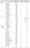
This study was conducted to determine the prevalence of S. enterica and STEC in zoo animals from Chile. The studied population included waterfowl and terrestrial mammals (Table 1), all of which were clinically healthy. A total of 316 fecal samples were collected through both rectal and cloacal swabbing, and these were later inoculated into Cary-Blair transport medium (Copan Diagnostics, USA). For Salmonella isolation, swabs were placed into buffered peptone water (Difco APT broth; Beckton, Dicknson and Company, USA) supplemented with 20 µg/mL novobiocin (Sigma, USA) and incubated for 24 h at 37℃. Aliquots of this suspension were then inoculated into modified semisolid Rappaport Vassiliadis basal medium (Oxoid, Brazil) supplemented with 20 µg/mL novobiocin and incubated for 24 or 48h at 41.5℃. Cultures were subsequently plated onto Xylose Lysine Deoxycholate agar (Difco XLD broth; Beckton, Dicknson and Company), and suspicious colonies were identified by biochemical tests and invA gene detection by PCR. Finally, S. enterica isolates were serotyped according to the Kauffman-White Scheme [8]. For isolation of STEC, swabs were placed into 5 mL buffered peptone water (Beckton, Dicknson and Company) and incubated for 24 h at 37℃. Aliquots of this suspension were then plated into MacConkey medium (Beckton, Dicknson and Company) and incubated for 24 h at 37℃. Next, 10 lactose positive suspicious colonies from each sample were analyzed by PCR for detection of stx1, stx2 and eae genes, as previously described [16]. The STEC reference strains C600J (stx1) and C600W (stx2) and the Enteropathogenic strain 2348/69 (eae) were used as positive controls [16]. Finally, strains were analyzed by biochemical tests to confirm their identity.
A χ2 test was performed to identify statistical associations between order, gender and age of sampled animals using the InfoStat (ver. 2010) software.
From the sampled population, S. enterica strains were isolated from 24 animals (7.5%) (Table 1), all of which belonged to the serotype Enteritidis. In this group, the order Artiodactyla had the highest abundance (p < 0.05), with a prevalence of 18%. In contrast, previous studies have reported less than 10% prevalence of Artiodactyla [2512], suggesting epidemiological variability between populations and reinforcing the need for ecological studies and selection of preventive measures for specific scenarios.
To the best of our knowledge, this is the first report of S. Enteritidis detected in the culpeo fox (Lycalopex culpaeus), black-capped capuchin (Sapajus apella) and Peruvian pelican (Pelecanus thagus). Additionally, this is the first description of STEC in Thomson's gazelle (Eudorcas thomsonii).
S. enterica is a priority pathogen for establishing surveillance programs in wild ruminants from Europe [4]. These animals represent sanitary risks for transmission of pathogens with costly control programs in humans and production animals [4]. Although several reports of Salmonella in artiodactyls have suggested that it causes an asymptomatic infection, mortality has also been described and could have ecological relevance in certain populations [56].
In this study, all S. enterica isolates corresponded to the serotype Enteritidis, which is the most frequent Salmonella serotype isolated from humans. Despite wild birds being considered reservoir hosts of Salmonella in Chile [13], no infected birds were observed in the present study, suggesting that the sanitary condition of zoo birds does not necessarily represent the infectious status of free-range populations for this pathogen. Moreover, these findings indicate that mammals from the zoo do not share transmission routes with zoo birds. For this reason, future genotypic analyses of bacteria must be more informative of their source in zoo facilities.
In the case of pathogenic E. coli, STEC strains were detected in 4.4% of samples (Table 1), all of which belonged to the order Artiodactyla (p < 0.05). Positive samples resulted in amplification of the stx1 and stx2 genes (individually or simultaneously), but not the eae sequence (Table 1). Among artiodactyls, the prevalence of STEC was 12.6%, which is lower than that reported in previous studies (20–50%) [1011]. However, target populations differ between studies.
Within Artiodactyla, a mammal order that includes all even-toed hoofed animals, 30.6% of animals were positive for at least one pathogen, having isolation rates of 18.0% and 12.6% for S. enterica and STEC, respectively. This was the most epidemiologically relevant order (p < 0.05) for detection of these enteric pathogens. The sex and age of animals were not associated (p > 0.05) with bacterial detection.
Artiodactyla usually carry bacteria in their gastrointestinal tract with no symptoms [1], which is supported by the results of this study. Moreover, they have been subjected to greater exposure to enterobacteria than other confined animals investigated to date. Because their pens are distributed in several sectors of the zoo, geographically associated contamination might be irrelevant. Moreover, their food is prepared under strict hygiene standards and their water is properly chlorinated. Therefore, other transmission routes or risk factors likely explain its infection status. Such potential routes include confinement conditions, quality and storage of raw foods, access of synanthropic wild animals (rodents, birds), contact with other captive animals, and direct feeding by visitors [7], although none of the animals from the petting zoo were positive. The isolation of bacteria from these potential carriers and the genotypic characterization of isolates would provide definitive evidence of transmission through such routes. Regardless of the source of infection, the detection of these zoonotic enterobacteria suggest a potential risk of transmission between workers and zoo animals, highlighting the need for awareness of risks and improvement of hygienic procedures, staff training and advertisements targeting visitors to optimize both recreational and educational activities of the zoo.
Figures and Tables
Acknowledgments
We thank Nora Navarro-Gonzalez (University of California, Davis) for critical review of the manuscript. We also thank Alda Fernández (Instituto de Salud Pública) for serotyping Salmonella isolates. This research received financial support from the Fondecyt project No. 11110398.
References
1. Bosilevac JM, Gassem MA, Al Sheddy IA, Almaiman SA, Al-Mohizea IS, Alowaimer A, Koohmaraie M. Prevalence of Escherichia coli O157:H7 and Salmonella in camels, cattle, goats, and sheep harvested for meat in Riyadh. J Food Prot. 2015; 78:89–96.

2. Branham LA, Carr MA, Scott CB, Callaway TR. E. coli O157 and Salmonella spp. in white-tailed deer and livestock. Curr Issues Intest Microbiol. 2005; 6:25–29.
3. Chaudhuri RR, Henderson IR. The evolution of the Escherichia coli phylogeny. Infect Genet Evol. 2012; 12:214–226.
4. Ciliberti A, Gavier-Widén D, Yon L, Hutchings MR, Artois M. Prioritisation of wildlife pathogens to be targeted in European surveillance programmes: expert-based risk analysis focus on ruminants. Prev Vet Med. 2015; 118:271–284.

5. Díaz-Sánchez S, Sánchez S, Herrera-León S, Porrero C, Blanco J, Dahbi G, Blanco JE, Mora A, Mateo R, Hanning I, Vidal D. Prevalence of Shiga toxin-producing Escherichia coli, Salmonella spp. and Campylobacter spp. in large game animals intended for consumption: relationship with management practices and livestock influence. Vet Microbiol. 2013; 163:274–281.

6. Foreyt WJ, Besser TE, Lonning SM. Mortality in captive elk from salmonellosis. J Wildl Dis. 2001; 37:399–402.

7. Gopee NV, Adesiyun AA, Caesar K. Retrospective and longitudinal study of salmonellosis in captive wildlife in Trinidad. J Wildl Dis. 2000; 36:284–293.

8. Grimont PAD, Weill FX. WHO Collaborating Center for Reference and Research on Salmonella. Antigenic Formulae of the Salmonella Serovars. 9th ed. Paris: Institut Pasteur;2007.
9. Jardine C, Reid-Smith RJ, Janecko N, Allan M, McEwen SA. Salmonella in raccoons (Procyon lotor) in southern Ontario, Canada. J Wildl Dis. 2011; 47:344–351.
10. Leotta GA, Deza N, Origlia J, Toma C, Chinen I, Miliwebsky E, Iyoda S, Sosa-Estani S, Rivas M. Detection and characterization of Shiga toxin-producing Escherichia coli in captive non-domestic mammals. Vet Microbiol. 2006; 118:151–157.

11. Pritchard GC, Smith R, Ellis-Iversen J, Cheasty T, Willshaw GA. Verocytotoxigenic Escherichia coli O157 in animals on public amenity premises in England and WalesWales, 1997 to 2007. Vet Rec. 2009; 164:545–549.

12. Renter DG, Gnad DP, Sargeant JM, Hygnstrom SE. Prevalence and serovars of Salmonella in the feces of free-ranging white-tailed deer (Odocoileus virginianus) in Nebraska. J Wildl Dis. 2006; 42:699–703.

13. Retamal P, Fresno M, Dougnac C, Gutierrez S, Gornall V, Vidal R, Vernal R, Pujol M, Barreto M, González-Acuña D, Abalos P. Genetic and phenotypic evidence of the Salmonella enterica serotype Enteritidis human-animal interface in Chile. Front Microbiol. 2015; 6:464.
14. Stevens MP, Humphrey TJ, Maskell DJ. Molecular insights into farm animal and zoonotic Salmonella infections. Philos Trans R Soc Lond B Biol Sci. 2009; 364:2709–2723.


 PDF
PDF ePub
ePub Citation
 Print
Print


 XML Download