

 PDF
PDF ePub
ePub Citation
 Print
Print


Introduction
During the past few decades, the genus Anaplasma within the order Rickettsiales has attracted a great deal of interest because of its pathogenicity in farm animals and its ability to infect humans. Anaplasmosis, which is caused by various species of Anaplasma, is a tick-borne infectious disease that affects wild and domestic animals worldwide [17]. Many types of ruminants (deer, sheep, goat and cattle), wild animals, small mammals, rodents and humans have been shown to be susceptible to the disease, although there are some differences in severity and symptoms between them [10].
Members of the genus Anaplasma include A. phagocytophilum, A. marginale, A. bovis, A. ovis, A. platys and A. centrale, all of which are obligate intracellular bacteria that infect a variety of cell types [3]. A. phagocytophilum, which infects neutrophils in humans and animals, causes human granulocytic anaplasmosis and tick-borne fever in animals. A. ovis, A. marginale and A. centrale are intraerythrocytic pathogens of ruminants. A. bovis is another leukocyte pathogen of ruminants that is usually found in monocytes [16]. Finally, A. platys shows unique tropism for the platelets of dogs, leading to infectious canine cyclic thrombocytopenia [12].
In China, wild and domestic ruminants play active roles as Anaplasma carriers and as infection reservoirs. A. phagocytophilum has been detected in rodents, sheep, rabbits, cattle, goats and ticks in different areas of China [720212223]. In addition, the presence of A. bovis and A. ovis in sheep and goats, as well as A. marginale in cattle has been reported in China [72025]. In a previous study, 262 field blood samples of goats collected from four cities in China were analyzed for the presence of A. phagocytophilum, A. bovis and A. ovis [7]. Molecular investigations have also been conducted in other provinces in China. In the current study, 1331 blood samples were collected from 22 counties in six provinces distributed across central, western and southwestern China. Here, we show that domestic ruminants in this area are commonly infected by distinct Anaplasma species.
Materials and Methods
Ethics statement
This study was conducted in accordance with the Chinese Laboratory Animal Administration Act of 1988. The research protocol was reviewed and approved by the Research Ethics Committee of Henan Agricultural University. The field studies did not involve endangered or protected species.
Sample collection and DNA extraction
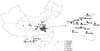
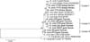
A total of 1331 EDTA-K2 blood samples were collected from asymptomatic domestic ruminants (621 sheep and 710 goats) from 2011 to 2014 during June and October. Samples were collected from 22 randomly selected livestock farms or domestic animal owners interspersed across six provinces of China (Fig. 1). One blood sample was taken from the jugular vein of each animal. DNA was extracted from all samples using a TIANamp Blood DNA kit (TIANGEN biotech, China) according to the manufacturer’s instructions.
In summer of 2011 (July) and 2012 (May, June and July), a total of 31 adult female ticks were collected from naturally infected sheep at four localities in Henan province, China. After collection, ticks were identified morphologically by light microscopy, then preserved in absolute ethanol. Genomic DNA was extracted and purified from the entire body of adult ticks using a Blood and Tissue Gen DNA Kit (CWBiotech, China) according to the manufacturer’s protocols. All DNA samples were stored at –20℃ until use for molecular analysis.
PCR amplification
Nested polymerase chain reactions (PCRs) were applied to detect the presence of A. phagocytophilum, A. bovis and A. ovis. In the first round of the reaction, genus-specific primers EE1/2 were utilized to amplify the Anaplasma spp. 16S rRNA gene [1]. The products were then used as templates for the second round of PCR using the A. phagocytophilum-specific primers SSAP2f and SSAP2r, which generate a product of 641 bp, and the A. bovis-specific primers AB1f and AB1r, which generate a product of 551 bp [5]. For A. ovis, the major surface protein 4 (msp4) gene was amplified as previously described [2]. The primer pair T1B and T2A targeting the 16S rDNA gene of ticks was used to verify the results of microscopic observation. For the 60 kDa heat shock protein (groEL) of A. phagocytophilum, nested PCR containing two primer sets (groEL-1F: 5’-TATAGCTAGCATAATTACCCAGAGC-3’, groEL-1R: 5’-GGTTAGTTCTGCTTTCGATGC-3’, groEL-2F:5’-TTATGTCTATGCGCCGTG-3’, groEL-2R: 5’-CGGACCTTGCCACATTTT-3’), which generates a product of 339 bp, was conducted. PCR amplification was performed as previously described [24]. The products were electrophoresed in a 0.8% agarose gel containing GelRed (Biotium, USA) and observed under UV light.
Sequence analysis
The PCR products of positive samples were sent for sequencing as previously described [24]. The sequence accuracy was verified by two-directional sequencing, and the assembled sequences were analyzed by a BLASTn search (National Center for Biotechnology Information, USA). Phylogenetic analyses were performed using the MEGA 5.05 software [6]. Phylogenetic trees were constructed using previous methods [1324].
Results
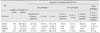
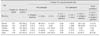
As shown in Table 1, of 621 sheep samples, 242 (39.0%) were positive for Anaplasma. The infection rates at different sampling sites varied from 36.2 to 78.1%. In Qinghai, only A. ovis infection (54.5%) was detected. The most commonly found co-infection was A. bovis and A. phagocytophilum combination, which reached 12.5% (4/32) in sheep from Shanxi, and had an overall infection rate of 6.4% (40/621). The simultaneous infection of three pathogens occurred in only 1.3% (8/621) of the sheep studied. Of 710 goats sampled from four provinces, Anaplasma DNA was identified in 323 (45.5%), among which only 15 (2.1%) were positive for A. phagocytophilum (Table 2). A. ovis and A. phagocytophilum co-infection was not detected in any goats. Simultaneously infection with three pathogens was found in 20 (2.8%) goats from Henan and Shaanxi. Among the 180 A. phagocytophilum 16S rRNA-positive animals, the groEL gene was amplified from 131 specimens (data not shown). Additionally, 31 tick samples (Haemaphysalis longicornis) were collected at the same time and tested for the presence of A. phagocytophilum/A. bovis/A. ovis DNA, and the results showed that 29% (9/31) were positive for Anaplasma (Table 3). Moreover, in all areas in which ticks positive for Anaplasma were reported, there were some positive cases of sheep/goats.
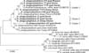
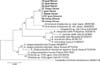
To investigate the genetic variability of Anaplasma spp. in sheep and goats from six provinces of China, positive samples representative of different hosts and geographic locations were sequenced, and 26 sequences including nine from A. phagocytophilum, six from A. bovis and 11 from A. ovis were obtained. A total of 613 bp of 16S rRNA of A. phagocytophilum and 541 bp of 16S rRNA of A. bovis, as well as 559 bp of the msp4 gene of A. ovis were analyzed. The nine A. phagocytophilum 16S rRNA sequences grouped into three sequence clusters (Fig. 2), which were designated 1–3 in this study. The similarity among the five isolates (LY2, YY, YY10, YY19 and LY24) in cluster 1 ranged from 99.5% to 100%, while the three clusters were found to be more divergent, sharing 98.5% to 99.7% homology. Cluster 2 (JY, ZY and JC) was located in a separate clade with 99% identity to strain CE18 (GenBank accession No. GQ450278) that was detected in red deer (Cervus elaphus) from Poland. Cluster 3 had just one isolate, GY, which was most different from other isolates. The nine partial groEL gene sequences obtained in this study, which had only several basepair differences among them, grouped together on a separate clade (Fig. 3). In addition, the results of a BLASTn search of the NCBI showed the highest similarity (92%) with an uncultured Anaplasma sp. (GenBank accession No. JN055360) isolate, which was obtained from a Japanese sika deer (Cervus nippon yasoensis).
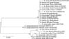
The 541 bp partial 16S rRNA genes of A. bovis were 100% identical and homologous with the sequences derived from several hosts (goat, cattle and deer) from different areas of China (GenBank Accession No. HQ913644, FJ169957 and KJ639883), as well as a deer from South Korea (GenBank accession No. EU682764) (Fig. 4). There was no evidence of any geographic segregation of A. bovis in this study. The 11 sequences of the msp4 gene of A. ovis formed three clusters, designated I–III (Fig. 5). The sequences within cluster I were 99.6% to 100% homologous with each other, while the sequences in cluster II showed 99.6% to 99.8% homology. The two isolates in cluster III showed 100% identity.
Discussion
Sheep and goats are natural reservoirs for a variety of pathogens including bacteria (e.g., A. phagocytophilum and Brucella abortus) [1921], parasites (e.g., Theileria spp. and Haemonchus contortus) [415] and viruses (e.g., peste-des-petits-ruminants virus) [14]. In China, the animal breeding industry and family farming are flourishing, which means that epidemiological investigations into Anaplasma spp. are of particular importance.
Previous studies on A. phagocytophilum infection in sheep and goats have been conducted in a forest area of northeastern China [22], four counties in southeastern China [7] and six counties in Xinjiang, northwest China [20]. The results of the present study in the six provinces of China showed that the overall infection rate of A. phagocytophilum was 3.6%, which was much lower than that found in livestock from Changbai Mountain in Jilin Province, where there was a prevalence of 6.7% [22], as well as in sheep and goats in four counties in southeastern China, where there was a prevalence of 6.1% [7]. It was also far lower than the detection rate of sheep and cattle from Xinjiang (17.6%) [20]. This may have been due to the sampling sites in these three studies all being in mountainous areas, while the investigated sites in this study were mostly plain areas. Despite PCR detection of A. phagocytophilum in the study areas, no clinical symptoms were observed in any of the investigated animals. This was in agreement with the findings of Zhou et al. [25], indicating that subclinical infections caused by A. phagocytophilum occurred in sheep and goats. Phylogenetic analysis of A. phagocytophilum using the nine sequences in this study as well as five other sequences previously obtained from ruminants in different areas of the world indicated that the isolates in present study were located in three different clusters. Overall, five sequences within cluster 1 and three isolates in cluster 2 were obtained from different sampling sites.
The groEL gene is often used for phylogenetic studies of A. phagocytophilum owing to its high genetic heterogeneity, and because two distinct lineages could be delineated in Europe by sequence analysis [811]. However, the partial groEL sequences obtained in this study appeared to be far from both two clusters mentioned above, implying a separate lineage or cluster may exist in China.
Investigation of A. bovis infection revealed that it was the dominant pathogen in goats from all sampling sites except Yunnan province. The overall infection rates in sheep and goats were 9.7% and 10.4%, respectively, which were similar to the prevalence of A. bovis in deer (9.0%) and Haemaphysalis longicornis ticks (12.0%) in Japan, but much lower than that of goats in China (49.6%) and Japanese cattle (53.5%) [579]. Phylogenetic analysis indicated that the six sequences obtained in the present study were 100% homologous and formed a clade with seven previously obtained sequences derived from different hosts and areas. The phylogenetic trees of A. phagocytophilum and A. bovis showed no relationship between host and cluster. There was also no evidence of any geographic segregation of the two organisms in this study.
The average infection rates of A. ovis were relatively high in both sheep (14.2%) and goats (18.2%); however, they were lower than that of sheep in Xinjiang [720]. Half of the sheep from Qinghai were infected with A. ovis, and none of these were positive for two other Anaplasma species. Infection with A. ovis was confirmed by sequencing of the msp4 gene, which has been shown to be reliable for phylogenetic studies of A. ovis [2]. The 11 sequences analyzed in the present study fell into three clusters. Additionally, three of the four isolates in cluster I from sheep and goats in central China fell into a clade with another isolate previously obtained from Hebei, which is also a central province of China (GenBank accession No. HQ456350). The sequences in cluster II were all obtained from goats. The two sequences in cluster III, which were derived from two goats in Yunnan, fell into a separate clade. Additionally, the results revealed geographic segregation to a certain extent, as well as a relationship between the host and cluster of A. ovis in the present study.
Co-infection of two Anaplasma species was detected in nearly all investigated sites. A. ovis was found to be dominant in sheep, while A. bovis was the dominant Anaplasma species found in goats, which was consistent with the results reported by Liu et al. [7]. Furthermore, 40 (6.4%) sheep and 62 (8.7%) goats were found to be positive for both A. phagocytophilum and A. bovis, which was much more common than other combinations. It is still not clear if this could be explained by the exclusion of different Anaplasma species; however, similar phenomena occurred among genotypes of A. phagocytophilum [18].

 XML Download