

 PDF
PDF ePub
ePub Citation
 Print
Print


Introduction
Paclitaxel is an anti-cancer drug known as taxol [2023] that stabilizes microtubules during cell division, thus arresting the process. As a result, paclitaxel has been used as an anticancer agent for treatment of breast, ovary, and lung cancer. However, since paclitaxel cannot select between cancer and normal cells, it has side effects including myelosuppression [515].
To date, few studies have investigated the effects of paclitaxel on immune cells. It has been reported that paclitaxel modulates the activity of various immune cells [8]. Specifically, paclitaxel binds to myeloid differentiation protein-2 (MD-2), which is an accessory protein of Toll-like receptor 4 (TLR4), the main receptor of lipopolysaccharide (LPS) [11]. As a result, paclitaxel can block TLR4-mediated nuclear factor (NF)-κB signaling of LPS on cells [25]. LPS is the primary causative agent of inflammation by Gram negative bacteria and sepsis [21]. However, the immunomodulatory activity of paclitaxel remains unclear. Paclitaxel induces the maturation of dendritic cells (DCs) associated with up-regulation of major histocompatibility complex class II molecules [9] and increases DC viability [14], while it makes splenic lymphocytes hyporesponsive to LPS [16].
In this study, we investigated whether paclitaxel modulates the activation and function of spleen cells activated by LPS as an inflammatory agent. The functional changes in LPS-treated spleen cells in response to paclitaxel focused on alteration of spleen cell proliferation, cell morphology, presence of apoptosis, and production of cytokines.
Materials and Methods
Animals and reagents
BALB/c or C57BL/6 mice were purchased from ORIENT BIO (Korea) and maintained in our animal facility. Mice used in this study were 7- to 12-weeks-old. All animal experiments conformed to the institutional guidelines of Jeju National University for laboratory animal use and care. LPS purified from Escherichia coli and paclitaxel were purchased from Sigma (USA) and dissolved in phosphate buffered saline. The concentration of dimethyl sulfoxide in culture medium was 0.1% (v/v).
Preparation of spleen cells
Spleen cells were prepared from the spleens of mice [10]. Briefly, spleens were smashed and treated with ammonium chloride potassium lysis buffer to remove red blood cells. The cells were then passed through a 70 µm cell strainer to obtain individual cells, which were cultured in a complete culture medium for lymphocytes, RPMI 1640 medium containing 10% fetal bovine serum, 2 mM L-glutamine, 0.1 mM non-essential amino acids, 1 mM sodium pyruvate, 100 IU/mL penicillin/streptomycin, and 50 µM 2-mercaptoethanol.
Cell morphology
To observe the effects of paclitaxel, the morphological changes of spleen cells were investigated. Briefly, cells were cultured at a concentration of 2 × 10⁶ cells/mL in 96- or 6-well culture plates in the absence or presence of 1.0 µg/mL LPS and 5.0 µg/mL paclitaxel for 3 days. The cell morphology was then observed and photographed using an inverted optical microscope (Olympus, Japan) with a digital camera.
Assessment of cellular viability
The viability of LPS and paclitaxel-treated spleen cells was measured by a cellular viability assay [112]. Briefly, spleen cells were seeded at a concentration of 2 × 10⁶ cells/mL in 96-well plates and treated with 5-fold serial dilutions of LPS (0, 0.008, 0.04, 0.2, 1.0, 5.0 µg/mL) and paclitaxel (5.0 µg/mL). After 3 days of culture, 3-[4,5-dimethylthiazol-2-yl]-2,5-diphenyltetrazolium bromide (MTT; Sigma) was treated at a concentration of 0.5 mg/mL for 4 hr. The insoluble violet crystal generated by viable cells was dissolved by 100 µL/well 10% sodium dodecyl sulfate solution for 2 h, after which the optical density of samples was measured at 570 nm using a microplate reader (Multiskan FC; Thermo Fisher Scientific, USA).
Measurement of cytokine production
A range of concentrations of LPS (0, 0.04, 0.2, 1.0 µg/mL) and paclitaxel (5 µg/mL) were added to 2 × 10⁶ cells/mL of spleen cells in 96-well culture plates. After 3 days, the culture supernatants were collected and used to quantify tumor necrosis factor-alpha (TNF-α) and interleukin-10 (IL-10), which are critical inflammatory and anti-inflammatory cytokines, respectively. For these assays, enzyme-linked immunosorbent assay (ELISA) using CytoSet antibody pairs (Thermo Fisher Scientific) was performed based on the manufacturer's instructions.
Flow cytometry analysis
Flow cytometry analysis was performed as previously described [713]. Briefly, spleen cells were cultivated in 6-well culture plates and treated with 1 µg/mL LPS and 5 µg/mL paclitaxel for 3 days. To check the membrane potential of the mitochondria, the cells were stained with 10 µg/mL rhodamine 123 (Sigma) for 30 min at room temperature. To measure apoptosis, the cells were stained with 2 µg/mL propidium iodide (PI). The cluster of differentiation (CD)25 and CD69 on the surface of the LPS- and paclitaxel-treated cells were stained with purified anti-mouse CD25 antibody, followed by phycoerythrin (PE)-labeled anti-rat IgM antibody, and PE-labeled anti-mouse CD69 antibody (all from BD Biosciences, USA). The stained cells were analyzed using FACSCalibur flow cytometer and the CellQuest software (both from BD Biosciences).
Results
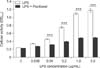
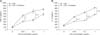
Paclitaxel inhibits the activity of LPS-treated spleen cells
To measure the cellular activity and proliferation rate, MTT assay was performed (Fig. 1). LPS alone increased the activity and proliferation of spleen cells in a concentration-dependent manner (0–5 µg/mL). However, paclitaxel significantly decreased the activity and proliferation of LPS-treated spleen cells (0.04–5 µg/mL). These results suggest that paclitaxel might prevent the effects of LPS on spleen cells.
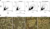
Paclitaxel decreases the population of large cells in LPS-activated spleen cells
We used flow cytometry analysis and a microscope to investigate the effect of LPS and paclitaxel on the cell size and morphology of spleen cells (Fig. 2). The cell size was measured by forward scattered light (FSC) of flow cytometry and the cell morphology was observed using an inverted optical microscope. Flow cytometry analysis (panel A in Fig. 2) revealed that paclitaxel alone induced minor changes in cell size compared to the control, whereas LPS markedly increased the population of large cells (with high FSC, in circle). Interestingly, paclitaxel decreased the population of large cells in LPS-treated spleen cells. Microscopic analysis (panel B in Fig. 2) revealed growing clusters (a black circle) and large single cells in LPS-treated spleen cells. However, spleen cells treated with paclitaxel and LPS contained almost no clusters and very few large single cells (a black arrow).
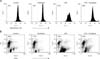
Paclitaxel increases death of LPS-treated spleen cells
Rhodamine 123 and PI staining were used to investigate cell viability and death of the spleen cells treated with LPS and paclitaxel. Rhodamine123 staining revealed that LPS increased the geometric mean fluorescence intensity (MFI) of spleen cells compared to the control, whereas paclitaxel did not. Paclitaxel decreased the MFI of LPS-treated spleen cells (panel A in Fig. 3). Given that MFI in Rhodamine 123 staining indicates the stability of mitochondrial membrane potential, these results suggest that paclitaxel may affect the viability of LPS-treated spleen cells, indicating increased cell death. Indeed, PI staining demonstrated that paclitaxel consistently increased the death (FSClowPIhigh cells) of LPS-treated spleen cells (panel B in Fig. 3).
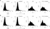
Paclitaxel decreases the expression of CD25 on LPS-treated spleen cells
LPS is a representative activator of B lymphocytes. To investigate the effects of paclitaxel on the expression of activation markers on spleen cells, we measured the expression level of lymphocyte activation markers, CD25 and CD69 (Fig. 4). Flow cytometry analysis revealed that paclitaxel did not affect the expression of either marker on spleen cells compared to the controls, whereas LPS increased the expression of both markers. Paclitaxel decreased the expression of CD25 on LPS-treated spleen cells.
Paclitaxel decreases the cytokine production of LPS-treated spleen cells
We measured cytokine production by ELISA to evaluate function. In this study, TNF-α and IL-10 were selected as representative inflammatory and anti-inflammatory cytokines, respectively. ELISA revealed that LPS enhanced the production of TNF-α and IL-10 in a concentration-dependent manner. Paclitaxel significantly inhibited LPS- induced production of both cytokines (Fig. 5).
Discussion
The anticancer drug, paclitaxel, has been used to treat breast, ovary, and lung cancer. This drug inhibits microtubule depolymerization, resulting in cell cycle arrest and death of cancer cells. However, few studies have investigated the effects of paclitaxel on immune cells, although an action mechanism of paclitaxel on the immune system has been demonstrated [222]. Paclitaxel binds to MD-2 protein of TLR4 and sequentially prevents the signal transduction of LPS, an activator of B cells. Indeed, paclitaxel converts immune cells hypo-responsive to LPS through modification of the signal transduction [16]. In this study, we demonstrated that paclitaxel inhibits the function and further induces death of LPS-activated cells.
To investigate the effects of paclitaxel on immune cells, we measured the activation and proliferation of spleen cells treated with paclitaxel and LPS by MTT assay, flow cytometry, and microscopic analysis. Paclitaxel significantly decreased LPS-induced effects on the proliferation and activation of spleen cells. Moreover, microscopic analysis revealed that some LPS- and paclitaxel-treated cells were large and swollen.
We suspected that paclitaxel may induce the death of LPS-treated spleen cells; therefore, Rhodamine123 [24] and PI staining were performed. The results revealed that paclitaxel decreased the number of large cells induced by LPS treatment and increased the number of PI-highly stained small cells (dead cells). Therefore, these results confirm that paclitaxel induced death of LPS-induced spleen cells, including apoptosis. Additionally, we analyzed the expression of CD25 and CD69, activation markers of lymphocytes. CD25, which is the alpha chain of interleukin-2 (IL-2) receptor, converts lymphocytes sensitive to IL-2, and is thus related to the proliferation of lymphocytes [17]. CD69 is an early activation marker for lymphocytes [6]. Paclitaxel also decreased the expression of activation markers in LPS-treated spleen cells. We speculated that paclitaxel can inhibit activation of the spleen cells by LPS, resulting in a reduced number of activated cells.
We next investigated if paclitaxel also affects cytokine production of LPS-treated spleen cells. ELISA demonstrated that paclitaxel decreased the production of TNF-α and IL-10, which are representative inflammatory [3] and anti-inflammatory cytokines [19], respectively. Both cytokines are closely related to the immune responses generated by LPS [4]; thus, these findings suggest that paclitaxel modulates LPS-induced immune responses by inhibiting the production of related cytokines. Therefore, we speculate that paclitaxel not only induces hypo-response of immune cells to LPS via the inhibition of signal transduction, but also prevents LPS-induced immune response through inhibition of activation and proliferation, induction of apoptosis, and inhibition of cytokine production in immune cells.
Based on these results, paclitaxel can be applied to treat diseases caused by hyper-activation of B cell. Indeed, LPS causes a variety of diseases, including the generation of auto-reactive B cells [18]. We expect paclitaxel to be particularly useful for treatment of currently incurable autoimmune diseases. To accomplish this, the therapeutic effects of paclitaxel should be verified, especially its inhibitory effects on antigen-specific IgG production of B cells through in vivo experiments. Moreover, it is necessary to investigate which subsets of spleen cells can be affected since spleen cells are composed of multiple subsets and lineages of immune cells.
In conclusion, the results of this study provide evidence that paclitaxel inhibits the activation and proliferation of spleen cells by LPS, as well as induces the death of activated cells. These results will help broaden the use of paclitaxel in basic science and clinical areas beyond as an anti-cancer therapeutic agent.
 XML Download