

 PDF
PDF ePub
ePub Citation
 Print
Print


Rapid and accurate diagnostic methods for avian influenza A virus (AIV) infection are necessary for the surveillance, outbreak management, and early infection control of the emerging influenza virus [12]. Several molecular diagnostic methods including reverse transcription polymerase chain reaction (RT-PCR) and real-time RT-PCR (RRT-PCR) have been developed for the rapid detection of AIV [612]. However, these techniques require sophisticated and expensive instrumentation and specialized personnel, limiting their effectiveness and availability.
Loop-mediated isothermal amplification (LAMP) is a novel nucleic acid amplification method [9] that has been widely applied to the detection of various human, animal, and plant pathogens [28]. Recently, reverse transcription LAMP (RT-LAMP) assay was successfully applied to the detection of AIVs [1013]. However, LAMP or RT-LAMP assay can be particularly vulnerable to carry-over DNA contamination because of its high sensitivity and productivity [34]. Therefore, if LAMP or RT-LAMP is used as a routine diagnostic method for pathogen detection, the possibility of false-positive reactions by carry-over contamination should be eliminated. False-positive reactions because of carry-over contamination can be avoided by using deoxyuridine triphosphate (dUTP)/uracil-DNA glycosylase (UNG)-based strategy [7]. This strategy has been widely applied in PCR-based amplification methods, but was not employed in LAMP-based methods until recently when an UNG-treated LAMP (uLAMP) assay was reported by Hsieh et al. [3].
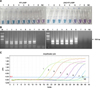
In this study, we developed a carry-over-contamination-free uRT-LAMP for the rapid detection of AIVs. To the best of our knowledge, uRT-LAMP assays have not yet been described for AIVs. Reference AIV strains (subtypes 1–16), two highly pathogenic AIV (HPAIV) subtypes H5N1 and H5N8 (Korean representatives), human influenza B virus (HIBV), and Newcastle disease virus (NDV) were used to evaluate the specificity of the assay (Table 1). Viral RNA was extracted using an RNeasy Mini kit (Qiagen, Germany) according to the manufacturer's instructions. Extracted nucleic acid was stored at -20℃ until further use. RT-LAMP assay for AIV detection was performed using AIV matrix gene-specific primer sets as previously described [5]. The amplification reaction was performed at 58℃ for 40 min, followed by heating at 80℃ for 5 min to terminate the reaction. After RT-LAMP, the positive results were visually confirmed by a colorimetric change from purple to sky blue in the reaction tubes without an additional detection process (Fig. 1). Because of the high mutation rate of AIV genes, it is difficult to design a multi-set of RT-LAMP primers to detect all subtypes of AIVs [1013]. Six primer sets (F3, B3, LF, LB, FIP, and BIP) for the proposed RT-LAMP that specifically target eight different regions highly conserved among all subtypes of AIVs were carefully designed by analyzing most of the AIV matrix gene deposited in the Influenza Sequence Database during 2012–2014 [15]. The RT-LAMP assay using these primers specifically detected all subtypes of AIVs tested, but not HIBV and NDV (Table 1), indicating the assay was highly accurate and specific for all subtypes of AIVs.
In this study, we adopted the dUTP/UNG strategy in RT-LAMP to prevent a carry-over contamination with pre-amplified RT-LAMP products. To remove dUTP-incorporated RT-LAMP products using UNG, dUTP must be substituted for dTTP in all LAMP products so that the Bst DNA polymerase can incorporate dUTP instead of dTTP in the uRT-LAMP reaction [34] Thus, the uRT-LAMP assay for AIV detection was performed in the above described reaction mixture containing 10 mM dUTP instead of 10 mM dTTP and 5 U of UNG (ArticZymes, USA), while other parameters were maintained. To evaluate the ability of uRT-LAMP assay to prevent carry-over contamination, the reaction was performed using 10-fold serially diluted uRT-LAMP products amplified from a previous reaction as a template. The concentration of the uRT-LAMP products ranged from 10 picograms to 10 attograms per reaction. After UNG treatment for 5 min at 25℃, as recommended by the manufacturer, the chief RT-LAMP reaction was performed for 40 min at 58℃ without opening the tubes. In RT-LAMP assay without UNG, amplification occurred in reaction tubes containing pre-amplified DNA at 10 picograms to 100 attograms/reaction (panel A in Fig. 1; lane 1-6). These results clearly indicate the risk of carryover contamination in RT-LAMP reactions, where even trace amounts of any contaminant can cause unwanted amplification. In contrast, UNG treatment in the uRT-LAMP prevented amplification when 1,000 and 100 attograms of carryover contaminated DNA were used (panel B in Fig. 1; lane 5 and 6), and LAMP-positive color change and ladder-like DNA bands were observed only in reaction tubes where contaminants were added at levels of 1 femmtogram or higher. The ability of uRT-LAMP to prevent contamination was identical to that reported in a previous study [3], indicating that the assay can prevent the typical aerosol-based contamination that occurs in RT-LAMP [311].
Next, to determine the analytical sensitivity of RT-LAMP and evaluate the effects of dUTP incorporation and UNG treatment on RT-LAMP, both RT-LAMP and uRT-LAMP assays were performed with 10-fold serially diluted viral RNA extracted from the Korean H5N8 HPAIV (A/broiler duck/Korea/Buan2/2014) at an initial viral titer of 108 median embryo infection dose (EID50)/0.1 mL. The results of RT-LAMP and uRT-LAMP were compared with those of previously reported RRT-PCR using the same viral RNA diluent as the template. The RRT-PCR for the detection of all AIV subtypes was performed using a one-step PrimeScript RT-PCR kit (Takara Bio, Japan) in a real-time PCR instrument (Applied Biosystems, USA) as previously described [6]. The results showed that the detection limit of RT-LAMP was a 107 dilution of the original viral RNA concentration, which is the same as that observed for RRT-PCR. The detection limit of uRT-LAMP (106 dilution) was 10-fold lower than that of RT-LAMP and RRT-PCR (Fig. 2). It is believed that this occurred owing to supplementation with UNG or substitution of dUTP in the reaction mixture as reported by Hsieh et al. [3]. Although the analytical sensitivity of uRT-LAMP showed a slight reduction in response to UNG treatment, it was identified as a valuable screening tool for AIVs because it can effectively prevent potential carry-over contamination. The reduction in detection limit should be improved through further studies.
The detection methods used with RT-LAMP are critical and constitute a rapidly developing field because of their practical application and commercial value [14]. Recently, a simple colorimetric assay was applied for visual detection in the LAMP assay by adding metal indicators to the pre-reaction mixture of LAMP [14]. In the present study, we used a colorimetric detection method with hydroxyl naphthol blue for RT-LAMP (Figs. 1 and 2), which rendered the LAMP assay more simple and user-friendly for application as a molecular diagnostic method, and a suitable method for smaller laboratories or as an on-site rapid diagnostic tool [214].
It should be noted that the sample set tested in this study is relatively limited to some reference strains and field isolates. Therefore, further validation with additional influenza isolates and clinical samples is needed to better define the usefulness of this assay. Further, continuous surveillance and genetic characterization of AIVs are required to guarantee the significance of primers used in the RT-LAMP assay.
In this study, we first developed and evaluated an uRT-LAMP for quick detection of AIVs. The developed method prevents false-positive reactions because of carry-over DNA contamination and allows visual detection of results by the naked eye. The uRT-LAMP assay can be applied for the rapid, user-friendly, and reliable detection of AIVs, thereby aiding efficient control of AIV infections and outbreaks.


 XML Download