

 PDF
PDF ePub
ePub Citation
 Print
Print


Introduction
Corneal ulceration is a common and clinically important ocular disease in dogs. In the case of large and deep corneal defects, various surgical managements have been attempted to promptly and effectively repair the cornea, including conjunctival pedicle graft [25], corneal-scleral transposition [24] and autogenous corneal graft [8]. In addition, preserved biological membranes including pericardium [2], intestinal submucosa [11], amniotic membrane [7] and renal capsule [3] are used in medicine and in veterinary general surgery for deep corneal defects and perforations. However, these methods for treatment of large corneal defects are not able to recover corneal transparency. Additionally, the indicated cases are restricted to certain types of techniques.
Transplantation of various corneal parts is an essential technique for the treatment of severe corneal damage in humans [6] that has been performed successfully in horses for therapeutic and tectonic reasons [9]. Furthermore, penetrating keratoplasty (PK), has been performed in the rabbit cornea in experimental models [22]. However, corneal transplantation is very limited in dogs because of insufficient donor corneas and frequent graft rejections characterized by corneal vascularization, graft failure, and subsequent corneal edema. Therefore, few studies have investigated canine corneal transplantation [20].
Deep anterior lamellar keratoplasty (DALK) removes and replaces the pathologic corneal stroma while preserving the host endothelium, which eliminates the risk of endothelial graft rejection and has a reduced effect on the endothelial cell count [14]. Thus, DALK is indicated for patients with a healthy endothelium to achieve a high success rate for corneal transplantation as an alternative procedure to PK [14]. Several surgical methods were used to bare the Descemet's membrane (DM) during DALK, including layer-by-layer manual dissection [28], hydro-delamination [27], viscoelastic dissection [19] and air injection [4]. The big-bubble technique (BBT) introduced by Anwar and Teichmann is a method that injects air, which forms a large bubble in the stroma to detach the DM during DALK [4]. The present study was conducted to establish the feasibility of corneal transplantation with BBT when performing DALK in dogs.
Materials and Methods
Animal studies and preparations
Three eyes from three healthy male beagles with normal corneas were used in this study. The donor corneas were obtained from dogs scarified in other experiments unrelated to this study. The animal use and experimental protocols were approved by the Institutional Animal Care and Use Committee (SNU-140520-1; Seoul National University, Korea).
Complete ophthalmic examinations were performed before the experiment with a rebound tonometer (TonoVet; Tiolat, Finland), Schirmer Tear Test (Schering-Plough Animal Health, USA), slit-lamp biomicroscope (Topcon SL-D7; Topcon, Japan) and indirect ophthalmoscope (Vantage; Keeler Instruments, USA) with a 30-diopter indirect lens (Classic BIO Lens; Volk Optical, USA). None of the beagles had any corneal diseases.
Surgical technique
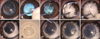
The surgical procedure was performed under general anesthesia with the BBT as described by Anwar and Teichmann [4]. Before surgery, the central corneal thickness (CCT) was measured with an ultrasonic pachymeter (Pachmate DGH 55; DGH Technology, USA). The central axial cornea was trephined 750 µm with an 8 mm diameter Barron radial vacuum trephine (Katena Products, USA) to create an incision of approximately 80% thickness (panel A in Fig. 1). A partial-thickness superficial anterior keratectomy was performed by dissection with a No. 66 lamellar blade (Katena Products) (panel B in Fig. 1). A small amount of air was introduced into the anterior chamber by intracameral injection at the limbus using a 26-G needle. A 30-G needle attached to a 4 mL filled syringe with the tip manually bent to approximately 30 degrees was introduced with its bevel down into the cornea stroma through the trephination groove and advanced to the center of the cornea (panel C in Fig. 1). At this point, air was injected gently, being forced through the posterior stromal lamella along the path of least resistance, causing the DM to separate from the deep stroma (panel D in Fig. 1). A blanched corneal stroma was incised with a 15° slit-knife (Alcon Laboratories, USA) to let the air escape and collapse the bubble (panel A in Fig. 1). A corneal dissector was carefully inserted and advanced into the cleavage plane that was created until its tip approached the opposite trephination groove. Corneal scissors were used to remove the remaining corneal tissue and expose the DM (panel F in Fig. 1). The donor cornea was gently stripped off the DM and endothelium with a cellulose sponge or forceps. The donor cornea was then punched from the endothelial side with an 8.5 mm-diameter Barron punch (Katena) (panel G in Fig. 1). This prepared donor corneal button was initially sutured onto the bare DM with 4 cardinal 10-0 nylon sutures at 3, 6, 9, and 12 clock-hour positions (panels H and I in Fig. 1). There was also a single running suture with 16 to 18 bites using same suture materials (panel J in Fig. 1). At the conclusion of the surgery, gentamicin and triamcinolone were injected subconjunctivally.
Post-operative care and evaluation
After surgery, atropine eye drop (1%, Isopto Atropine; Alcon, Belgium) was applied two times a day for 3 days and levofloxacine eye drop (0.5%, Cravit; Santen, Japan) was administered every 6 h for 7 days. Cyclosporine ointment (2 mg/g, Optimmune; Schering-Plough, France) was administered every 12 h until 30 days post operatively. All corneal sutures were removed under the topical anesthesia with proparacaine hydrochloride (0.5%, Alcaine; Alcon, New Zealand) at day 21 post-surgery.
The eyes were examined by slit-lamp biomicroscopy (SL-D7), tonometry (TonoVet), and indirect ophthalmoscopy (Vantage) to evaluate corneal condition and graft rejection at 7, 14, 21, 28 and 150 days post-surgery. During these periods, menace response, dazzle reflex, and pupillary light reflex (PLR) were evaluated, and intraocular pressure (IOP) and CCT were measured. In addition, blepharospasm, corneal edema, corneal vascularization, and haze development at the central cornea and suture line were examined clinically. The level of corneal haze was evaluated using the following previously reported clinical grading system [12]: grade 0, completely clear cornea; grade 0.5, trace amount of haze observed with careful oblique illumination; grade 1, mild obscuration of the iris details; grade 2, more prominent haze not interfering with the visibility of fine iris details; grade 3, moderate obscuration of the iris and lens; and grade 4, complete opacification.
At the same time, the CCT was measured using the ultrasonic pachymeter (Pachmate DGH 55; DGH Technology).
The dogs were euthanized at day 150 after surgery, and the eyes enucleated and fixed in 10% formalin. Sections were stained with hematoxylin and eosin and examined by light microscopy. The results of CCT and IOP are expressed as the mean ± SD. Statistical analyses were performed using SPSS for windows (ver. 20; IBM, USA). One was analysis of variance (ANOVA) with Bonferroni's post-hoc assessment was used to test for significance when comparing the CCT. P values < 0.05 were considered significant.
Results
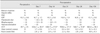
The mean CCT of the three eyes was 817 ± 23 µm before surgery. The central cornea was trephined to a depth of 750 µm for approximately 80% of the corneal thickness. DALK was performed for the three eyes successfully without any DM tearing. After the surgical procedure, no rejection of the corneal implants was observed in the eyes for five months postoperatively (Table 1). The menace response, dazzle reflex, and PLR were normal in all experimented eyes during this period. In addition, the IOP was in the normal range and did not show any significant changes. The fluorescein dye staining test was positive in two cases at 7 days after surgery, and the blepharospasm was examined in the two cases at the same examination time. Because the corneal ulcer was located near the suture line, the ulcer and blepharospasm were related with the spur of the suture materials. No abnormal responses were observed after suture removal.
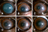
A corneal haze was developed from 7 days after surgery at the central transplanted cornea and around the suture line (Table 1). The corneal haze of central corneal part was reduced from 21 days after surgery. However, the corneal haze around the junction of the donor and recipient cornea and suture lines increased at 21 days after surgery and remained throughout the experimental period. The central portion of the transplanted cornea remained transparent while a corneal haze developed around the transplanted margin (Fig. 2). Stromal edema of the donor cornea was present from immediately after surgery until three weeks after surgery in all three beagles (Fig. 3), then began to decrease from four weeks after surgery. The corneal thickness was significantly increased at the day 7, 14, 21, and 28 after surgery compared to the CCT of pre-operative examined values (Table 1). The menace response was normal even though the transplanted corneas were edematous.
After the sutures were removed, the corneal haze around the implant margin decreased, and the corneal haze near the suture line disappeared. The marginal haze at the junction between the donor and recipient corneas decreased to almost nothing at 150 days after the operation (Fig. 2). At 150 days after surgery, a new spotted haze developed in the central part of the cornea that appeared as a deposited mineral spot. The spotted haze was found in the deep stroma near DM on the slit image (panel F in Fig. 3).
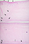
Upon histopathological examination, the stroma and epithelium of the donor cornea had normal structures (Fig. 4). A modified arrangement of the corneal stroma was confirmed in the junction between the donor and recipient corneas. In addition, keratocyte and epithelial proliferation and a distortion in the stromal structure were detected at the junction of the transplant. There were more severe pathological changes in the recipient cornea than in the donor parts. Some particles of the donor's DM in the cornea were observed in the stroma layer (Fig. 4).
Discussion
The BBT for DALK has been shown to be a valuable method as an alternative to PK with the purpose of reshaping the cornea profile in human patients with keratoconus by removing and replacing most of the corneal ectasia [13]. Thus, DALK is performed without substituting the host's healthy endothelium, and it could reduce the risk of immunological rejection [13]. In addition, DALK has been performed experimentally on rabbits [27] and horses [18]. In this study, BBT with DALK was performed on the canine cornea to establish the feasibility of using this technique in dogs. The shape and transparency of the transplanted cornea were successfully maintained, and 5 months after the surgery there still was no rejection in any of the experimental eyes.
Corneal grafts showed a higher success rate than other organ transplants. To the best of the authors' knowledge, there have been no reports of BBT in dogs to date. BBT, which is normally conducted for human ophthalmology, has not been a feasible procedure in canine eyes [16]. However, according to our results, BBT is not necessarily impossible, although it appears to require a skilled surgeon who knows the technique. A small portion of the stromal layer of the recipient cornea after BBT remained in our experiments, similar to a report by Leiva et al. [16]. However, when the thickness of the remaining stroma in our experiment was estimated by histopathological examination, its thickness was relatively lower in this study compared to the previous report. Similar to our results, very small parts of the stroma remained, which caused the development of corneal haze in studies of human ophthalmology [21].
In addition, part of DM was left in the stromal layer, which determines the junction between the recipient cornea and the donor cornea. Part of the membrane may remain during the process of removing DM from the donor cornea. DM incarceration was reported in human ophthalmology under microscopic examination [15]. The stromal structures of the human cornea after air injection appear similar to those of the dog cornea [5]. Therefore, the results of human ophthalmology support our speculation about the incarcerated part of donor DM.
In our results, the swelling of the stroma of the transplanted cornea was confirmed during the first three weeks after surgery. Edema did not progress and normalized three weeks after surgery. In clinical examinations, an eye that has rejection after corneal transplantation shows conjunctival hyperemia, anterior chamber reaction, keratic precipitates, and graft edema [22]. Corneal transplant rejection is a process in which a corneal graft that has been clear for 5 to 7 days in horses [10] or 2 weeks in humans suddenly develops graft edema in conjunction with signs of anterior segment inflammation [23]. Corneal edema could be produced as part of a sudden rejection of the transplant. However, in this study, corneal edema was present in all of the transplanted corneal parts from the first examination immediately after surgery. Thus, we believe that the corneal edema developed during the surgical procedure, especially in the procedure for donor cornea preparation. When another examination was conducted three weeks after surgery, the corneal edema had resolved itself in all of the cases.
A corneal haze appeared near the junction of the transplant and the suture lines, and overgrowth of the epithelium was observed in these areas. In particular, these reactions were severe on the recipient side of the cornea, but the epithelial cells of the central part of the donor cornea maintained their normal morphology. Overgrown epithelial cells were also more detected on the inside of the recipient cornea. Corneal epithelial breach can be caused by exposed suture knots and a loose suture, which are some of the predictors for the occurrence of corneal haze during the post-operative period [27]. These factors give rise to irritation, leading to a subsequent corneal ulcer and corneal vascularization. Therefore, these reactions could be predisposing factors to graft rejection [27]. In these cases, the haze may invade the adjacent host cornea, which is closer to the vascularization [13].
Much equipment has been developed for human ophthalmology to enable delicate surgical techniques, such as excimer lasers and femtosecond lasers [117]. This equipment has been used to reduce corneal haze, as well as other post-surgical complications. In the veterinary field, BBT can be used to maintain the vision of patients that have a wide range of corneal diseases while preserving the healthy endothelium. Furthermore, considering the quality of life of the animal, BBT is a better surgical method for restoring vision.
In conclusion, corneal transplantation using DALK with the BBT could be performed in a dog that has a large corneal defect and vision loss with a healthy endothelium.

 XML Download