

 PDF
PDF ePub
ePub Citation
 Print
Print


Introduction
Highly pathogenic avian influenza (HPAI) viruses cause economic losses and pose a threat to public health [15]. Avian influenza viruses (AIV) have segmented genomes, and antigenic drift and shift are important mechanisms for producing rapid diversity [16]. H5N1 is the representative subtype of HPAI in Asia and has evolved into over 32 clades distinguished by their hemagglutinin (HA) genes [13].
In Korea, there have been four outbreaks of H5N1 HPAI [6]. The A/chicken/Korea/Es/2003 (H5N1) caused the first outbreak in 2003 and 2004. This virus clustered with the A/Duck/China/E319-2/03 (H5N1), which is designated as clade 2.5. The virus involved in the second outbreak, A/chicken/Korea/Is/2006 (H5N1), belongs to the Qinghai-like H5N1 HPAI viruses [79]. In the third outbreak, the causative virus was A/chicken/Korea/Gimje/2008 (H5N1) from clade 2.3.2, which was similar to A/Muscovy duck/Vietnam/1455/06. The fourth outbreak, caused by A/duck/Korea/Cheonan/2010 (H5N1), occurred in 2010–2011. In this outbreak the strain was clustered into clade 2.3.2.1 and was similar to the A/Whooper swan/Mongolia/21/10 (H5N1) [5]. This virus has been found in various wild bird species such as the mallard, Baikal teal, mandarin duck, whooper swan, and Eurasian eagle owl [5].
On January 16, 2014, the fifth outbreak was caused by a strain of H5N8 in Korea. The first reported case was in a breeder duck farm in Gochang in Jeonbuk province. On January 17, another infection was reported in broiler ducks in Buan (near Gochang), and flocks of Baikal teal carcasses were found in the Donglim reservoir in Gochang [10].
Here, we obtained nineteen H5N8 viral isolates from wild Baikal teal in the Donglim reservoir in Gochang, Korea, where the 2014 outbreak was first reported [7]. To obtain further genetic information regarding these viruses, all eight gene segments were sequenced and characterized by molecular and phylogenetic analysis.
Materials and Methods
Sampling and virus isolation
Nineteen viruses were isolated from cloacal and tracheal swab samples of 27 wild Baikal teals that were found dead at Donglim reservoir in Gochang, Jeonbuk province, Korea on January 20, 2014. Virus isolation was performed in 9- to 11-day-old specific-pathogen-free embryonated chicken eggs. After 72 h of incubation at 37℃, the eggs were chilled, and allantoic fluids were harvested and tested for hemagglutinin activity according to the WHO manual [17]. Necropsy and virus isolation were performed under biosafety level 3 conditions at Konkuk University in Korea.
Genome sequencing
RNA was extracted using a RNeasy kit (Qiagen, USA) according to the manufacturer's instructions. AIV-positive specimens were further characterized by complete genome sequencing. The eight genes of AIV were amplified by multiplex RT-PCR using previously described primers [1]. Next, 2 µg of the RT-PCR amplicons of all eight gene segments was used for preparation of the Ion Fragment sequencing library (Life Technologies, USA) according to the manufacturer's instructions. Briefly, amplicons were loaded onto beads, and emPCR or emulsion PCR was conducted prior to sequencing with the Ion 318 Chip on the Ion Torrent Personal Genome Machine. De novo and directed assembly of genome sequences was performed using the Geneious R7 program [4]. Nucleotide sequences for gene segments have been deposited in GenBank under accession No. KJ756562 to KJ756713.
Phylogenetic analysis
The basic local alignment search tool, BLAST (National Center for Biotechnology Information, USA) was used to search for homologous genes. Phylogenetic analysis was conducted using the MEGA 6 program and inferences were made using the neighbor-joining method from 1,000 bootstrap values. For phylogenetic analysis, the nucleotide sequences used in this study were deposited in GISAID (Friedrich-Loeffler-Institut Germany's Federal Research Institute for Animal Health, Germany) and in GenBank (National Center for Biotechnology Information, USA). The viral isolates were named according to their sample numbers: for example, A/Baikal teal/Korea/D2402/2014 (H5N8) was 2402.
Results
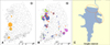
Nineteen viruses were isolated from cloacal and tracheal swab samples of a flock of Baikal teal from Donglim reservoir. The sampling area was mainly in the north-east area of Donglim reservoir (panel C in Fig. 1), where 89 Baikal teal, seven bean goose, one common coot, and one whooper swan were found dead.
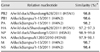
Subsequently, all eight gene segments of the 19 different viruses were sequenced by high-throughput sequencing using the Ion Torrent PGM platform. The number of obtained sequencing reads from each sample was between 1,207 and 48,200 (average of 15,736 reads). Furthermore, mapping of the reads to the reference genome (A/baikal teal/Korea/Donglim3/2014 (H5N8); GenBank accession No. KJ413847-KJ413854) was performed at a depth of 124.43–11177.50 (Supplementary Fig. 1). The reads covered 99.88–100% of the total genome, which suggests that all eight genes were completely sequenced. Homology analysis of the nineteen viral genome sequences showed that most of the genes shared high nucleotide sequence identity of 99.7% to 100% (Supplementary Fig. 2). All 19 viral isolates were homologous to viral isolates from Eastern China (Table 1), with 97% to 99% similarity at the nucleotide level. These viruses were also homologous to previous Korean isolates of H5N8 from Buan (GenBank accession No. KJ413839-KJ413846) and Donglim (KJ413847-KJ413854), with 99.6% similarity. Deduced amino acid sequence analysis of the complete genome sequence compared to sequences of Buan and Donglim strains, revealed that viruses sequenced in this study had 1 to 8 variations (Table 2).
Compared to the sequences of the nineteen viral isolates, the PB2 and HA genes in A/wild duck/Shandong/628/2011 (H5N1) showed 98.8% and 97.2% to 97.3% similarity, and NP had 98.9% to 99.0% similarity with the respective sequence in the A/wild duck/Shandong/1/2011 (H5N1). The PB1, PA, NS, and M genes of the nineteen viruses showed 98.6%, 98.2% to 98.4%, 98.7% to 98.8%, and 98.7% to 98.9% similarity to the respective nucleotide sequences of A/duck/Jiangsu/1-15/2011 (H4N2), whereas the NA genes were closely related to those of A/duck/Jiangsu/k1203/2010 (H5N8) with 97.9% to 98.1% similarity.
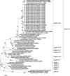
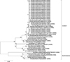
Phylogenetic analysis of the HA genes indicated these viruses belonged to H5 clade 2.3.4.4. (Fig. 2). The HA genes of the viral isolates in this study were located in the same cluster as the H5 Eastern China isolates, such as A/wild duck/Shandong/628/2011 (H5N1). These HA genes also formed a branch with other Korean H5N8 isolates available in GenBank: A/breeder duck/Korea/Gochang1/2014 (Gochang1), A/broiler duck/ Korea/Buan2/2014 (Buan2), and A/baikal teal/Korea/Donglim3/ 2014 (Donglim3). The HA genes had 99.6% similarity to those in Buan2 and Donglim3 and 96.4% to 96.6% similarity with Gochang1 (GenBank accession No. KJ413831–413838). The NA genes were 99.8% similar to those in Buan2 and Donglim3 and had 97.8% to 97.9% similarity with those in the Gochang1 isolates. Phylogenetic analysis of the NA genes indicated that these viruses belonged to the N8 subtype of the Eurasian lineage, and they clustered with the H3N8 isolates (Fig. 3). Phylogenetic analysis of the six internal genes indicated that these 19 strains were reassortant viruses with genes derived from H5N2, H4N2, H5N5 and H5N8 viruses from eastern China.
All of the viruses in this study have a highly pathogenic motif with multiple basic amino acids, such as PLRERRRKRG at the HA cleavage site (Table 3), and they have Q222 and G224 at the receptor-binding sites of the HA gene, similar to H5N8 viruses. The other amino acid sequences were similar to those of clade 2.3.4.4 viruses, with the exception of K156A in antigenic site A and S94T in antigenic site D. Although the neuraminidase protein contained I314V and the matrix 2 protein contained an S31N substitution, other mutations resulting in oseltamivir and amantadine resistance were not evident (Table 4). Other amino acid residue changes known to be relevant for virulence or transmission included 627E and 701D in the PB2 protein (Table 5). The NS1 proteins had a length of 237 amino acids and the ESEV PDZ-binding motif (PBM) with additional RGNKMAD amino acids in the C terminal region [11].
Discussion
On January 17, 2014, an outbreak of HPAI H5N8 was reported in the Donglim reservoir in Gochang, Korea. AIV infections in wild waterfowl are usually mild or asymptomatic [2]; however, this virus caused deaths in the Baikal teal (Anas Formosa) [10]. In addition, during the outbreak period, H5N8 infection was identified in carcasses, feces, and cloacal and tracheal swabs of wild birds such as the bean goose (Anser fabalis), common coot (Fulica atra), and mallard (Anas platyrhyncos). In this study, we sequenced and analyzed the complete genome sequences of 19 H5N8 viral isolates using high-throughput sequencing with an Ion Torrent PGM platform and provided detailed information regarding their molecular characteristics.
Two opinions exist regarding the origin of the H5N8 viruses, Gochang1 and Buan2. In March 2014, Lee et al. [10] suggested that the H5N8 viruses were reassorted in Eastern China and introduced by migratory birds. In May 2014, Wu et al. [18] described a novel reassortant H5N8 virus isolated in 2013 that is the precursor of Gochang1. However, Ku et al. [6] suggested that Buan2 may have reassorted in Korea because the HA gene of A/waterfowl/Korea/S005/2014 (H5N8) has 97% homology to the closest matching gene in GenBank. Phylogenetic analysis of our H5N8 viruses showed that they are genetically similar to viruses isolated from Eastern China since 2010 [3]. The PB2, HA, NP, and NA genes were from H5 subtype viruses, and four other genes were from the H4N2 strain A/duck/Jiangsu/1-15/2011 (KC282877-78, KC282882-83). According to Zhao et al., this H4N2 virus was a natural reassortant with regard to the HA gene from A/wild duck/Korea/CSM4-28/2010 (H4N6) and seven other genes from HPAI H5N2 strains, including A/duck/Eastern china/1111/2011 and A/goose/Eastern China/1112/2011 [19]. Therefore, the 19 viral isolates in this study were likely derived from HPAI H5 reassorted viruses, and their origin appears to be in China. Additionally, H5N8 viruses identified in Korea were genetically similar to H5N8 viruses in clade 2.3.4.4 reported in Europe, Japan, and North America since late autumn 2014 [8].
The HA of the H5N8 viruses contains Q226 and G228, which indicates that these viruses preferentially bind to avian-like α-2,3 sialic acid linkages on host receptors. The NA of the H5N8 viruses has the I314V substitution, which is a molecular marker for oseltamivir resistance [10]. However, other molecular markers of oseltamivir resistance (I117V, E119V, D198N, H274Y, R292K and N294S) and zanamivir resistance (V116A, R118K, E119G/A/D, Q136K, D151E, R152K, R224K, E276D, R292K and R371K) [12] were not observed (N2 numbering). The PB2 protein contained the mutations 627E and 701D, and the NS1 protein contained the ESEV motif at the C-terminus. In particular, when the 19 H5N8 viruses were compared with Buan2, some point mutations became evident and were presumed to be host adaptations resulting from neutral evolution of the Baikal teal. PB2 of 2402 (KJ756575) showed the most changes, with variations in 11 nucleotides corresponding to six amino acid changes: A59G, R70I, G682R, V690L, G693E, and F694I. G693E may alter the nuclear localization signal (NLS) [14], whereas the potential function of the other substitutions is unknown.
In summary, 19 H5N8 viruses were isolated from Baikal teal in Donglim reservoir in Gochang, Korea at a time when an H5N8 outbreak was occurring in both poultry and wild birds. Phylogenetic analysis showed that the H5N8 outbreak strains were related to highly pathogenic H5 and H4N2 reassortant viruses that originated from Eastern China. Although the route of virus spread still needs to be clarified, our results revealed a close relationship between HPAI isolates identified in 2014 from Korea and Eastern China. Therefore, enhanced surveillance of AIV from poultry farms and wild bird populations could increase the epidemiological understanding of HPAI and facilitate the design of prevention strategies.
 XML Download