

 PDF
PDF ePub
ePub Citation
 Print
Print


Newcastle disease, which is caused by avian paramyxovirus serotype 1 (APMV-1), also known as Newcastle disease virus (NDV), causes one of the most devastating poultry diseases and leads to huge economic losses [1]. NDV belongs to the Avulavirus genus of the Paramyxoviridae family [2], which can be divided into two distinct classes (I and II) within a single serotype. Class II viruses are more prevalent than Class I viruses and divided into nine genotypes, designated I–IX. Based on the virulence of disease in chickens, NDVs have been categorized into lentogenic, mesogenic, and velogenic strains. In addition, NDV are classified into five pathotypes based on the disease induced in chickens under laboratory conditions, viscerotropic velogenic NDV (vvNDV), neurotropic velogenic NDV, mesogenic NDV, lentogenic respiratory NDV, and asymptomatic enteric NDV [3].
In addition to NDV, highly pathogenic avian influenza (HPAI) is a devastating poultry disease that negatively impacts the economy of the poultry industry. In previous studies, the feather tropism of HPAI subtype H5N1 was observed in experimentally infected birds [5678]. Feathers that easily detach from infected birds have the potential to transmit pathogens. Resistance to NDV in the environment permits spread of the virus [3], but little is known about persistence of NDV in feather pulps derived from infected birds. In this study, we investigated viral replication in feather pulps of chickens inoculated with vvNDV and evaluated the potential risk of viral transmission.
Ten nine-week-old specific pathogen-free white leghorn chickens (Namduck Sanitec, Korea) were used in this study. Chickens were challenged intramuscularly with 105.5 EID50 of virulent NDV genotype VII virus strain Kr-005/00 under biosafety level 2-enhanced conditions. Clinical manifestations (depression, diarrhea, and neurologic signs) and mortality were observed on a daily basis. To determine vvNDV replication, oropharyngeal and cloacal swabs and feather pulp samples were collected 0, 2, 3, and 5 days post infection (dpi) and suspended in 1 mL of phosphate-buffered saline (PBS). Three feather pulp samples were taken from the wings of each bird and pooled. Of this suspension, 200 µL were used for RNA extraction using an RNeasy Mini Kit (Qiagen, USA) according to the manufacturer's instructions. The content of the NDV viral genome was quantified based on the cycle threshold value using matrix gene-based real-time reverse transcription PCR as previously described [4]. For histopathology and immunohistochemistry (IHC), feather pulps were fixed in 10% neutral-buffered formalin, processed, embedded in paraffin, sectioned, stained with hematoxylin and eosin, and examined by light microscopy. Feather pulps were processed via IHC to detect viral nucleoprotein. Unstained sections were subjected to heat mediated antigen retrieval by microwaving for 3 min in 10 mM sodium citrate buffer. Following antigen retrieval, samples were blocked in PBS containing 5% normal horse serum. The primary antibody, made in rabbits, was an anti-peptide against nucleoprotein diluted 1:8,000. The slides were incubated with biotinylated goat anti-rabbit IgG antibody (Vector Laboratories, USA) and then with Streptavidin-HRP (Vector Laboratories) at room temperature. The reaction was revealed using a diaminobenzidine.
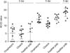
Chickens showed severe depression and reached 100% mortality within 5 days post inoculation (mean death time = 4.0), confirming proper virus inoculation. No virus was detected from swabs and feathers before the challenge. At 2 dpi, viral RNA was detected in the respiratory tract, digestive tract, and feather samples of the chickens. Viral replication was positive for oropharynx, cloaca, and feather pulp in 70%, 80% and 60% of the chickens, respectively (Fig. 1). At 3 dpi, all 10 chickens were positive for viral RNA detection, with the highest frequency observed in the oropharynx, cloaca, and feather pulp. At 5 dpi, all feathers detached from bodies of chickens were positive for viral replication. Upon histopathological examination, infected chickens at 3 dpi exhibited focal necrosis of feather epidermal cells, moderate amounts of heterophilic and lymphocytic infiltration in the inner feather pulp and acute heterophilic perivascular cuffing around small capillaries at the epidermal junction (panel B in Fig. 2). We expect that if the animal had survived several more days, larger vessels in the dermis would have perivascular cuffs with lymphocytes and heterophils. In addition, viral nucleoprotein was detected in a group of feather epidermal cells at 5 dpi (panels C and D in Fig. 2).
To the best of our knowledge, this is the first report that shows vvNDV genotype VII strain can efficiently replicate in the feather pulp of chickens. This raises the possibility that feathers may play an important role in viral transmission. Feathers can easily fall off the bodies, blow away, or be reduced to dust, suggesting that feathers of NDV infected poultry can be a potential source of transmission, along with their feces and respiratory secretions. In addition, feathers can be easily collected from live or dead birds, and thus serve as suitable samples for diagnosis of NDV in chickens.
 XML Download