

 PDF
PDF ePub
ePub Citation
 Print
Print


Introduction
Intercostal thoracotomy is widely performed to explore the thoracic cavity and surgically manage thoracic diseases including vascular anomalies, mediastinal diseases, cardiac diseases, and diseases within ipsilateral lung lobes in dogs and cats [6,10,16]. Thoracotomy is one of the most painful surgical procedures currently performed [3,7,13]. Pain after intercostal thoracotomy has been associated with surgical trauma (transecting muscles, spreading the ribs, and neurovascular compression with suture during closure), mental state of the patient, and the anesthetics used; this can lead to hypoventilation, increased morbidity, prolonged hospitalization, and delayed recovery [14,15]. Therefore, analgesia is necessary for patients undergoing intercostal thoracotomy to reduce complications associated with pain.
Intrapleural and intercostal anesthetics, epidural morphine, and systemic opioids have been routinely used for analgesia with varying efficacy [2,7,14]. Alternately, cryoanalgesia, localized freezing of the intercostal nerves, can offer both short- and long-term pain relief [11]. For this technique, a probe tip - 50 to - 70℃ is applied to peripheral nerves to induce second-degree nerve lesions [11]. Transcostal sutures can be placed so as not to entrap the caudal neurovascular bundles during intercostal thoracotomy closure [15]. The sutures are threaded through holes in the ribs instead of circumcostal placement [15].
A muscle-sparing thoracotomy, which preserves the latissimus dorsi, scalenus, serratus ventralis muscles, and pectoral muscles, can be used to decrease postoperative pain-related morbidity without compromising adequate organ exposure. To the best of our knowledge, there are no published data comparing the muscle-sparing intercostal thoracotomy with the traditional technique. Therefore, the purpose of the present study was to compare pain, duration of approaching and closure, and surgical exposure associated with intercostal thoracotomy between the muscle-sparing and traditional techniques in 20 dogs. We hypothesized that sparing the muscles would provide a less painful recovery after intercostal thoracotomy, proper time for surgical approach and closure, and an excellent surgical window without compromising exposure of the target organs.
Materials and Methods
Animals
Twenty privately owned adult dogs (weight range, 1.3 to 50 kg) were used for this prospective clinical trial. Ten dogs underwent the muscle-sparing technique and the other 10 were subjected to the traditional technique. All animals had an intercostal thoracotomy for reasons including patent ductus arteriosus ligation, lung lobectomy, and mass resection. Preoperative assessment included measurement of baseline heart rate and respiratory rate while resting and before anesthesia. The dogs were randomly assigned to the muscle-sparing technique (n = 10) or traditional technique (n = 10) groups. Ten dogs in the muscle-sparing group underwent a left intercostal thoracotomy at the third (n = 1), fourth (n = 6), or fifth (n = 3) intercostal space. Ten dogs in the traditional technique group were subjected to a left intercostal thoracotomy at the third (n = 1), fourth (n = 7), or fifth (n = 2) intercostal space. The same surgeon (HY Yoon) performed all procedures for the 20 dogs.
Preoperative management
The dogs were premedicated for surgery with glycopyrrolate (0.01 mg/kg, IM, Tabinul injection; Hana Pharm, Korea) and acepromazine (0.05 mg/kg, IM, SEDAJECT injection; Samu Median, Korea) followed by induction of anesthesia with propofol (6 mg/kg, IV, Provive injection; Claris Lifesciences, India). The dogs also received cefazolin (20 mg/kg, IV, CKD Cefazolin injection; ChongKunDang Pharm, Korea) at the time of anesthetic induction. The dogs were intubated and anesthesia was maintained with isoflurane (Ifran Solution; HANA Pharm, Korea) and oxygen. Normal saline was administered intravenously at a rate of 10 mL/kg/h until completion of the surgical procedure. All dogs were ventilated by intermittent positive pressure ventilation (15 cm H2O peak pressure and 5 cm H2O peak positive end expiratory pressure). End tidal CO2 was monitored with a capnograph (Datex Ohmeda FM CO2 Monitor; Datex-Ohmeda, Finland) to ensure adequate ventilation.
Muscle-sparing technique
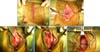
For the muscle-sparing group, skin, subcutaneous tissue, and cutaneous trunci muscle incisions extended from 2 cm ventral to the rib head to near the sternum (panel A in Fig. 1). The ventral border of the latissimus dorsi muscle was bluntly dissected using Integra Miltex Vantage Metzenbaum scissors (Intergra LifeSciences, Germany) and a saline-moistened index finger, cranially, caudally, and dorsally as far as possible (approximately from the third to the seventh rib; panel B in Fig. 1). The latissimus dorsi muscle was retracted dorsally using a Senn-Miller retractor (Professional Hospital furnishers, Pakistan; panel C in Fig. 1). The scalenus muscle was detached from the rib (panel C in Fig. 1). Serrations of the serratus ventralis muscle were split at the intended intercostal space (panel D in Fig. 1). A Senn-Miller retractor was then placed for pectoral muscles retraction (panel D in Fig. 1) and the intercostal muscles were transected. A Balfour retractor (Professional Hospital furnishers) was placed for rib and muscle retraction. The side blades were used for rib retraction, and a center blade was used to retract the latissimus dorsi and serratus ventralis muscles (panel E in Fig. 1). Closure was preformed by apposition of the ribs followed by musculature attachment to the detached areas with routine placement of subcutaneous and skin sutures using 3-0 polyglycolic acid (Safil; B. Braun Melsungen, Spain) and 3-0 nylon (Nylon; Namhae, Korea) respectively. Negative pressure was re-established by a thoracostomy tube (All-silicone thoracic catheter; Sewoon medical, Korea) placed in the thorax.
Traditional technique
For the traditional technique group, skin, subcutaneous tissue, and cutaneous trunci muscle incisions extended from 2 cm ventral to the rib head to near the sternum. The incisions were deepened through the latissimus dorsi muscle. The scalenus, pectoral, serratus ventralis, and intercostal muscles were then transected. A Finochietto retractor (Solco Biomedical, Korea) was used for rib retraction. Closure was performed by apposition of the ribs and routine placement of musculature, subcutaneous, and skin sutures using 3-0 polyglycolic acid (Safil; B. Braun Melsungen) and 3-0 nylon (Nylon; Namhae) respectively. Negative pressure was re-established with a thoracostomy tube placed in the thorax.
Postoperative management
Postoperatively, all dogs in both groups received butorphanol (0.2 mg/kg, IV, BUTOPHAN injection; Myungmoon Pharm, Korea) before extubation. The animals recovered in an isolated environment.
Numerical pain scoring
The pain score for each dog was determined (Table 1) based on a modified pain scoring technique previously described [17]. The following behavior patterns were observed for pain evaluation: gait, behavior, appetite, agitation, and vocalization. Pain scores (0 to 2) were assigned by an observer (HY Yoon) using data from the pain assessment criteria on the day of surgery immediately before anesthesia and 2 h after surgery. The same observer assigned pain scores 1, 2, 3, 4, 5, 6, and 7 days after surgery. Behavior patterns were assessed before any other observations or manipulations planned for the day were performed. Heart rate, respiratory rate, and wound palpation were then monitored. A numerical pain score was calculated for each observation point based on heart rate, respiratory rate, behavior patterns, and wound palpation. Heart rate and respiratory rate were scored based on comparisons to preoperative measurements.
Measurement of the duration of approaching and closure
Approaching time was measured from when the skin incision was made until a Balfour retractor or Finochietto retractor was placed. Time for closure was measured from apposition of the ribs until the skin was sutured. The extent of intrathoracic organ exposure for surgery was evaluated for each technique after placement of a Balfour or Finochietto retractor.
Statistical analysis
All data are expressed as the mean ± standard deviation (SD). A Mann-Whitney test was used to compare mean values for weight, age, and duration for approaching and closure between the two groups. Comparison of pain scores was made with a Satterthwaite t-test or pooled t-test. Data analysis was performed with SAS software (ver. 9.1; SAS Institute, USA). A p value ≤ 0.05 was considered significant.
Results
Animals
The mean ± SD weight of the dogs was 10.5 ± 15.8 kg for the muscle-sparing group and 7.3 ± 9.3 kg for the traditional technique group. There was no significant (p = 0.868) difference in weight between the two groups. The mean ± SD age of the dogs was 4.5 ± 3.6 years for the muscle-sparing group and 5.7 ± 4.8 years for the traditional group. There was no significant (p = 0.866) difference in age between the two groups.
Numerical pain scores
The mean ± SD numerical pain scores before anesthesia along with 2 h or 1, 2, 3, 4, 5, 6, and 7 days after surgery are presented in Table 2. There were significant differences in numerical pain scores at 2 h as well as 1, 2, 3, 4, 5, 6, and 7 days after surgery when the two groups were compared (p < 0.0001).
Duration of approaching and closure
The mean ± SD approaching time was 8 min and 50 sec ± 53 sec for the muscle-sparing technique, and 9 min and 5 sec ± 1 min and 49 sec for the traditional method. There was no significant difference (p = 0.725) in approaching time between the two methods. Mean ± SD time for closure was 10 min and 39 sec ± 1 min and 8 sec for the muscle-sparing technique, and 11 min and 4 sec ± 1 min and 39 sec for the traditional technique. There was no significant (p = 0.590) difference in time for closure between the two groups.
Adequate exposure for surgical procedures
Compared to the traditional method, the muscle-sparing technique also achieved the desired exposure without compromising exposure of target organs when the center blade of a Balfour and Senn-Miller retractor was placed for retraction of the latissimus dorsi and serratus ventralis muscles or pectoral muscles, respectively.
Discussion
Currently available approaching options for intercostal thoracotomy are transection of the thoracic muscles such as the latissimus dorsi, scalenus, pectoral, and serratus ventralis muscles, and sparing the thoracic muscles [9,12]. Transection of the thoracic muscles, known as the traditional technique, is associated with substantial postoperative pain that has been described as the main factor for increased morbidity, delayed recovery, and longer hospitalization [15]. Our results suggest that the muscle-sparing technique is superior to the traditional method due to less pain during recovery in the first 7 days after intercostal thoracotomy. Additionally, the muscle-sparing technique is as effective as the traditional method for providing an appropriate time for approaching and closure during intercostal thoracotomy and adequate exposure for surgery.
Muscles of the thorax, including the trapezius, latissimus dorsi, serratus ventralis, serratus dorsalis, scalenus, pectoralis, longissimus thoracis, and iliocostalis, form five layers that lie alongside and above one another [4]. Nerves that serve the thorax include the long thoracic nerve, dorsal thoracic nerve, lateral thoracic nerve, ventral thoracic nerve, and caudal pectoral nerve, as well as cutaneous and muscular branches of the thoracic nerves such as dorsal, lateral, and ventral cutaneous branches along with proximal and distal muscular branches [5]. Transecting the thoracic muscles and nerves has been described as the predominant factor that causes pain after an intercostal thoracotomy [17]. For the traditional technique, the thoracic muscles should be transected to enter the thoracic cavity, a process that incurs nerve injury. In particular, the latissimus dorsi, serratus ventralis, scalenus, and pectoralis muscles as well as the long thoracic nerve running on the serratus ventralis, dorsal thoracic nerve with innervation of the latissimus dorsi, lateral thoracic nerve lying between the latissimus dorsi and deep pectoral muscles, caudal pectoral nerves with innervation of deep pectoral muscle, and many cutaneous and muscular branches of the thoracic nerves should be incised to perform thoracotomy at the third, fourth, or fifth intercostal space.
Several attempts have been made to decrease pain associated with intercostal thoracotomy in dogs [11,15,17]. Thoracoscopic pericardectomy and open pericardectomy have been performed, and comparisons of postoperative pain with morbidity have been made [17]. In these comparisons, thoracoscopic pericardectomy was found to have a few advantages over open pericardectomy including decreased pain and reduced postoperative morbidity. However, some procedures still necessitate an intercostal approach. Transcostal sutures have been recommended for thoracotomy closure [15]. For this, transcostal closure is accomplished by drilling 5~6 holes in the fifth rib and passing sutures through the holes and around the fourth rib. This method spares the neurovascular bundle; however, the procedure can be time-consuming and cause pain due to drilling the holes in the rib. In a 2001 experimental study, localized freezing of the intercostal nerves was performed to evaluate the effect of cryoanalgesia on post-thoracotomy pain and the structure of intercostal nerves in dogs [11]. Cryoanalgesia provided post-thoracotomy pain relief and did not cause any long-term histological damage to the intercostal nerves. In the present investigation, a muscle-sparing technique was performed by dissecting rather than incising the muscles. This method decreased pain experienced during recovery, and allowed proper time for surgical approaching and closure. A muscle-sparing technique could also be helpful for cases in which surgical procedures require an intercostal approach instead of a thoracoscopic approach.
A few postoperative pain studies comparing thoracic surgical procedures based on a numerical pain scale using behavioral observations, blood glucose and plasma cortisol concentrations, heart rate, respiratory rate, and pain threshold tests have been performed in veterinary medicine [3,8,15,17]. Postoperative pain assessment using blood glucose and plasma cortisol levels as well as pain threshold tests incur additional expenses. Economic considerations are significant when planning the management of veterinary patients. In the present study, objective measurements of pain such as heart rate and respiratory rate along with numerical pain scores based on wound palpation and behavior patterns such as gait, appetite, agitation, and vocalization were used for pain evaluation.
In previous studies comparing thoracic surgical procedures, short-term follow-up (53 h and 24 h after surgery) has been performed for postoperative pain assessment; therefore, only comparisons of pain experienced immediately after surgery have been described [15,17]. In the present study, postoperative pain was evaluated until 7 days after surgery. Dogs in the muscle-sparing group had significantly reduced pain scores at 2 h as well as 1, 2, 3, 4, 5, 6, and 7 days after surgery. In a 2003 human prospective study, postoperative pain was evaluated until 8 days after surgery in patients who underwent a muscle-sparing thoracotomy or standard posterolateral thoracotomy [1]. It was reported that the muscle-sparing thoracotomy resulted in less postoperative pain 8 days after surgery. Compared to the traditional method, the muscle-sparing technique we performed could provide more effective pain management, shorter periods of pain management, and shorter hospital stays after intercostal thoracotomy.
There are no reports in the veterinary literature on muscle-sparing thoracotomy as a surgical method. Despite this lack, actual cases requiring the procedure may be more than what is reported. This may be due to misconceptions regarding longer duration of approaching and closure, and inadequate exposure for the surgical procedure. Additionally, surgeons may be reluctant to perform this technique due to a lack of information about clinical outcomes of muscle-sparing thoracotomy in the literature. For the muscle-sparing thoracotomy performed in the present study, approaching and closure were completed within 8 min and 50 sec, and 10 min and 39 sec, respectively. These times are not different compared to those required for the traditional technique. A human study performed in 2003 also indicated that there is no difference in surgical approaching time between a muscle-sparing thoracotomy and standard posterolateral thoracotomy [1]. The latissimus dorsi muscle originates from the spinous processes of the lumbar vertebrae and the last seven or eight thoracic vertebrae, and lies caudal to the shoulder on the dorsal half of the lateral thoracic wall [4]. The deep pectoral muscle is a broad muscle lying ventrally on the thorax and extends between the sternum and humerus [4]. In the present investigation, sharp dissections along the ventral fascial attachments of the latissimus dorsi muscle and dorsal border of the deep pectoral muscle were made to form wide and vertically long intercostal spaces or for ventral exposure, respectively. Scalenus muscle detachment from the rib and serration splitting of the serratus ventralis also enabled middle and dorsal exposure of the intercostal spaces, respectively. Compared to the traditional method, the muscle-sparing technique also achieved the desired exposure. The similar time required for approaching and closure as well as adequate exposure needed for surgery without compromising the exposure of target organs suggest that the muscle-sparing technique is acceptable for intercostal thoracotomy.


 XML Download