

 PDF
PDF ePub
ePub Citation
 Print
Print


Introduction
Brucellosis is a zoonotic disease caused by the Gram-negative genus Brucella [3]. Brucellosis has been widely disseminated throughout the world. In animals, brucellosis causes epididymitis in males and infertility or abortion in pregnant females. In humans, brucellosis causes inflammation and undulating fever [18]. However, there is no effective or approved Brucella vaccine for humans. Therefore, new low residual virulence marker vaccines that offer high levels of protection are required.
The live vaccine, Brucella (B.) abortus strain 19 (S19), is a spontaneously attenuated mutant [6] that deletes the erythritol catabolic gene. S19 had been used to prevent the infection of B. abortus. Some studies have shown that S19 provided protection against cattle abortion. S19 is also effective at protecting animals against B. abortus infections. S19 is an effective vaccine in animals that has been widely applied [15]. However, the vaccine is less effective, can induce abortion and milk excretion, and interferes with classical serological diagnostic tests [15]. Accordingly, one potential approach to these problems is development of a marker vaccine through deletion of virulence genes from these parental vaccine strains with good immunogenicity and vaccine efficacy. However, a great deal of research is needed to develop live vaccines against B. abortus that are superior to S19.
Lipopolysaccharides (LPS) are important virulence factors that have special pathogenicity. The LPS of Brucella has three structural regions: the O-antigen, the core oligosaccharide and lipid A. At present, Brucella LPS encodes 32 virulence factors [5812]. WbkA, which is one of these virulence factors, encodes N-formyl-glutamine transferase, which is essential to biosynthesis of the Brucella LPS O antigen. The deletion or expression of the truncated form of wbkA may affect the antigenic structure of Brucella. Early studies have shown that B. melitensis 16MΔwbkA is a rough mutant, and deletion of the wbkA flanking transposase improves the stability of B. melitensis Rev 1 vaccine. In this report, we describe the construction of a B. abortus 2308ΔwbkA mutant (2308ΔwbkA) and evaluate its role in intracellular multiplication in RAW 264.7 macrophages and protection of mice with the goal of producing a candidate vaccine against B. abortus 2308.
Materials and Methods
This study was approved by the Institutional Committee of the Post Graduate Studies and Research at Shihezi University (China). All efforts were made to minimize animal suffering.
Bacterial strains, plasmids, cells and mice
B. abortus strain 2308 (S2308) and the vaccine strain S19 were obtained from the Center of Chinese Disease Prevention and Control (Beijing, China). Brucella was cultured in tryptone soy agar (TSA) or tryptone soy broth (TSB; Oxoid, UK). E. coli strain DH5α was grown on Luria-Bertani (LB) medium. Plasmid pGEM-7Zf+ was purchased from Promega (USA). The RAW 264.7 murine macrophage was purchased from Cell Resource Center, IBMS, CAMS/PUMC (Beijing, China). Six-week-old BALB/c female mice were obtained from the Experimental Animal Center of Academy Military Medical Science (Beijing, China). All experimental procedures and animal care was performed in compliance with institutional animal care regulations.
Construction of the 2308ΔwbkA
The deletion mutant 2308ΔwbkA was constructed with pGEM-7Zf+ as follows. Two pairs of primers with restriction sites at the 5' ends were designed for amplification of the upstream (1100 bp) and downstream (1086 bp) arms of the S2308 wbkA, in which the Xba I, Kpn I, Kpn I, and Sac I (italicized) sites were integrated into both PCR fragment ends. The designs of the primers were based on B. abortus 2308. The primer sequences were as follows: wbkA-N-F, TCT AGA TTA CAG ATG AGC AAT GGA ACC; wbkA-N-R, GGT ACC TCC TTC TAT GAA GCT AAT TGT TTG; and wbkA-C-F, GGT ACC ATT ACT TGC GAA TAT CGG TCG C; wbkA-C-R, GAG CTC GCG TCA TTG GTC TCT TTG CAC. One pair of primers with restriction sites at the 5' ends was designed for amplification of the SacB DNA fragment (1477 bp), which is a selectable marker gene from Bacillus subtilis. The primer sequences were as follows: SacB-F, GAG CTC GGG CTG GAA GAA GCA GAC CGC TA, SacB-R, GAG CTC GCT TAT TGT TAA CTG TTA ATT GTC C. The two homologous arms and the SacB DNA fragment were sequentially cloned into the multi-cloning sites of pGEM-7Zf+ (wbkA-N cloned at the Xba I/Kpn I sites, wbkA-C at the Kpn I/Sac I sites and SacB at the Sac I site) to generate the suicide plasmid, pGEM-wbkA-SacB (pWS). Competent S2308 were electroporated with pWS and potential wbkA deletion mutant 2308ΔwbkA was isolated in the presence of 100 µg/mL ampicillin for the first screening and 5% sucrose for the second screening. The mutant was further confirmed by PCR amplification with primers wbkA-I-F (GAA AAC TAC CTC GCA GCC GT) and wbkA-I-R (AGC GGA TGA AAC GGT TGT AG), located in the upstream and downstream portion of the homologous arm of wbkA, respectively. PCR products were sequenced for confirmation. The deletion mutant was further confirmed by PCR amplification and RT-PCR sequencing, as previously described [20].
Complementation of 2308ΔwbkA
To restore the wbkA activity, PCR amplification fragments primers wbkA-N-F and wbkA-N-R, and the products were ligated with pMD19-T simple vector (Takara Bio, Japan) to generate pMD-wbkA, which was then transformed into E. coli DH5α. Plasmid pMD-wbkA was isolated from these transformants and analyzed by agarose gel electrophoresis following restriction digestion, after which it was electroporated into competent 2308ΔwbkA. The transformants were then selected on TSA containing 100 µg/mL ampicillin. The transcription restoration of wbkA in the complementary strain (2308-wbkA) was further confirmed by PCR and RT-PCR.
Evaluation of 2308ΔwbkA attenuation intracellular survival in RAW264.7 murine macrophages
RAW264.7 murine macrophages were seeded in 6 well plates and infected with 2308ΔwbkA, S19, 2308-wbkA or parental strain S2308 at a multiplicity of infection of 150, as previously described [13]. Culture plates were centrifuged at 350 g for 5 min at room temperature, then placed in a 37℃, 5% CO2 incubator. At 45 min post-incubation, the cells were washed three times with phosphate buffered saline (PBS) and then incubated with 50 µg/mL of gentamicin (Invitrogen, USA) for 1 h to kill extracellular bacteria. Subsequently, the culture was replaced with Dulbecco's modified Eagle's medium (DMEM; Gibco BRL, USA) containing 25 µg/mL of gentamicin (time zero). At different time points post-infection, the supernatant was discarded, cells were lysed by PBS containing 0.1% (v/v) TritonX-100, and the live bacteria were enumerated by plating on TSA plates. All assays were performed in triplicate wells, and the results represent the means from at least three separate experiments.
Evaluation of 2308ΔwbkA attenuation virulence in mice
Six-week-old female BALB/c mice were inoculated intraperitoneally (i.p.) with 1 × 10⁶ CFU of 2308ΔwbkA, S19, 2308-wbkA or S2308. Infected mice were held in microisolator cages in biosafety level 3 facilities. Bacterial counts in the spleen were calculated for five mice of each group at 3, 7, 14, 21 and 28 days post-inoculation. Mice were euthanized by CO2 asphyxiation and their spleens were removed aseptically. The spleens were then homogenized in 1 mL PBS containing 0.1% (v/v) TritonX-100, 10-fold serially diluted, and then plated on TSA. The plates were incubated at 37℃ and the number of CFU counted after three days. All assays were performed three times with similar results [22].
Protection efficiency evaluation induced by 2308ΔwbkA in mice
Groups of six-week-old female BALB/c mice (n = 10 per group) were injected i.p. with 1 × 10⁶ CFU (200 µL) of 2308ΔwbkA (experimental vaccine group), S19 (reference vaccine control group) or 200 µL PBS (unvaccinated control group). The mice were challenged i.p. with 1 × 10⁶ CFU per mouse (200 µL) of virulent strain S2308. Mice (n = 5/time point per group) were euthanized at 2 and 4 weeks post challenge, and bacterial CFUs in the spleens were determined as described above. The mean value for each spleen count was obtained after logarithmic conversion. Log units of protection were obtained by subtracting the mean log CFU for the experimental group from the mean log CFU for the control group, as previously described [111]. The experiments were repeated three times.
Evaluation of antibody
Six-week-old female BALB/c mice were randomly divided into three groups. Group 1 and 2 were inoculated i.p. with 1 × 10⁶ CFU of 2308ΔwbkA or S19, respectively, and group 3 was inoculated i.p. with PBS as a negative control. Serum samples were obtained from immunized mice at 3, 7, 14, 21 and 28 days post-immunization, and IgG levels were determined by enzyme-linked immunosorbent assay (ELISA) as previously described [9]. Serum samples were diluted with sample diluent buffer (1: 5). The levels of IgG in the supernatants were measured using an ELISA Quantikine Mouse Kit (R&D Systems, USA) according to the manufacturer's instructions. All assays were performed in triplicate and repeated at least three times.
Cytokine production assay
IFN-γ detection was carried out as previously described [19]. Specifically, IFN-γ was determined using an ELISA Quantikine Mouse Kit (R&D Systems) according to the manufacturer's instructions. All assays were performed in triplicate and repeated at least three times.
Cloning, expression, and purification of recombinant protein
The WbkA open reading frame was amplified by PCR of DNA from S2308, after which the amplified DNA fragment was cloned into the pET-32a vector (Novagen, USA) and expressed in E. coli BL21 as N-terminally His-tagged recombinant protein. The expression of the recombinant protein was analyzed by SDS-PAGE (12%). The recombinant protein, WbkA, was then purified by affinity chromatography with Ni2-conjugated Sepharose (GE Healthcare Life Sciences, USA).
Western blotting
The recombinant protein WbkA cell lysates were analyzed by Western blotting as previously described [21]. The membrane with recombinant protein WbkA cell lysates was then incubated with a primary Brucella-vaccinated sera and sheep anti-mouse IgG (horseradish peroxidase conjugated; EarthOx Life Sciences, USA) secondary Ab. The membrane was developed using an enhanced HRP-DAB substrate color kit (TIANGEN Biotech, China).
WbkA indirect ELISA (iELISA)
Serum samples were obtained from Brucella-infected mice. Antibody responses to the purified recombinant WbkA protein were estimated via WbkA-based iELISA, as previously described [14].
Results
Construction and complementation of the B. abortus 2308ΔwbkA mutant
The wbkA gene deletion mutant of B. abortus strain 2308 (2308ΔwbkA) was successfully obtained. A 433 bp DNA fragment was amplified from 2308ΔwbkA, but a 1131 bp DNA fragment was amplified from S2308 with primers wbkA-I-F and wbkA-I-R, indicating that wbkA was correctly knocked out. These findings were subsequently confirmed by sequencing (data not shown). The complementary strain was constructed by integration of wbkA at the disrupted locus of 2308ΔwbkA. RT-PCR showed that wbkA was not transcribed in 2308ΔwbkA, but was restored in 2308-wbkA (data not shown). Taken together, these findings indicate that both 2308ΔwbkA mutant and complementary strain 2308-wbkA were correctly constructed.
2308ΔwbkA mutant is attenuated relative to B. abortus 2308 based on survival in RAW 264.7 murine macrophages
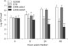
RAW 264.7 murine macrophages were infected with 2308ΔwbkA, S19, 2308-wbkA and S2308, after which their survival capability in macrophages was determined. Macrophages were infected with the three strains at 1:150, and the surviving bacteria were calculated. At 4 h post-infection, no differences in the amount of bacteria were observed among strains (Fig. 1). By 8 h post-infection, there was a 1.08 log (p < 0.05), 0.87 log (p < 0.05) and 0.27 log (p > 0.05) decrease in the number of 2308ΔwbkA bacteria compared to that of S2308 and 2308-wbkA or S19. At 24 h post-infection, there was a 3.19 log (p < 0.01), 3.05 log (p < 0.05) and 1.11 log (p < 0.05) decrease in the number of 2308ΔwbkA bacteria compared to that of S2308, 2308-wbkA or S19 bacteria, respectively, indicating that the numbers of 2308ΔwbkA were significantly lower than in these strains. At 48 h post-infection, decreases of 4.23 log, 4.13 log and 1.27 log were observed inside the macrophages (p < 0.01) (Fig. 1). These results showed that 2308ΔwbkA mutant had a decreased survival capability in RAW 264.7 murine macrophages relative to S2308, 2308-wbkA and S19, indicating that 2308ΔwbkA was attenuated compared with B. abortus 2308 based on survival in RAW 264.7 murine macrophages.
2308ΔwbkA mutant is attenuated compared with B. abortus 2308 based on survival in BALB/c mice
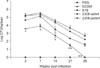
To determine the survival capability in BALB/c mice, animals were inoculated i.p. with 1 × 10⁶ CFU of 2308ΔwbkA, S19, 2308-wbkA and S2308. When compared with S19, 2308-wbkA and S2308, splenic CFU in 2308ΔwbkA infected mice were significantly reduced (p < 0.01) at days 3, 7, 14, 21, and 28. At 28 days post-inoculation, 2308ΔwbkA had been cleared from the spleens of mice (Fig. 2). Taken together, these results showed that 2308ΔwbkA mutant was greatly attenuated in BALB/c mice.
2308ΔwbkA induces immune protection against challenge with B. abortus 2308
To determine the protection efficiency of 2308ΔwbkA, mice were vaccinated i.p. with 1 × 10⁶ CFU of 2308ΔwbkA, S19 or PBS. At 4 weeks post-vaccination, mice were challenged with 1 × 10⁶ CFU (200 µL) of virulent strain S2308. The mice immunized with 2308ΔwbkA exhibited significantly fewer splenic Brucella than non-immunized mice at 2 (2.22 log units) and 4 (1.76 log units) weeks post-challenge (p < 0.05) (Table 1). As expected, S19 also induced significant protection at 2 (1.83 log units) and 4 (1.34 log units) weeks after challenge, and the protective efficacy of S19 was lower than that in the 2308ΔwbkA-vaccinated mice. The 2308ΔwbkA showed similar protective efficacy to that of S19 (Table 1). Taken together, these results indicate that 2308ΔwbkA can provide similar protection efficacy against challenge as the S19 vaccine.
2308ΔwbkA induces both humoral and cytokine responses
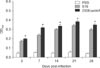
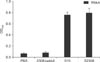
Serum samples from mice inoculated with 2308ΔwbkA, S19 or PBS were obtained from immunized mice at selected intervals post-immunization to monitor total IgG levels by ELISA. For mice inoculated with 2308ΔwbkA and S19, the total IgG levels peaked at 21 days post-inoculation. The 2308ΔwbkA-vaccinated mice expressed slightly higher IgG levels than the S19-vaccinated mice (p > 0.05) and significantly higher IgG levels than the PBS-infected mice (p < 0.05) (Fig. 3).
To characterize the cellular immune response, the IFN-γ levels in the splenocytes of the 2308ΔwbkA and S19 vaccinated mice were evaluated at 28 days post-vaccination. The splenocytes of immunized mice were isolated and stimulated with heat-killed S2308, complete RPMI 1640 medium (Gibco BRL) (negative control) or concanavalin A (positive control). The 2308ΔwbkA-vaccinated mice expressed slightly higher IFN-γ levels in the splenocytes than the S19-vaccinated mice (p > 0.05) and significant higher levels than the PBS-injected mice (p < 0.05) (Fig. 4).
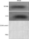
Differentiation of 2308ΔwbkA
Serum from mice inoculated with 2308ΔwbkA, S2308, S19 or PBS were collected to determine whether the WbkA protein can be used as a diagnostic antigen. Western blotting showed that antibodies against the WbkA protein could be detected in the serum of S2308- or S19-inoculated mice, and produced specific bands. However, the antibodies could not be detected in the serum of 2308ΔwbkA- or PBS-inoculated mice (Fig. 5). Furthermore, antibodies against the WbkA protein were detected by iELISA. Serum from S2308- or S19-inoculated mice showed positive reaction and serum from 2308ΔwbkA- or PBS-inoculated mice showed negative reaction according to iELISA using WbkA as antigen. The results of iELISA were similar to those obtained Western blotting (Fig. 6). Taken together, these results indicate that WbkA protein had good reactogenicity and could be used to differentiate vaccination from natural infection by WbkA-iELISA after confirmation of brucellosis infection using LPS-based serological tests.
Discussion
The development of an efficacious vaccine against brucellosis has long been a challenge to scientists. Most currently available vaccines have several limitations, such as residual virulence, interference of serodiagnosis and splenomegaly [24717]. One of the drawbacks to the rational development of new Brucella vaccines is limited virulence. Furthermore, serological interference has limited development of an effective vaccine. The ideal vaccine must be protective and should allow for differentiation of natural and vaccinated infection [15]. LPS are important virulence factors with unique pathogenicity. Brucella LPS encodes 32 virulence factors, including WbkA. To investigate whether 2308ΔwbkA maintains protective efficacy, we constructed B. abortus 2308ΔwbkA mutant and its virulence and protection efficacy were evaluated in cells and mice.
The 2308ΔwbkA was constructed to confirm that the reduced survival capability of the mutant was directly related to the deleted wbkA gene. As shown in this study, 2308ΔwbkA was defective for survival in RAW 264.7 cells and BALB/c mice, and it was cleared faster than S19, within 28 days. This finding showed that the lack of splenomegaly in inoculated mice increased the safety of 2308ΔwbkA as a vaccine.
An ideal Brucella live attenuated vaccine strain must be non-virulent towards the host and induce higher levels of protection. Therefore, we performed the protection experiment in BALB/c mice. We found that the 2308ΔwbkA mutant could provide slightly higher protection than S19.
The cytokine and humoral immune responses have been evaluated for the degree of protection conferred by 2308ΔwbkA. Brucella infects the host cells and mainly causes cellular immunity. Th1 immune responses characterized by production of IFN-γ are associated with protective immunity to Brucella. IFN-γ is a critical cytokine required for macrophage bactericidal activity that is produced by T lymphocytes and is a potent macrophage-activating factor [1016]. IFN-γ plays an important role in killing intracellular Brucella. IFN-γ is antibacterial, and this study evaluated the antimicrobial capacity and cellular immunity of the host. Our results indicated that 2308ΔwbkA induced higher levels of IFN-γ than S19. In addition, one weak challenge, 2308ΔwbkA, induced anti-Brucella containing a high level of IgG. These results showed that immunization with 2308ΔwbkA elicits a Th1 response.
Brucella LPS was the most important antigen during immune response in brucellosis. Current serological diagnostic tests include the standard tube agglutination test, Rose Bengal plate test, and iELISA using LPS antigens of smooth Brucella. However, LPS-based serological tests cannot easily differentiate between the serum of vaccinated and infected animals. Therefore, we used WbkA protein as a diagnostic antigen and detected the antibody profiles in different sera. The results indicated that the antibodies against the WbkA protein could be detected in infected serum, but not in that from 2308ΔwbkA-immunized animals. These results showed that WbkA could be used as a diagnostic antigen to be differentiated from vaccination serum. Furthermore, 2308ΔwbkA vaccination was detected by iELISA. The results revealed that S2308- and S19-infected mice were positive, whereas 2308ΔwbkA-infected mice were negative. Therefore, 2308ΔwbkA allows for differentiation between infected and vaccinated animals.
In this study, 2308ΔwbkA mutant may be another ideal live vaccine candidate for S2308 because of its low virulence in macrophage RAW264.7 and BALB/c mice. Moreover, it provided protection similar to that provided by the S19 vaccine strain. The humoral responses indicated that 2308ΔwbkA could elicit an anti-Brucella-specific IgG response. Moreover, the WbkA protein is an ideal diagnostic antigen for differentiation of immunization from infection by WbkA-iELISA. In future studies, comprehensive protection experiments must be conducted to determine whether the measurable immune responses in systemic compartments confer detectable protection against Brucella infection. In addition, further testing in livestock will determine whether 2308ΔwbkA will be a promising live vaccine candidate.
 XML Download