

 PDF
PDF ePub
ePub Citation
 Print
Print


Introduction
Meningoencephalitis (ME) results from concurrent inflammation of the meninges (meningitis) and brain (encephalitis) [22]. ME can be classified as granulomatous meningoencephalomyelitis, necrotizing meningoencephalitis, or necrotizing leukoencephalitis based on histopathologic features [28]. However, in veterinary practice, this classification is challenging because it requires biopsy and histopathologic examination. Thus, a simple and rapid strategy for identifying the causative agent of ME would be useful for selecting an appropriate therapeutic regimen.
Multiple infectious agents can cause ME in dogs. The major infectious agents include viruses (canine distemper virus [CDV]), fungi (Blastomyces [B.] dermatitidis, Cryptococcus [C.] neoformans, Histoplasma capsulatum, and Coccidioides spp.), bacteria (Borrelia [B.] burgdorferi, Bartonella spp., and Ehrlichia [E.] canis), and parasites (Neospora [N.] caninum and Toxoplasma [T.] gondii) [4151719232531]. The pathogenesis of ME can also involve immune-mediated or idiopathic etiologies that provoke aseptic inflammatory responses in the central nervous system (CNS).
Rapid and reliable detection of the causative pathogens of ME is essential for implementation of timely and effective therapeutic strategies. Routine techniques in veterinary diagnostic laboratories include bacterial and fungal cultures, immunohistochemistry, antigen-capturing enzyme-linked immunosorbent assay (ELISA), and polymerase chain reaction (PCR) [4151719232531]. These methods are laborious, expensive, and time-consuming, and their sensitivity depends on specimen quality, timing of sampling, and prior antimicrobial therapy [7]. Furthermore, different assays for detecting the various pathogens have variable sensitivities and specificities, leading to biased outcomes [3]. Recent studies have used multiplex quantitative real-time PCR (mqPCR) as a diagnostic tool for multifactorial diseases because this approach can simultaneously detect multiple pathogens with good diagnostic performance and is faster than conventional methods [829].
The present study was conducted to develop an mqPCR-based diagnostic approach for ME (hereafter, ME mqPCR) that simultaneously detects eight infectious agents (B. dermatitidis, C. neoformans, N. caninum, B. burgdorferi, Bartonella spp., T. gondii, E. canis, and CDV) and to assess the prevalence of these pathogens among cases of canine ME in South Korea.
Materials and Methods
Biological materials and vectors
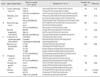
Type strains of C. neoformans (ATCC 32045), Staphylococcus aureus (ATCC 29213, 33592, BAA-44, BAA-41, and BAA-1683), Escherichia coli (ATCC 25922), and Malassezia pachydermatis (ATCC 14522) were purchased from the American Type Culture Collection (ATCC; USA). Field isolates of B. burgdorferi (n = 2), T. gondii (n = 2), E. canis (n = 1), E. ewingii (n = 1), Anaplasma phagocytophilum (n = 2), candidatus Mycoplasma haematoparvum (n = 1), S. pseudintermedius (n = 5), Candida spp. (n = 3), Malassezia spp. (n = 5), Microsporum canis (n = 3), Microsporum gypseum (n = 2), Aspergillus spp. (n = 3), canine parvovirus (n = 3), canine coronavirus (n = 3), and CDV (n = 6) were identified at Chungbuk National University Veterinary Diagnostic Laboratory. Recombinant plasmid vectors for B. dermatitidis, C. neoformans, N. caninum, B. burgdorferi, Bartonella spp., T. gondii, E. canis, and CDV were used to assess the analytical specificity and as positive controls. Nucleic acids (DNA and RNA) extracted from whole blood in two dogs were used as negative controls. All field isolates were identified by culture, conventional PCR, or antigen-capturing ELISA and verified by DNA sequencing. The vectors were constructed by amplifying synthetic oligonucleotides based on sequences obtained from the GenBank database (National Center for Biotechnology Information, USA) using the primers listed in Table 1 and cloned into a T-Blunt PCR Cloning Kit (SolGent, Korea) according to the manufacturer's instructions. Correct insertion into the vector was verified by sequencing. Each recombinant vector was prepared in diethylpyrocarbonate-treated distilled, deionized water as a series of 10-fold serial dilutions and then used to assess the analytic sensitivity of the test.
Nucleic-acid extraction
Total nucleic acid was extracted from the samples using the MagMAX Total Nucleic Acid Isolation Kit (Applied Biosystems, USA) according to the manufacturer's instructions. Briefly, 200 µL of each sample were added to a bead tube containing zirconia beads, followed by 235 µL of lysis/binding solution. The bead tube was then placed on an Ambion Vortex Adapter and beaten with a Vortex-Genie 2 (Scientific Industries, USA) at maximum speed for 15 min. After beating, the tubes were centrifuged at 16,000 × g for 3 min, and the supernatant was then carefully transferred into clean microcentrifuge tubes. Following another centrifugation step at 16,000 × g for 6 min, 115 µL of the supernatant was transferred to a 96-well, deep-well microplate containing 20 µL of paramagnetic beads and 65 µL of 100% isopropanol. The plate was shaken for 1 min on an orbital multi-well-plate shaker at maximum speed without spilling the sample, after which it was placed on a magnetic stand for 5 min and the supernatant was carefully discarded. The bead pellet was then washed twice each with Wash solution I and II (150 µL). Next, the plate was vigorously shaken for 2 min, after which the resultant pellet was resuspended in 50 µL of elution buffer. Finally, the extracted total nucleic acid in elution buffer was stored at -80℃ until use in PCR.
Primers and probes
Primer and probe sequences used for ME mqPCR were based on published reports [1011121316202730] and are listed in Table 1. All primers and probes were synthesized and purchased from Bioneer (Korea), except probes for Minor Groove Binder (Applied Biosystems). A 65 bp oligonucleotide was used as an internal control (IC) to monitor false negatives resulting from failure of PCR due to the presence of inhibitory substances. The IC contained a non-specific 16S ribosomal RNA gene sequence flanked by the uvrC gene sequence of Mycobacterium (M.) bovis to minimize cross-reactivity with M. bovis. The IC (0.001 µM) was spiked into each reaction, which resulted in a positive signal with threshold cycle (CT) values between 36 and 38.
mqPCR panel
Primers and probes were categorized as mqPCR A, B, C, and D (Table 1). Each mqPCR reaction included two primer/probe sets for the target pathogen, one primer/probe set for the IC, and a reference dye. To minimize cross-interference of the dyes for each probe, a combination of FAM and HEX was used for the target probes. Cy5 was used for the IC, and ROX was used as the reference dye. PCR amplification was conducted using an Eco Real-Time PCR system (Illumina, USA). The Path-ID Multiplex One-Step RT-PCR kit (Applied Biosystems) was used for PCR A, and the QuantiTect Multiplex PCR (Qiagen, USA) kit was used for PCR B, C, and D according to the manufacturer's recommended protocols. Each 20 µL reaction contained 0.4 µM of each primer, 0.2 µM of each probe, and 5 µL (for PCR A) or 2 µL (for PCR B, C, and D) of the template. Cycling conditions were as follows: reverse transcription for 10 min at 45℃ (omitted for PCR B, C, and D), 10 min (for PCR A) or 15 min (for PCR B, C, and D) initial activation at 95℃, and then 35 cycles of 15 sec at 95℃ and 60 sec (for PCR A) or 90 sec (for PCR B, C, and D) at 60℃. A CT of 35 or lower was considered positive for a given pathogen.
Limit of detection measurement
To compare and optimize the PCR conditions, analytical sensitivity was assessed for each pathogen in triplicate as a singleplex assay and the multiplex panel by using the corresponding recombinant vector. The singleplex PCR assay was performed using the same regents and PCR conditions as the multiplex panel. The reaction for E. canis was performed using two mqPCR kits (Path-ID Multiplex One-Step real time-PCR kit; Applied Biosystems; QuantiTect Multiplex PCR; Qiagen) with the recombinant vector as a template to determine whether the two kits had comparable sensitivity.
Cerebrospinal fluid (CSF) samples
A total of 47 CSF samples collected from dogs with ME or submitted to Chungbuk National University between January 2010 and August 2013 and six CSF samples collected from healthy dogs were included in this study. All dogs with ME presented with non-specific neurological symptoms, such as seizure, incoordination, or altered consciousness, and had no laboratory abnormalities consistent with a metabolic disorder or radiographic abnormalities (i.e., complete blood count, serum biochemistry profile, urinalysis, X-ray and abdominal ultrasonography) that would suggest non-CNS disease as the cause of symptoms. The criteria for case selection included: (1) evidence of focal or multifocal brain lesions during neurological examination without signs of spinal cord lesions or lesions in the peripheral nervous system; (2) abnormal result of CSF examination indicating inflammation and tissue necrosis (reference interval: < 5 white blood cells/µL, total protein < 0.3 g/L); and (3) evidence of focal or multifocal intracranial lesions upon computed tomography (CT) or magnetic resonance imaging (MRI) [56142129]. After diagnosis, the residual CSF samples were used for the mqPCR assay.
Results
Analytical specificity and sensitivity
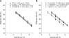
All mqPCR assays detected only the target pathogen and did not produce any non-specific amplification products or products of other pathogens in repeated experiments. In addition, there were no false positives due to cross-talk between dye signals within each reaction. Standard curves for all multiplex assays generated from 10-fold serial dilutions of each template showed correlation coefficients (R2) ranging from 0.970 to 0.995 and slopes of 3.41-3.59 (Fig. 1), indicating good linearity of PCR amplification.
Next, the performance of the mqPCR panel and singleplex PCR for each of the eight pathogens were compared directly to identify any negative effects of multiplexing on PCR detection. The analytical sensitivity (i.e., limit of detection) of the PCR was estimated using serially diluted recombinant vectors with known copy numbers (molecules per µL). The estimated mqPCR detection limits of the mqPCR panel were 3.8 for CDV, 3.7 for E. canis, 3.7 for Bartonella spp., 3.8 for B. burgdorferi, 3.7 for B. dermatitidis, 3.7 for C. neoformans, 38 for N. caninum, and 3.7 for T. gondii (Table 2). The results of singleplex and multiplex PCR were identical except for CDV, and CT differences between PCR reactions were not significant, demonstrating that multiplexing did not have a significant negative effect on sensitivity of the PCR.
The CT value obtained for E. canis using the Path-ID Multiplex One-Step RT-PCR kit (Applied Biosystems) was slightly lower than that obtained with the QuantiTect Multiplex PCR kit (Qiagen); however, the difference in CT was less than 1, suggesting that the kits had comparable performance.
CSF sample analysis
Of the 47 CSF samples obtained from dogs diagnosed with ME, seven (15%) were positive for the target pathogen. The most common pathogens were C. neoformans (3/7), B. dermatitidis (2/7), and B. burgdorferi (2/7). In contrast, conventional CSF analysis and culture was negative for all pathogens. None of the samples were positive for more than one pathogen. For the seven samples with discrepant results between the mqPCR panel and routine test methods, agarose gel electrophoresis, sequencing, and comparison with sequences available in the GenBank database confirmed the presence of the identified pathogen. Among the six CSF samples from healthy dogs, all were negative for the eight pathogens of the mqPCR panel, suggesting that mqPCR did not falsely detect normal CSF constituents as target materials.
Discussion
In the present study, a panel of four mqPCR assays (one qRT-PCR and three qPCRs) that can simultaneously detect eight pathogens (B. dermatitidis, C. neoformans, N. caninum, B. burgdorferi, Bartonella spp., T. gondii, E. canis, and CDV) was optimized and applied to CSF samples. The analytic sensitivity of the panel was as good as each singleplex assay, and there was no interference among primer/probe sets, suggesting that the panel could be substituted for the singleplex assay. The panel optimized in this study was also designed for use with one-step extraction and PCR procedures in a 48-well format. Accordingly, simultaneous testing of a large number of samples can be achieved at a lower cost, with less time and effort than routine diagnostic techniques, such as individual PCR, cultures, microscopic examination, and ELISA.
ME is a common inflammatory CNS disease in dogs [22]. Histopathology can be used to classify ME based on the tissue manifestations of the disease. However, histopathological diagnosis has little relevance for clinical outcome because treatments are not specific for the ME subtype. A previous study employed PCR to examine the presence of viral agents that cause human ME in a small number of canine ME cases and found that none were positive for human ME viruses [26]. In contrast, the present study focused on eight known neurologic pathogens that have caused outbreaks in Korea and confirmed their relatively high prevalence among dogs with ME (7/47). Therefore, it is possible that the prevalence of neurologic infections causing ME in dogs has been underestimated. Although the cases of ME that tested positive for pathogens were not confirmed by histopathology, one case of infection with Cryptococcus spp. exhibited dramatic clinical improvement after a two-month treatment with oral fluconazole. Moreover, after observation for one year, there was no relapse of neurological signs, suggesting the importance of early pathogen detection for the selection of an effective treatment strategy. In the present study, five of the seven cases of infection were with a fungal agent that could not be eradicated by conventional antibiotics or anti-inflammatory agents, demonstrating the importance of proper diagnosis.
Although uncommon, fungal infections in dogs pose a diagnostic and therapeutic challenge [19]. Symptoms of fungal infections vary depending on the location and distribution of the lesion, making it difficult to distinguish them from non-infectious inflammatory or neoplastic disorders based on clinical manifestations [19]. For example, Cryptococcus spp., the most common fungal pathogen in this study, tends to infect the cerebellum, resulting in non-specific neurological symptoms, such as seizures and obtundation [219]. B. dermatitidis, which is endemic to North America, is seldom associated with CNS infection [1] and usually presents with lymphadenopathy or pulmonary infiltration [17]. B. dermatitidis infection is rare among humans in Korea [24]; however, it was found to be the second most common pathogen in the present study. Interestingly, these cases did not exhibit typical clinical signs, indicating that appropriate laboratory tests are critical for achieving a definitive diagnosis. This is the first report to implicate B. dermatitidis in canine ME in Korea.
Lyme disease is a zoonotic tick-borne disease caused by B. burgdorferi that affects various organs in dogs, including the skin, lymph nodes, aorta, and kidney [49]. Although the bacterium also infects the meninges and choroid plexus, CNS involvement is not pathognomonic of Lyme disease [18]. In the present study, two cases positive for B. burgdorferi had a history of ixodid tick infestation. In both cases, the locations of CNS lesions included the choroid plexus, which can be associated with neurologic signs in dogs afflicted by neuroborreliosis. However, it was not possible to confirm whether the lesions were caused by B. burgdorferi because the owners declined additional tests and therapeutic trials.
In conclusion, an mqPCR-based test panel for eight canine neurologic pathogens was developed and applied to 47 CSF samples in this study. The panel with high analytic sensitivity confirmed that seven (15%) samples were positive to the target pathogen, suggesting the necessity for screening of infectious agents in dogs with ME. Because treatment strategies for different histopathologic subtypes of ME are not specific, initial screening for infectious agents using the developed panel followed by CT or MRI would be useful for selecting an appropriate treatment in a timely manner.

 XML Download